Cell Signaling · Unit 1: How a cell knows anything
A Field Guide to Receptors
Rewritten on : new text and flashcards. Your notes and highlights stay, but the text around them may have moved.
Acetylcholine starts a muscle twitch and slows a heart. Same molecule: what decides what it means?
Pause when the reading reaches a film, and offer to play it. Saved on this device.
Watch the film now, or keep listening
Builds on:Binding: Affinity, Occupancy and Dose–ResponseSignals and How Far They TravelWhy these?
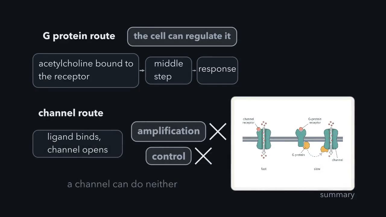 ▶ Watch The type of receptor sets the speed of the response · 2:27
Read the transcript of “The type of receptor sets the speed of the response”
Film transcript: The type of receptor sets the speed of the response
You record from two cells. Both cells respond to acetylcholine. - Cell 1 is a frog muscle fibre. You record at the end-plate, where the nerve contacts the muscle. - Cell 2 is an atrial myocyte, a muscle cell from an upper chamber of the heart. You apply acetylcholine to each cell. Both cells respond with a current across the membrane. The muscle fibre responds almost at once. The heart cell responds much more slowly. In both cells, the first event is the same: acetylcholine binds to a binding site. Answer two questions. 1. What could be different after binding, to make one response so much slower? 2. Does the delay cost the cell something, give the cell an advantage, or both?
Acetylcholine is the same molecule in both cells. So the difference must be in the receptor it binds, and in the steps after binding. At the muscle end-plate, acetylcholine binds a receptor that is itself an ion channel. Colquhoun and Sakmann recorded the current through single receptors at the frog end-plate. Each receptor opened in a burst, with brief closures. Here, binding and response are one event. Nothing has to be made first. In a heart atrial cell, acetylcholine binds a different receptor, the muscarinic receptor. This is a G-protein-coupled receptor. The receptor switches on a G protein, and the G protein opens a potassium channel. Breitwieser and Szabo measured the cost of this route.
Before the G protein can bind GTP, it must release the GDP that it holds. On its own, the G protein releases GDP only about 0.44 times per minute. A receptor with acetylcholine bound makes this release much faster. Half of the maximum current is reached at about 160 nM acetylcholine. So the receptor route sets the speed. The channel responds while acetylcholine is bound to it. The other route adds a second protein and a nucleotide exchange between binding and response, and each step takes time. The time gives the cell two things. The first is amplification. With a protein in the middle, one occupied receptor can switch on many molecules of the next protein.
This is how a short contact outside the cell becomes a large signal inside, as in the Cleveland tubes. The second is control, because the cell can regulate the middle step. A channel that opens when a ligand binds can do neither.
Acetylcholine makes a skeletal muscle fibre twitch and slows a heart. It is the same molecule in both cells, so what is different?
The difference is the receptor, the protein that binds a signalling molecule (the ligand) and passes the news on. Receptors come in a few designs, and the design does much to set what the cell does, how fast it answers and how long the answer lasts.
Three questions and one idea
Three questions run through the lesson: how information crosses a membrane the ligand cannot cross, why one ligand excites one cell and inhibits another, and why responses take from under a millisecond to hours.
The one idea to carry through this lesson. Wiring sets what; steps set when. What a receptor is wired to (a pore, a G protein, a kinase, a gene) decides the effector, the protein that makes the output, and so what the signal does: which ion moves, which enzyme runs. How long the steps between binding and that output take, above all the slowest, sets when it starts; more steps usually mean more waiting, but one slow step can outweigh several fast ones. The steps that keep running after the ligand leaves set how long it lasts. Each step costs time, and a step that is an enzyme also multiplies the signal. Acetylcholine wired to a cation pore excites muscle within a millisecond; wired through a G protein to a potassium channel, it starts opening that channel tens of milliseconds later and slows the heart within a beat or two.
The membrane problem: three ways across, and one way around
Most ligands, such as neurotransmitters, peptide hormones and growth factors, are water-soluble, so the double sheet of fat that is the plasma membrane keeps them outside. A receptor has to change what happens inside without letting the ligand through. There are three main ways, plus one family whose ligand comes inside: four architectures.
- Open a pore. The receptor is an ion channel. The ligand binds, the pore opens, and ions flow.
- Switch on a G protein. One chain crosses the membrane seven times. Binding on the outside rearranges the bundle, and the new shape makes the receptor an exchange factor: it pries GDP out of a G protein, and because the cell holds far more GTP than GDP, the empty site fills with GTP. A G protein is a switch protein, off while it holds GDP and on while it holds GTP.
- Bring single-pass chains together. Each chain crosses the membrane once. The ligand pulls two or more chains into contact, or changes how the chains of an existing pair sit, so the enzyme parts inside meet or are recruited.
The fourth way goes around the problem. Steroid hormones such as estradiol dissolve in fat and slip through the membrane, so their receptors wait inside the cell. A few receptors use still other routes.
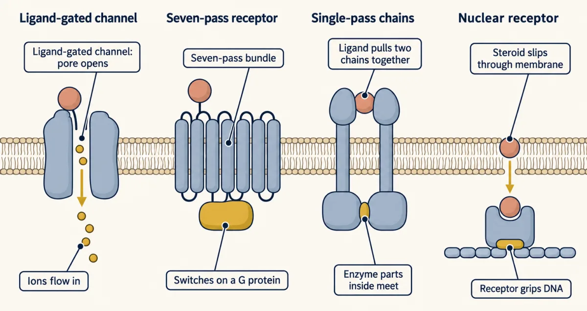
The map: four architectures
| Architecture | Where the ligand binds | What binding does | Examples | First output, and how soon |
|---|---|---|---|---|
| Ligand-gated ion channel | Outside, on a multi-subunit pore | Opens the pore | Nicotinic acetylcholine receptor; AMPA and NMDA glutamate receptors | Current begins in about a millisecond or less for the fastest; NMDA receptor current rises over about 10 ms |
| Seven-pass (G-protein-coupled) | Outside, in or near the helix bundle | Changes the bundle’s shape, so the receptor switches on a G protein | Muscarinic acetylcholine receptor, beta-2 adrenergic receptor, metabotropic glutamate receptors (mGluRs) | An effector current or second messenger: tens of milliseconds to seconds |
| Single-pass, enzyme or enzyme-recruiting | Outside, on the outer domain | Brings two or more chains together, or rearranges a pair, so kinase domains, or the kinases and adaptors they carry or recruit, act | EGF receptor, TGF-beta receptors, cytokine receptors, Fas | Receptor or associated kinases activated and partners recruited: seconds to minutes; changes in gene expression: tens of minutes to hours |
| Nuclear | Inside the cell | Changes the receptor’s shape, so at its DNA sites it recruits coactivators, proteins that help switch a gene on | Estrogen receptor | New RNA within minutes; new protein: tens of minutes to hours |
Ligand-gated channels. The muscle acetylcholine receptor, the nicotinic type, is five subunits arranged around a central pore. The ions that move are the signal.
Seven-pass receptors. These are the G-protein-coupled receptors, or GPCRs (GPCRs and trimeric G proteins). The effector the G protein reaches may be a channel, or an enzyme that makes a second messenger, a small molecule such as cAMP that carries the signal on inside the cell.
Single-pass receptors. The part outside the cell is the ectodomain. Receptor tyrosine kinases carry a kinase, an enzyme that attaches phosphate to other proteins, in the part inside the cell. Receptor serine/threonine kinases, such as the TGF-beta receptors, add phosphate to serine and threonine instead, and one receptor type phosphorylates the other. Cytokine receptors have no kinase of their own: a Janus kinase (JAK) kinase stays bound to the inner part of each chain, and the ligand brings two of these kinases together. Death receptors, such as Fas, recruit adaptor proteins that switch on enzymes that take the cell apart. For the kinase-linked receptors the core move fits in one sentence: the ligand brings kinase domains together, or rearranges a pair that already exists, and one switches on the other or each switches on its partner (receptor tyrosine kinases).
Nuclear receptors. A typical nuclear receptor is one chain with two working parts: a pocket that closes around the hormone and a region that grips a specific stretch of DNA. There is no second messenger and no kinase cascade, because the protein that received the signal acts directly on the gene that answers. Their ligands include estradiol, thyroid hormone and retinoic acid. Once estradiol is bound, the estrogen receptor holds its DNA and recruits the machinery that reads the gene; thyroid hormone and retinoic acid receptors work differently (see Going deeper).
One ligand, two receptors: acetylcholine
An ionotropic receptor is itself an ion channel; a metabotropic receptor acts through a G protein and other steps inside the cell. Acetylcholine uses both, and so does glutamate, the brain’s main excitatory transmitter (see the table).
The wiring. At the muscle end-plate, where the motor nerve meets the fibre, acetylcholine binds the nicotinic receptor. Binding opens the receptor’s own pore, and nothing has to be made first. In the heart, acetylcholine binds the muscarinic receptor (the M2 type), a G-protein-coupled receptor (GPCR), which switches on a G protein. The activated G protein rearranges or dissociates, making its βγ pair available to bind a potassium channel called GIRK (G-protein-gated, inwardly rectifying K⁺ channel) and open it. The single-cell recordings come mostly from atrial muscle cells; the same route in pacemaker cells slows the heart rate.
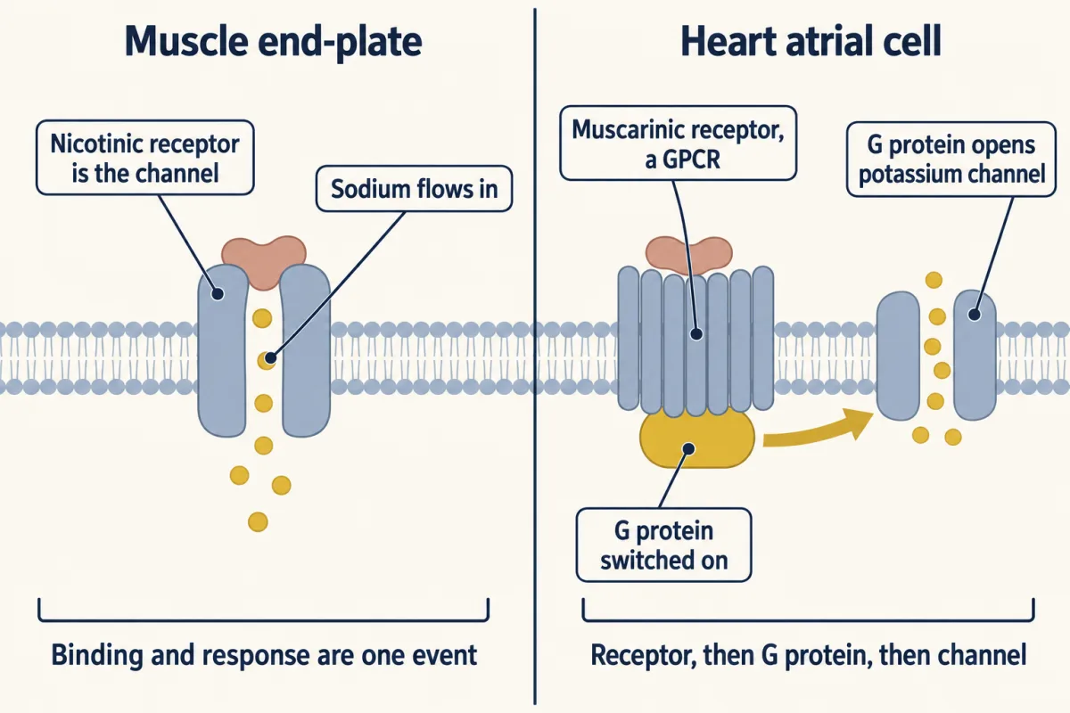
Predict. The nicotinic pore lets sodium and potassium through about equally. The muscarinic route opens a channel selective for potassium. An open channel pulls the membrane voltage toward its reversal potential, the voltage at which the net current through it is zero. Muscle rests near −90 mV; between beats, a pacemaker cell climbs from about −60 mV toward a threshold near −40 mV; the potassium equilibrium potential is close to −90 mV. Which way does each route move the voltage, relative to threshold, the voltage at which the cell fires?
Check your prediction
In opposite directions. The nicotinic channel’s current reverses near 0 mV: sodium flows in and potassium flows out, with a little calcium in. From −90 mV the net current is inward, carried mostly by sodium, and the fibre depolarizes toward a reversal potential well above threshold. The potassium channel’s current reverses at the potassium equilibrium potential, below the pacemaker cell’s voltage. Potassium flows out, the membrane is pulled down, and the cell takes longer to climb to threshold, so the next beat comes later. (This G protein, Gi, also lowers cAMP, which adds to the slowing.)
So the sign comes from the wiring, which ions the effector passes, read against their gradients in that cell: a current that reverses above threshold excites, and one that reverses below the cell’s voltage inhibits. The same ion can do either. A GABA-gated chloride channel inhibits most adult neurons but depolarizes immature ones, which hold more chloride inside. Now the timing.
Predict. Give each cell a brief pulse of acetylcholine. Which current starts sooner, and which outlasts the pulse?
Check your prediction
The end-plate current starts sooner and ends sooner:
| End-plate current (nicotinic) | Atrial current (muscarinic, through GIRK) | |
|---|---|---|
| Direction | Inward, drawn downward | Outward, drawn upward |
| Onset after acetylcholine arrives | Well under a millisecond | A delay of tens of milliseconds |
| Rise | Well under a millisecond | Over hundreds of milliseconds |
| After acetylcholine leaves | Decays in about a millisecond | Decays over seconds |
Timing alone suggests a G protein in the heart, but it does not prove one. The proof uses a non-hydrolysable GTP analogue, a GTP look-alike that a G protein can bind but cannot split, so a G protein that takes it up stays on.
Predict. Put the analogue inside both cells, give acetylcholine, then wash the acetylcholine away. What happens to each current?
Check your prediction
The atrial current persists for minutes after the acetylcholine is gone: G proteins switched on with the analogue cannot switch off. The end-plate current is unchanged: its pore has no nucleotide site and no G protein in its path. A tool that acts on a middle step changes only the route that has it.
Add up the waits
The film at the top says the receptor type sets the speed; it does so by setting the fewest steps a route can take. Each step has to finish before the next can start, so each one adds a wait.
time to the output ≈ the sum of the waits at every step, from binding to effector
Each wait is an average for one molecule; a whole cell can answer sooner, as the worked problem shows.
The channel: two steps. Acetylcholine must bind both sites, and the pore must open. In a cleft holding a few tenths of a millimolar acetylcholine, binding takes some tens of microseconds, and the muscle receptor opens within a few tens of microseconds after that. The total is well under a millisecond. Rates like these come from Colquhoun and Sakmann’s recordings of single receptors at the frog end-plate, where each receptor opened in a burst with brief closures.
The muscarinic route: four steps. Binding, exchange, G-protein rearrangement or dissociation, and βγ opening GIRK. No second messenger is made, which makes this one of the fastest G-protein routes, yet with a nucleotide exchange in the middle it cannot respond in under a millisecond. The exchange is the slow step. Breitwieser and Szabo estimated, from recordings in heart atrial cells, that unaided a G protein releases GDP only about 0.44 times per minute: a mean wait of 1/0.44 ≈ 2.3 minutes, about 136 seconds. A receptor with acetylcholine bound makes release much faster.
Worked problem. Suppose the agonist-bound receptor speeds GDP release 1,000-fold (an illustrative round number).
- New rate: 0.44 × 1,000 = 440 per minute, about 7.3 per second.
- Mean wait for one G protein: 1/7.3 ≈ 0.14 s, about 140 ms.
- But a cell has many G proteins, and the current starts once some are on. With a constant rate k, the fraction switched on by time t is 1 − e−kt. Setting that to 10% gives t = −ln(0.9)/k ≈ 0.105/7.3 s ≈ 0.014 s. A tenth of the G proteins are on within about 14 ms. (This treats current as rising with the fraction of G proteins on; in reality each GIRK channel needs several βγ pairs, which delays the current a little more.)
So the mean wait sets the pace, and the first small fraction sets the onset. Of the steps behind the atrial current’s real onset, tens of milliseconds or more, the exchange step is the slowest and a main contributor to that delay. At the 100-fold of the MCAT practice item, a tenth of the G proteins take about 140 ms, which is why the real acceleration must be large.
Predict. The N-methyl-D-aspartate (NMDA) receptor is a channel with no relay step, yet its current rises over about 10 ms, far slower than the muscle channel. Does that break the rule?
Check your prediction
No. The NMDA receptor’s own gating is slow, and a slow step counts whichever protein it happens in. The waits at each step, slow gating included, set the clock.
The slow routes. A single-pass kinase receptor adds pairing or rearrangement, phosphorylation and recruitment, hence seconds to minutes. A nuclear receptor skips every relay step, but its output is new RNA and then new protein.
What the steps buy
The extra time buys three things.
- Gain. A catalytic step multiplies the signal: one occupied receptor can switch on many G proteins, and one active adenylyl cyclase can make many messenger molecules. A directly gated channel has no catalytic relay: one receptor opens one pore, although that pore can pass thousands of ions.
- A control point. The cell can turn the signal up or down at every middle step. A directly gated channel has no middle relay point, though the cell can still tune the receptor itself, for example by phosphorylation or desensitization.
- Duration. A response lasts until its slowest off step finishes. The nicotinic response lasts only as long as the transmitter stays, and acetylcholinesterase keeps that stay short. Not every channel is this brief: an NMDA receptor holds on to glutamate and keeps reopening for hundreds of milliseconds. A G-protein route lasts until the G protein hydrolyses its GTP, which on its own takes seconds to tens of seconds, and any messengers it made are destroyed. RGS proteins (regulators of G-protein signalling) speed the hydrolysis, so they help set how fast the atrial current, and with it the heart rate, recovers when the vagus nerve goes quiet (turning the signal off).
The film at the top names two of these buys, amplification and control. Its closing “neither” means neither a catalytic relay nor a middle relay point; directly gated channels still provide ion-flux gain and can be regulated at the channel itself.
Predict. A drug blocks RGS proteins in atrial cells. Which changes more: how soon the acetylcholine current starts, or how long it lasts after acetylcholine is removed?
Check your prediction
How long it lasts. RGS proteins act on the off step, GTP hydrolysis, so blocking them leaves G proteins on longer and slows recovery. Onset depends mainly on the on steps, binding and exchange, so it changes less, although in heart cells RGS proteins also speed the rise somewhat, so blocking them can slow onset a little too.
Design problem
Three jobs. For each, choose a receptor architecture and name the sign, onset and offset of the response it gives.
- Relay every motor-nerve impulse to a muscle fibre, at up to about 50 impulses a second, one muscle action potential per impulse.
- Slow the heart’s pacemaker within a beat or two, by an amount that can be tuned, and let it recover over seconds.
- Commit a stem cell to a new identity that outlasts the signal.
Saved in this browser only.
One way to do it
Job 1: a ligand-gated cation channel, the nicotinic receptor. Sign: excitatory, because its current reverses near 0 mV, well above threshold. Onset: well under a millisecond. Offset: about a millisecond after acetylcholine leaves. At 50 impulses a second the impulses are 20 ms apart, so each response is over before the next arrives. With no enzymatic gain, the nerve must release acetylcholine in quantity, and the gap must be cleared fast.
Job 2: the M2 receptor, a G protein and GIRK. Sign: inhibitory, because outward potassium current pulls the cell toward the potassium equilibrium potential. Onset: tens of milliseconds, well inside a beat of about a second. Offset: seconds, set by GTP hydrolysis, which RGS proteins tune. The middle step is the dial: how many G proteins are on sets how much the rate slows.
Job 3: a nuclear receptor, such as the retinoic acid receptor. Hours are fine; duration matters. A brief pulse of ions leaves a lasting trace only if it sets off other signals, whereas a receptor on the genes starts a program that the new regulatory proteins it makes can keep going after the hormone is gone. A fat-soluble signal such as retinoic acid reaches its receptor directly. A single-pass kinase receptor feeding a gene program would also work and is common in fate decisions, but it adds relay steps between ligand and gene.
Back to the opening question
The two cells wire the same binding event to different outputs. In muscle it opens a cation pore: fast, brief, excitatory. In the heart a G protein opens a potassium channel: tens of milliseconds later, lasting seconds, inhibitory. The molecule does not dictate a single response; the receptor and the cell’s downstream wiring supply it.
Where the idea stops
- Each step costs time, unless gain makes up for it. Adding a step usually delays the start, yet gain can buy some speed back. In an ideal unsaturated two-stage enzyme cascade driven by a step input, before depletion or shutoff matters, early output is proportional to gain × time². The time to reach a fixed threshold therefore falls as one over the square root of the gain: 100 times more gain crosses it 10 times sooner. Rod phototransduction is a high-gain G-protein-coupled receptor (GPCR) cascade whose dim-flash response in mammalian rods peaks within about 100 to 200 ms.
- Binding time depends on concentration, and potency, how much ligand a route needs, is a separate question from speed. In atrial cells the acetylcholine current is half-maximal near 160 nM, the number the film quotes; that is a potency, not a speed (binding and dose-response).
- The rule stops at the first output in one cell. It does not cover desensitization, crosstalk between branches, or what the cell does with the output.
- Steroids are not only slow. Estradiol also has reported rapid actions at the cell surface, which the classical nuclear route does not explain.
Going deeper
How the map was drawn: sequences first
Until the 1980s a receptor was known by what it bound and did; a few had been purified, but most sequences were out of reach. In 1982 Shosaku Numa’s group in Kyoto read the sequence of one acetylcholine receptor subunit from the messenger RNA of the electric organ of the ray Torpedo, a battery of modified muscle cells packed with receptor. It was the first complete sequence for a neurotransmitter receptor. Two years later they read a voltage-gated sodium channel from the electric eel, and receptors and channels could be compared.
The next sequences showed families. In 1982 Ora Rosen’s laboratory showed that insulin switches on a tyrosine kinase that travels with its receptor and phosphorylates tyrosines on the receptor itself. Axel Ullrich’s group cloned the epidermal growth factor (EGF) receptor in 1984, and in 1985 his group and William Rutter’s each reported the insulin receptor. The EGF receptor resembled erbB, a viral cancer gene, and the insulin receptor’s enzyme part belonged to the same tyrosine kinase family.
By 1958 Elwood Jensen had seen that a rat’s uterus and vagina keep radioactive estradiol, unchanged, while other tissues clear it, so something there seemed to catch the hormone. David Toft and Jack Gorski extracted that protein in 1966. In March 1986 Pierre Chambon’s laboratory in Strasbourg and Geoffrey Greene’s in Chicago reported its sequence within weeks of each other. Both noticed that part of it resembled v-erbA, a viral gene later shown to be derived from a thyroid hormone receptor. The estrogen receptor had relatives.
Counting receptors
The human genome has about 800 G-protein-coupled receptors (GPCRs), and roughly half detect smells. Robert Lefkowitz and Brian Kobilka shared the 2012 Nobel Prize in Chemistry for working out how these receptors are built and how they work, and a later lesson is theirs. About 1,300 human proteins are estimated to cross the membrane once, many of them receptors; estimates vary with the prediction method and the definition. Humans have 58 receptor tyrosine kinases and 48 nuclear receptors.
How a ligand brings kinase domains together
Families differ in how the ligand puts two inner kinase parts side by side. Many growth factors are dimeric, built from two halves, and many of their receptors were long thought to sit in the membrane as single chains. A dimeric ligand such as nerve growth factor (NGF) can hold two receptor chains at once. A single-chain ligand such as EGF binds one receptor and changes the shape of its ectodomain, exposing a surface that grips a second receptor. Either way, two kinase domains end up side by side, and in many receptor tyrosine kinases they then phosphorylate each other. In the EGF receptor, one kinase domain first switches on its partner by contact, much as a cyclin switches on its kinase, and the receptor tails are phosphorylated after that. The insulin receptor starts out as a pair, and insulin changes how its two halves sit against each other, which has the same effect. How much receptor tyrosine kinase activation comes from ligand-induced pairing and how much from a shape change within a pair that already exists is still open, and the answer may differ between family members.
Other families differ in the details. Several cytokine receptors, such as the erythropoietin (EPO) and growth-hormone receptors, were long described as pre-formed pairs that the ligand reorients; single-molecule work suggests that at normal receptor levels they may instead be single chains that the ligand brings together, and the question is still debated. Death receptors such as Fas and the tumor necrosis factor (TNF) receptor are engaged by trimeric ligands and work as trimers and larger clusters. In the TGF-beta receptors, the ligand assembles type II and type I receptors, and the type II kinase, active on its own, phosphorylates the type I receptor, not each on each (TGF-β and Smads).
Other routes across, and nuclear receptors before the hormone
Some receptors use none of the three moves across the membrane. Notch is cut by enzymes when its ligand binds, and the freed inner piece travels to the nucleus itself, so the receptor becomes its own messenger (Notch).
Nuclear receptors do not all wait for the hormone in the same place. The glucocorticoid receptor waits in the cytoplasm, held by chaperones, until hormone binds. Thyroid hormone and retinoic acid receptors sit on DNA even without their ligand, bound to corepressors, and the hormone swaps those for coactivators. Thyroid hormone also needs transporters, such as MCT8, to cross the membrane. Nuclear receptors covers the family.
Your notes
Select any text in the lesson to highlight it or add a note to it. Your highlights and notes are saved in this browser and collected in your notebook.
This browser is not letting the site save anything (a private window or strict privacy settings), so highlights and notes will be lost when you leave the page.
How we measure it
Radiolabelled ligand tracing
You attach a radioactive atom such as tritium to a hormone, inject it into an animal, and count the label in each tissue. A tissue that holds the label while others clear it contains something that binds the hormone, and you can check whether what it holds is still the original molecule.
cDNA cloning from an enriched tissue
You copy the messenger RNA of a tissue into DNA, find the clone for your protein, and read its sequence. Choosing a tissue where the protein is abundant, such as a ray's electric organ, made the first receptor and channel sequences possible. The sequence reveals the protein's architecture and its relatives.
Single-channel recording
A glass pipette seals onto a small patch of membrane so that the current through one receptor can be measured. It shows each receptor opening and closing in real time, which is how the muscle acetylcholine receptor was seen to open in bursts.
Sequence comparison
You search a new sequence against known ones. A match between EGF receptor peptides and the viral protein erbB, and between the estrogen receptor and v-erbA, showed that receptors belong to families before anyone knew how each one worked.
Practice questions on this lesson (3) → MCAT-style, original, each with an explanation
Where this lesson fits
Before thisReview first
- Binding: Affinity, Occupancy and Dose–ResponseThis course · Lesson 3
The lesson's potency point (half-maximal near 160 nM) uses affinity and occupancy, which that lesson teaches.
- Signals and How Far They TravelThis course · Lesson 1
It sets out why a ligand's signal must be received at the cell surface and passed on, which this lesson maps by receptor type.
After thisStudy next
- GPCRs and Trimeric G ProteinsThis course · Lesson 12
It takes the seven-pass receptor, GDP-for-GTP exchange and the G-protein switch from this map and works through them in full.
- Receptor Tyrosine KinasesThis course · Lesson 18
It expands the single-pass kinase architecture, where the ligand brings two kinase domains together.
- Nuclear Receptors: Receptors That Are Transcription FactorsThis course · Lesson 31
It develops the fourth architecture, receptors that act directly on genes, including the thyroid and glucocorticoid cases.
Suggested by an AI model from the lessons themselves, then checked. How we use AI
Finish line
Test yourself
9 questions
Practice and MCAT-style, with explained answers.
Next · Lesson 5
Circuits: Amplification, Feedback, Adaptation and Switches
How can one photon change a rod's current without letting the signal spread or run forever?
Continue →