Cell Biology · Skim version
Stem Cells and Tissue Renewal
Read the full lesson → Skim the whole course →
The guiding idea
The lining of a mouse's small intestine is replaced every few days, yet it lasts for life. Stem cells do it by making new specialized cells while keeping up their own numbers. How would you prove which cells are the stem cells, and what keeps them that way?
A stem cell is defined by two jobs tested over time, making a tissue's specialized cells and keeping up its own numbers (self-renewal), not by how it looks or which marker it carries.
From the film: How a Stem-Cell Pool Persists While Its Clones Come and Go
- In the mouse gut, what descendants do over time—not a marker alone—identifies a stem-cell population.
- A permanent lineage mark follows descendants even after they stop expressing Lgr5.
- In the normally renewing mouse small intestine, Lgr5-high stem cells appear to compete for limited crypt-base positions.
- The stem-cell pool can stay balanced while most clones vanish and one may take over.
From the film: What Keeps a Cell a Stem Cell?
- A stem-cell niche is the local environment whose contact and signals bias whether a daughter stays a stem cell.
- In the mouse gut, Wnt is strongest at the crypt base and promotes self-renewal; BMP is strongest higher up and suppresses it.
- Cells that move up, away from the base signals, become more likely to leave the stem-cell state.
- Single Lgr5 cells grew into organoids when a matrix and added factors stood in for the niche.
- Injury asks whether cells can regain a tissue stem-cell state; reprogramming asks how far resetting can go.
-
What the lineage trace proves—and what it does not
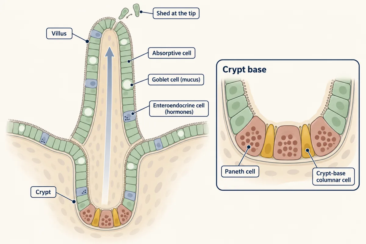
Cells are born in the crypt, move up the villus as they differentiate, and are shed at the tip. At the crypt base sit long-lived Paneth cells, which move down rather than up, with slender crypt-base columnar cells squeezed between them. AI-generated illustration -
Put neutral drift to work
-
The niche: what limits a stem-cell position
-
After injury, other cells can refill the stem pool
-
Resetting beyond one tissue: Gurdon to Yamanaka
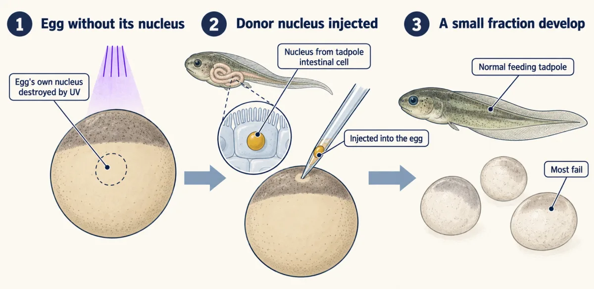
In Gurdon's Xenopus experiment, a nucleus from a feeding tadpole's intestinal cell, put into an egg whose own nucleus was destroyed by ultraviolet light, sometimes directed a normal tadpole: most eggs failed, but a small fraction became normal feeding tadpoles. AI-generated illustration -
Going deeper
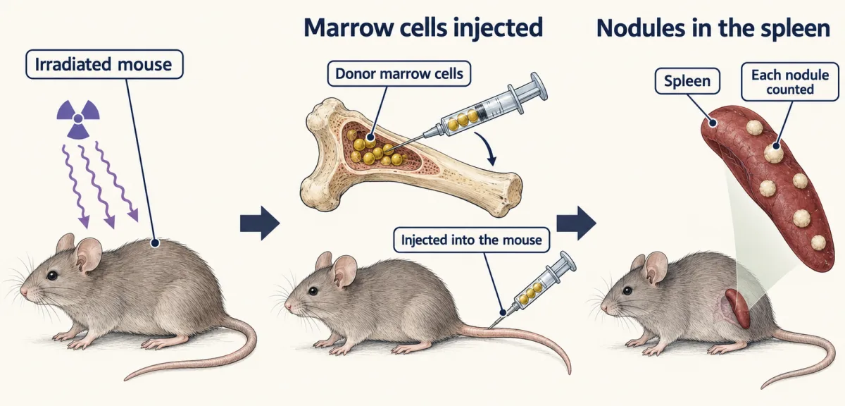
In Till and McCulloch's spleen colony assay, nodule counts in an irradiated mouse rose in proportion to the marrow cells injected, which made stem cells countable. AI-generated illustration
Where this lesson fits
Before thisReview first
- Epithelia and Tissue OrganizationThis course · Lesson 23
The gut lining's villi, crypts and cell types are described there, and this lesson assumes that layout.
- Cyclin-CDK Control of the Cell CycleThis course · Lesson 17
Self-renewal and transit-amplifying divisions rest on how cells decide to divide or stop.
- Cell Death and ClearanceThis course · Lesson 21
Shedding and clearance of cells at villus tips is part of why the lining needs constant renewal.
After thisStudy next
- Cell Identity, Cell State and DiseaseThis course · Lesson 25
It uses stem-cell renewal and cell state to ask why mutated cells in healthy tissue stay or spread.
- Designing a Cell Biology ExperimentThis course · Lesson 26
It practices the same skill of designing tests that show requirement or sufficiency, as in the niche problem.
Suggested by an AI model from the lessons themselves, then checked. How we use AI