Developmental Neuroscience · Skim version
Activity Before Experience: Spontaneous Retinal Waves
Read the full lesson → Skim the whole course →
The guiding idea
Weeks before vision begins, the retina already fires in waves. What are they for?
Timing is the address: a target cell keeps the inputs that fire in step with its other strong inputs, and because the inputs compete, the ones it keeps crowd out the rest.
From the film: Timing Is the Address
- Timing is the address: a target cell keeps the inputs that fire in step with its other strong inputs, and they crowd out the rest.
- Near neighbours in one eye fire almost together in a wave, while the other eye's waves match only by chance.
- Hebb's rule alone only strengthens inputs, so separating the eyes also needs competition between inputs.
From the film: Block It, Tilt It
- Blocking spikes with tetrodotoxin in fetal cats kept the two eyes' projections mixed, so spikes are needed.
- In young ferrets, the more active eye's projection expanded and the other's shrank, so relative activity shifts territory.
- Making waves more frequent in both eyes left the layers normal, so territory follows the difference between the eyes, not the total.
From the film: Correlation, Not Just Spikes
- In beta2 knockout mice, plenty of uncorrelated spiking in the first week left the termination zones broad.
- Normally patterned activity, not just spiking, is needed early for the map to sharpen.
- In the frog tectum, a third eye makes stripes where none normally exist, and keeping them needs NMDA receptors.
-
The problem: a rough first draft
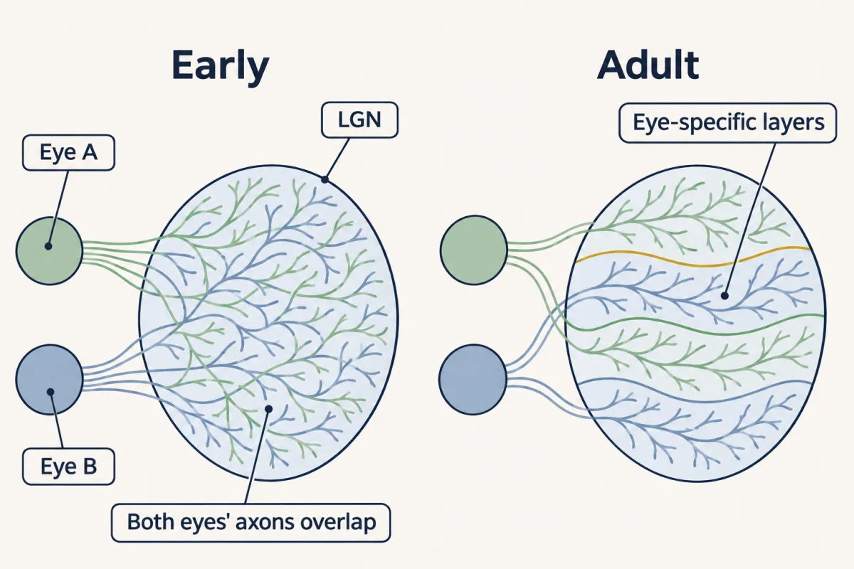
Early in development the axons from the two eyes overlap in the LGN; in the adult they end in separate eye-specific territories, which in cats, ferrets and primates are distinct layers. Getting from one to the other is refinement. The drawing is schematic. AI-generated illustration -
Why activity? Two neighbours look the same to a gradient
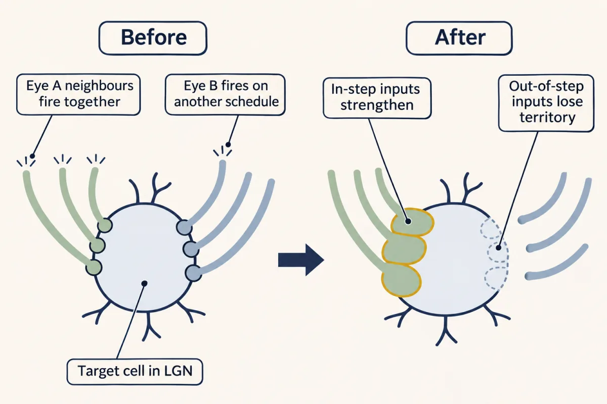
Timing is the address. Inputs from near neighbours in one eye fire together, and with competition the in-step inputs take over the cell while the out-of-step ones lose territory. Strengthening alone would leave the cell binocular. This is a sketch of the rule, and which eye wins a given cell depends on small early differences. AI-generated illustration -
What the waves look like
-
What makes a wave, briefly
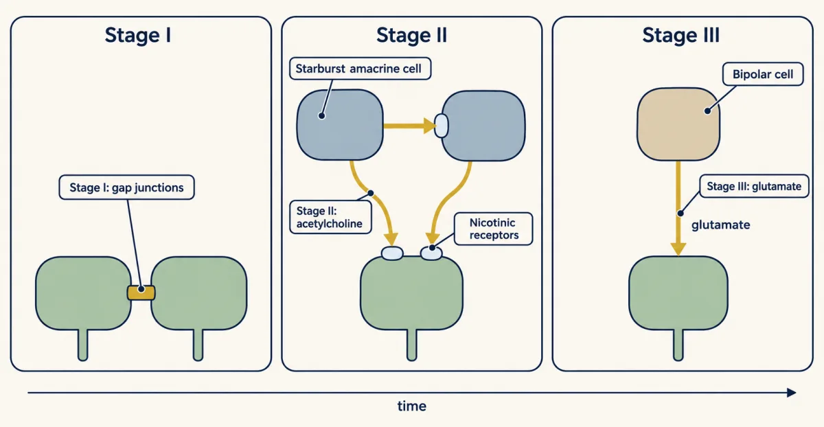
The dominant driver of retinal waves changes with development in the mouse: gap junctions (with nicotinic receptors) first, then acetylcholine from starburst amacrine cells, then glutamate from bipolar cells. The panels show selected contributions, not complete circuits; the stages overlap, embryonic waves are less well studied, and every stage gives correlated firing among neighbouring cells. AI-generated illustration -
Block it: silence everything
-
Tilt it: make the eyes unequal
-
Disrupt the correlation, keep plenty of spikes
-
Add an eye: the three-eyed frog
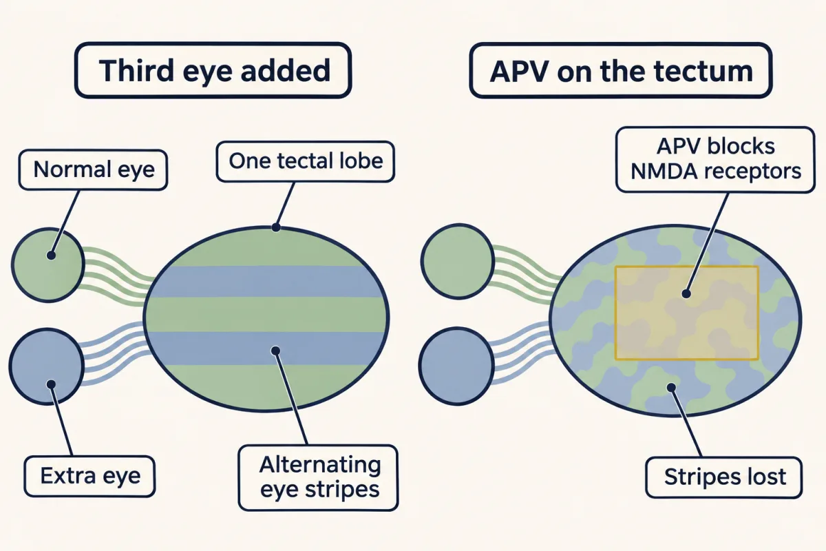
Two eyes sent into one frog tectal lobe form alternating stripes, where none normally exist. APV, which blocks NMDA receptors, desegregated the stripes in three-eyed tadpoles, and they returned after it was removed: in the frog tectum, keeping the stripes needs NMDA receptor activity. Whether the mammalian LGN uses the same detector is less clear. AI-generated illustration -
Putting it together
-
Going deeper
Where this lesson fits
Before thisReview first
- Topographic Maps: Sperry's Eye and Eph GradientsThis course · Lesson 26
Eph and ephrin gradients draw the rough first map that waves then refine, and this lesson builds directly on that.
- The Neurotrophic Hypothesis TestedThis course · Lesson 29
Inputs competing for a limited supply of survival factor is the same argument as competition for synaptic territory here.
- Competition: How Neurons Win a TargetThis course · Lesson 32
You need the idea that a small early lead becomes a win under competition, which the Hebb-plus-competition model reuses.
After thisStudy next
- Ocular Dominance and the Critical PeriodThis course · Lesson 39
It tests how relative activity between the two eyes shifts cortical territory, taking the competition idea into the critical period.
- Opening and Closing a Critical PeriodThis course · Lesson 40
It asks what opens and closes the window in which patterned activity can refine the wiring.
Elsewhere on the siteFilms, simulations and method films in other courses
- Depletion and the paired-pulse ratioSimulation · Neuroscience
Hebb's rule, strengthening a synapse when input and target fire together, is the core rule used here.
- A Synapse That Could LearnFilm · Neuroscience
It shows how Hebb's rule was tested through long-term potentiation, the synaptic change that correlation drives.
Suggested by an AI model from the lessons themselves, then checked. How we use AI