Developmental Neuroscience · Unit 3: Coordinates: patterning the tube
Local Organizers: The Midbrain-Hindbrain Boundary
Rewritten on : new text, figures, flashcards and film. Your notes and highlights stay, but the text around them may have moved.
How does one protein on a bead make a second midbrain grow, built back to front?
Pause when the reading reaches a film, and offer to play it. Saved on this device.
Watch the film now, or keep listening
Builds on:The Organizer: Spemann and MangoldHead to Tail: Hox Genes, Retinoic Acid and RhombomeresTools for Following Cells and GenesWhy these?
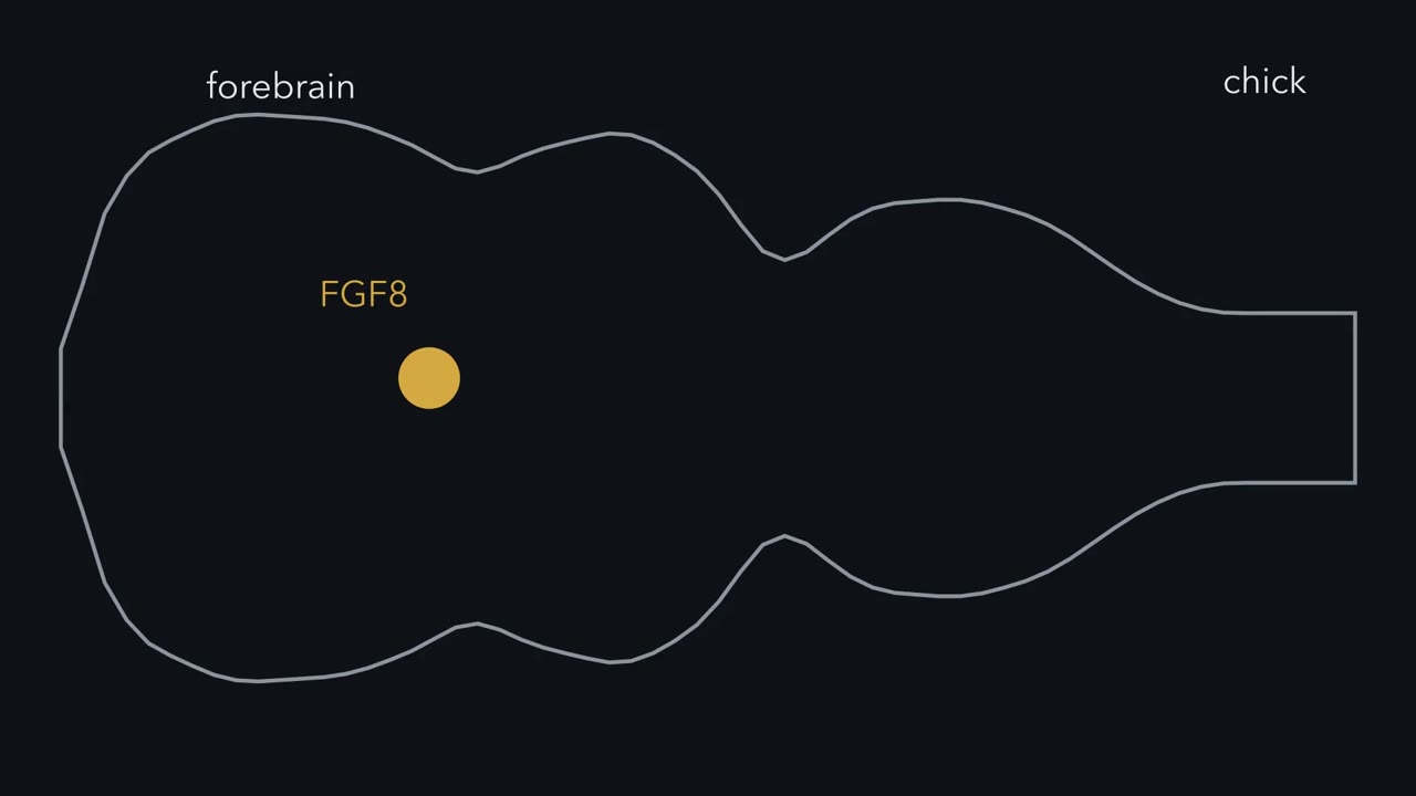 ▶ Watch Where the Line Falls · 2:48
Read the transcript of “Where the Line Falls”
Film transcript: Where the Line Falls
Push a small bead into the back of a chick embryo's forebrain. It is soaked in one protein, FGF8. A second midbrain can grow there, built back to front. Who tells cells what to build? Hox genes give the hindbrain and spinal cord their addresses. The midbrain and forebrain have no Hox code at all. Front at the left: forebrain, then midbrain, then a narrowing called the isthmus. Behind it, the roof of rhombomere 1 gives rise to the cerebellum. A local organizer is a small signaling center inside the tube. It tells nearby cells what to build. The best studied sits at the isthmus. It comes down to one idea. A line becomes a source. Where Otx2 meets Gbx2, the boundary cells become a signaling source.
FGF8 is the chief signal. On the simplest reading, how much signal a cell gets tells it which part to build. The repressor it carries sets which region it can become. In the roof of the tube, that means midbrain in front and cerebellum behind.
Start with the line. Otx2 is on in the forebrain and midbrain. Gbx2 is on in the front of the hindbrain. Each represses the other. A cell with a little more Otx2 makes less Gbx2. Its neighbour does the opposite. So a blurred edge can sharpen, and the line sits where the two meet. How sharp depends on how strong and switch-like the repression is. Predict. In a mouse, force Otx2 on in the front of the hindbrain. Where should the line go? What happens to the cerebellum?
The line moves back, with its band of FGF8. The cerebellum is smaller. Broccoli, Boncinelli and Wurst showed this in 1999. Millet and colleagues did the reverse. Gbx2 made in the back of the midbrain moved the line forward.
This is a move-it test. Change where the players are, and see if the pattern follows. The line follows the repressors. Right at the line, cells make secreted signals: FGF8 just behind it, Wnt1 just in front. This line is more than a fence. Takeaway: two repressors place the line. Next, is that line a source?
From the film
- A local organizer is a small signaling center that tells nearby cells what to build.
- A line becomes a source: where Otx2 meets Gbx2, boundary cells make signals such as FGF8 and Wnt1.
- On the simplest reading, the signal a cell gets tells it which part to build, and the repressor it carries sets which region it can become.
Push a small bead soaked in one protein, FGF8, into the back of a chick embryo’s forebrain, and a second midbrain can grow there, built back to front.
That result, from 1996, is the end of a story this lesson follows. By the time the neural tube has closed, its cells need to know where along the tube they sit, so that some build forebrain, some midbrain, some hindbrain. Who tells them? And why would one bead build a midbrain that faces the wrong way, with the parts that belong at its back, next to the hindbrain, sitting at its front?
Beyond the Hox border
In the lesson on Hox genes and the hindbrain you met one answer. Hox genes, switched on in a nested order along the tube, give each segment of the hindbrain and spinal cord an address. But Hox expression stops near the front of the hindbrain. The midbrain and forebrain have no Hox code at all.
First, the parts. Picture the young tube as a flat strip, front at the left.
| Region, front to back | What it is | What it gives rise to |
|---|---|---|
| Forebrain | the front of the tube; its back part, the caudal forebrain, is the diencephalon | the diencephalon gives, among other parts, the thalamus and prethalamus |
| Midbrain | the next region back; its roof is the tectum | on top the tectum (in mammals, the colliculi), below it the tegmentum |
| Isthmus | a narrowing of the tube where midbrain meets hindbrain | the site of the local organizer of this lesson; its own derivatives are small, and the cerebellum arises from the roof just behind |
| Hindbrain | a row of segments called rhombomeres, numbered from the front; the front of the hindbrain, including rhombomere 1, is the metencephalon | the roof of rhombomere 1 gives rise to the cerebellum |
Predict. The midbrain still needs an address. Suppose you had to design one for the front of the tube, using a secreted signal instead of a Hox code. Where would the signal come from, and what would a cell have to read to know which part to build?
Check your prediction
One design that works: put a source of signal inside the region you want to pattern, at a fixed place. The signal falls off with distance, so how much a cell receives tells it how far it sits from the source. A cell’s own transcription factors then decide what it makes of that signal, so the same signal can mean different things on two sides of the source. Embryos use centers of this kind, called local organizers.
An organizer, in embryology, is a group of cells that changes what its neighbours become. The first one found was Spemann’s organizer, the lip of the newt gastrula that can induce a whole second body axis, nervous system included (the lesson on the organizer). A local organizer (also called a secondary organizer) is smaller and later: a small signaling center inside the neural plate or tube that patterns the tissue around it. Think of it as a loudspeaker standing at a property line. It does not build the houses, but it tells the houses nearby what to build. The best studied sits at the isthmus.
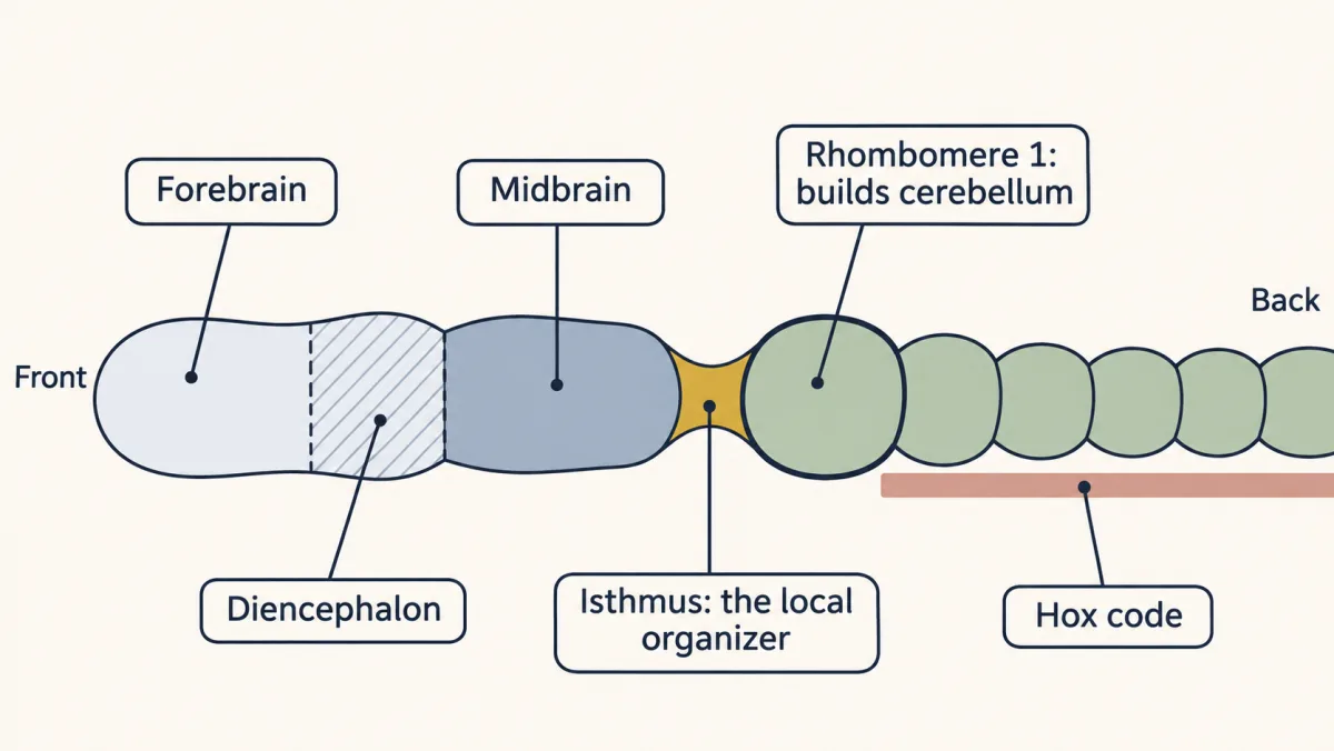
One idea carries the lesson. It uses three names met properly below: Otx2 and Gbx2, two transcription factors that repress each other, and FGF8, a secreted growth factor.
The one idea to carry through this lesson. A line becomes a source. Where Otx2 meets Gbx2, the boundary cells become a signaling source, with FGF8 as the chief signal. On the simplest reading, how much signal a neighbouring cell receives tells it which part to build, and the repressor it carries sets which region it can become: midbrain in front; behind, rhombomere 1, whose roof builds the cerebellum.
What the rule covers.
- Which tissue and when. The midbrain and rhombomere 1 while they are being patterned, before the parts take shape. Timed deletions of Fgf8 suggest that structures nearer the isthmus need the signal longer (Sato and Joyner, 2009).
- Which step. Where the line falls and which way the parts face. It does not cover how the isthmic genes first come on: they start in a broad region, in the mouse even in embryos lacking both Otx2 and Gbx2 (Li and Joyner, 2001), and the two repressors then refine them into bands at the line. Nor does it cover the border at the line that cells seldom cross, or the exact wiring that keeps the source on.
- “Distance” is shorthand for how much signal a cell receives, in level and in duration. And Otx2 and Gbx2 set what tissue can become, not everything it becomes, nor are they fixed: FGF8 feeds back on them (see the bead section).
- Other organizers follow related logic with other signals (see the end of the lesson).
Where the line falls
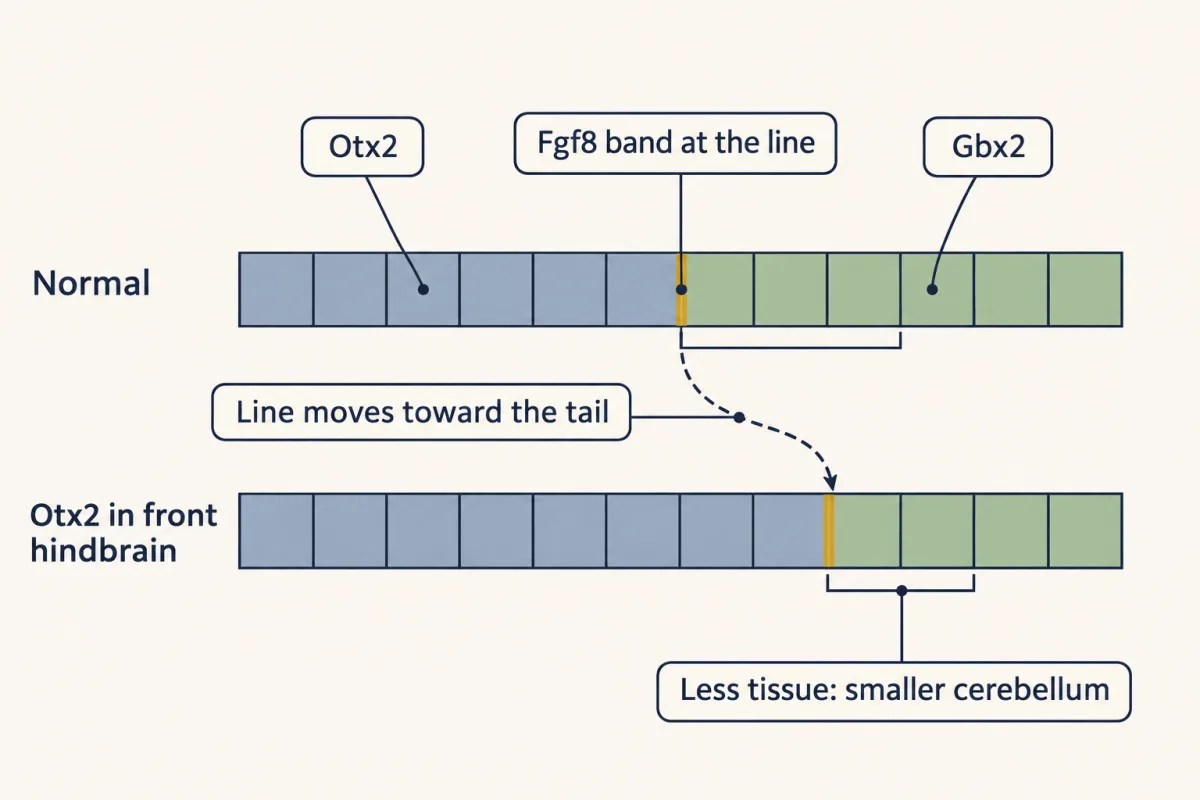
Start with the address system itself. Two transcription factors divide the front of the neural tube between them. Otx2 is on in the forebrain and midbrain. Gbx2 is on in the front of the hindbrain, including rhombomere 1. They do not do everything at the boundary, but they position it: in the neural plate, the isthmus normally forms where their domains meet.
The two are not just neighbours. Each represses the other. A cell that starts with a little more Otx2 makes less Gbx2, which lets it make still more Otx2, and its neighbour across the line does the opposite. So cells near the line come down firmly on one side or the other, and an edge that began blurred, with the two domains briefly overlapping, becomes sharp. (How sharp depends on how strong and how switch-like the repression is; see Going deeper.)
Predict. In a mouse, force Otx2 on in the front of the hindbrain, the territory that would normally carry Gbx2 and build the cerebellum. Where should the line go? What should happen to the cerebellum?
Check your prediction
The line should move back with the edge of Otx2, and the cerebellum should shrink, because less tissue is left behind the line. In 1999 Broccoli, Boncinelli and Wurst did this by putting the Otx2 coding sequence into the En1 gene, so that Otx2 was also made in territory that would become anterior hindbrain. The boundary, and the band of Fgf8 that marks it, moved toward the tail, and the cerebellum was smaller. In a paper published alongside it, Millet and colleagues in Alexandra Joyner’s laboratory did the reverse. They made Gbx2 in the back of the midbrain, and the boundary moved forward. The line follows the edge of each repressor.
This is a move-it test: keep the players, change where they are, and ask whether the pattern follows. You will meet the same logic, under other names, in the lesson on the neurotrophic hypothesis, where changing where nerve growth factor (NGF) is made tells permissive from instructive.
The line becomes a source
A boundary that only separated two regions would be a fence. This one does more. Right at the line, cells switch on genes for secreted signals:
- Fgf8, a growth factor made in a band just behind the line, in rhombomere 1.
- Wnt1, a signal made in a ring just in front of it, in the back of the midbrain.
Across the line, a broader band of cells makes the transcription factors Engrailed-1 and -2 and Pax2 and Pax5. Engrailed is a homeodomain transcription factor, a cousin of the Hox proteins whose gene sits outside the Hox clusters. It is needed for the midbrain and cerebellum to form. It is graded across the midbrain, highest next to the isthmus, and that gradient later gives the tectum its polarity, a topic for the lesson on topographic maps. The gradient fits the idea that the tissue around the line reads something that comes from it, though by itself it does not show that.
So here is the claim: the line is a source, and its neighbours take their identity from it. Two more kinds of experiment test it. A factor is sufficient if adding it where it is normally absent produces the structure. It is necessary if removing it takes the structure away. You need both: add the source somewhere new, and take it away.
Add it: a graft, then a bead
Read the transcript of “One Bead Builds a Midbrain”
Film transcript: One Bead Builds a Midbrain
Is the line really a source? Add it somewhere new, and watch what the neighbours do. A factor is sufficient if adding it where it is normally absent produces the structure. First, tissue. Alvarado-Mallart's group often grafted quail brain into chick embryos, since quail cells look different under the microscope. In 1991, Martínez, Wassef and Alvarado-Mallart moved tissue from just behind the junction into the forebrain. Forebrain cells next to the graft turned on Engrailed. Later they developed as midbrain. The junction could instruct its neighbours.
What was the message? In 1996, Crossley, Martínez and Gail Martin soaked beads in FGF8. They placed them in the diencephalon, the back of the forebrain. An extra midbrain formed. Predict. The real midbrain still has its own isthmus behind it. Which way will the new midbrain face?
It was built in reverse, a mirror image. Parts that normally lie nearest the isthmus formed nearest the bead. One source is enough to reverse the order. Here is a toy model, with made-up numbers, with a second source behind it: the bead and the real isthmus send out the same signal. A cell cannot tell which source a molecule came from. It gets the sum. The sum falls from the bead to the midpoint, then rises toward the real isthmus. In a normal midbrain, cells near the isthmus get the most signal, and they build the back parts. Cells next to the bead get a lot too. So they build back parts as well. Farther from the bead, the signal falls, so the parts run toward front of midbrain.
Between the bead and the midpoint, the order runs the wrong way round. One caution. The tissue around the bead switched on Engrailed-2, and in later bead experiments its own FGF8 and Wnt1. That induced organizer probably helped pattern the midbrain too. Predict. In the 1996 experiment, the bead made midbrain. But FGF8 normally comes from the side that builds cerebellum. Why midbrain?
The diencephalon is forebrain, so its cells carry Otx2 and can respond. At this dose, the response was midbrain. Whether the response is midbrain or cerebellum also depends on the strength and duration of FGF8. With a strong or lasting source, the rule bends. In later bead experiments, FGF8 switched Otx2 off and Gbx2 on. Then isthmus and cerebellum-like tissue formed. Takeaway: the line is a source. The signal tells a cell which part. The repressor sets which region it can become.
From the film
- An FGF8 bead in the chick caudal forebrain induced an extra midbrain built as a mirror image.
- This shows the line can act as a source, since adding it somewhere new instructs the neighbouring cells.
- The result may also depend on an organizer the bead induced, and on the dose and duration of FGF8.
The first sufficiency test used tissue, not molecules. Alvarado-Mallart’s group in Paris grafted pieces of young bird brain into new positions, often quail into chick, since quail cells can be told apart from chick cells under the microscope. In 1991 Martínez, Wassef and Alvarado-Mallart reported that metencephalic tissue from just behind the midbrain-hindbrain junction, grafted into the forebrain of a 2-day chick embryo, kept its own identity. The surprise was in the host. Forebrain cells next to the graft turned on Engrailed and later developed as midbrain. The junction could instruct its neighbours.
What was the message? Fgf8 was a candidate, since it is made right at the boundary. In 1996 Philip Crossley, Salvador Martínez and Gail Martin soaked small beads in FGF8 protein and placed them in the diencephalon of chick embryos. The paper reports that FGF8 protein had the same midbrain-inducing effect as isthmic tissue, and that in embryos allowed to develop further an extra midbrain formed.
Predict. The bead sits in the diencephalon, in front of the real midbrain, which still has its own isthmus behind it. Which way should the new midbrain face? Which of its parts should sit next to the bead?
Check your prediction
It was built in reverse, a mirror image of the normal midbrain: parts that normally lie nearest the isthmus formed nearest the bead, and parts that lie farther forward formed farther away.
One source is enough to reverse the order. In a normal midbrain, the cells that receive the most signal sit at the back, next to the isthmus. Cells next to the bead also receive a lot, so they read “close to the isthmus” and build back-of-midbrain parts; as the bead’s signal falls with distance, the parts run toward front-of-midbrain. This holds for signal below the level that reprograms the tissue. Right at a strong source, as at the isthmus itself, tissue takes isthmic character, and the back of the midbrain lies a little farther out.
The real isthmus adds a second source behind, and a toy model shows what it does. Put the bead at 0 micrometres and the real isthmus at 300, and give each the same steady gradient, C = C0 × e−x/λ with λ = 50 micrometres, where x is the distance from that source. A cell cannot tell which source a molecule came from, so it receives the sum. The numbers are illustrative, not measurements.
| Distance from the bead (µm) | Bead’s signal | Real isthmus’s signal | Total signal |
|---|---|---|---|
| 0 | 1.00 | 0.00 | 1.00 |
| 50 | 0.37 | 0.01 | 0.37 |
| 100 | 0.14 | 0.02 | 0.15 |
| 150 | 0.05 | 0.05 | 0.10 |
| 200 | 0.02 | 0.14 | 0.15 |
| 250 | 0.01 | 0.37 | 0.37 |
| 300 | 0.00 | 1.00 | 1.00 |
The total falls from the bead to the midpoint, then rises toward the real isthmus. The second source does not cause the reversal; it sets where the two midbrains meet, at the point of lowest total signal. With two equal sources that point falls halfway; a weaker bead would move it toward the bead.
One caution. The bead switched on Engrailed-2 in the tissue around it (Crossley and colleagues, 1996), and in later bead experiments also Fgf8 and Wnt1 (Martínez and colleagues, 1999), a sign that it had started an isthmus-like program. The new midbrain was probably patterned by that induced organizer as well as by the bead’s own protein, so the result does not show that the bead’s gradient alone set the order.
Predict. FGF8 is normally made just behind the line, on the side that builds cerebellum. Yet in the 1996 experiment the forebrain tissue around the bead built midbrain, not cerebellum. Why? (Ask which repressor the diencephalon carries.)
Check your prediction
The diencephalon is part of the forebrain, so its cells carry Otx2 and are competent to respond to FGF8. In that Otx2-positive tissue, at the dose and duration used in 1996, the response was midbrain. Otx2 versus Gbx2 is one of several factors, with the form of FGF8, its dose, its duration and the tissue’s competence, that decide between midbrain and cerebellum.
A strong or lasting source can reprogram the tissue. In later bead experiments FGF8 switched Otx2 off and Gbx2 on, and isthmus and cerebellum-like tissue formed (Martínez and colleagues, 1999). Later chick misexpression work found that the stronger form, FGF8b, is the one that can turn midbrain roof into cerebellum (Sato, Araki and Nakamura, 2001).
Take it away: started, then lost
Read the transcript of “Started, Then Lost”
Film transcript: Started, Then Lost
Adding the source is half the case. What happens if you take it away? In the mouse, gene targeting can remove one gene. The change is made in embryonic stem cells. Wnt1 is made in a ring just in front of the line. Mice lacking it lose most of the midbrain and much of the cerebellum. Predict. In these mice, does Engrailed never come on? Or does it come on, and then fade?
Engrailed comes on. Then it is lost, and the tissue disappears. Wnt1 is needed to keep Engrailed on, not to turn it on. Call this signature started, then lost. So supplying Engrailed should help. In 1996, Danielian and McMahon made En1 from Wnt1's own control elements, in the mutants. Early development of the region was restored. A signal keeps a transcription factor on. The transcription factor keeps the territory. FGF8 gives the same signature. In acerebellar, a zebrafish FGF8 mutant, boundary genes switch on, and then fade. Mice that lose FGF8 early from the region lack midbrain and cerebellum. Milder reductions spare part of both.
In these two mutants, the program does not fail to start. It fails to last. So the signals are needed to keep the program running. In the standard model, built on other experiments too, the transcription factors help keep the signals on. Remove a key part, and the rest fade. So the source has been tested three ways. Move it, add it, and take it away. Takeaway: a line becomes a source. It must stay on long enough for each part. Remove it too early, and parts are lost or changed.
From the film
- In Wnt1-null mice Engrailed comes on and then fades, so Wnt1 is needed to keep the program on, not to start it.
- Fgf8 loss gives the same started-then-lost signature, in zebrafish and in mice.
- The signals are needed to keep the program running, and in the standard model the transcription factors help keep the signals on.
Sufficiency is half the case. In the mouse, a gene can be removed by gene targeting: the gene is altered in embryonic stem cells, which are then used to make mice carrying the change. For each signal at the line the question is the same: what happens if you knock it out?
Wnt1 came first. In 1990 Andrew McMahon and Allan Bradley, and independently Kirk Thomas and Mario Capecchi, reported mice lacking Wnt1. They lose most of the midbrain and much of the cerebellum.
Predict. Wnt1 is made in a ring just in front of the line. In Wnt1-null mice, does Engrailed never come on in the region, or does it come on and then fade? Sketch Engrailed against time for normal and mutant embryos, and say what each shape would tell you about Wnt1’s job.
Check your prediction
Engrailed turns on in the region and is then lost, and the tissue disappears. (Its RNA is seen by in situ hybridization, a labelled probe that stains a gene’s RNA where it is made.) Had Engrailed never come on, one possibility would have been that Wnt1 starts the program. It came on, so Wnt1 is not needed to turn Engrailed on; its loss, and the rescue below, say Wnt1 is needed to keep it on. Call this signature started, then lost. The same shape returns for neurons in the lesson on the neurotrophic hypothesis: made, then lost.
If a main job of Wnt1 here is to keep Engrailed on, then supplying Engrailed should bring the region back. In 1996 Paul Danielian and McMahon made En1 from the Wnt1 gene’s own control elements in Wnt1 mutants, and early development of the missing midbrain and anterior hindbrain was restored. A signal keeps a transcription factor on, and the transcription factor keeps the territory.
Fgf8 gives the same signature. In zebrafish, the mutant acerebellar carries a broken Fgf8 gene. Its boundary genes switch on and then fade, so in the fish Fgf8 is needed more to maintain the program than to start it. Mice with no Fgf8 fail at gastrulation, so the brain question needs an allele that reduces the gene (a hypomorph) or removes it only in that region (a conditional deletion). Mice that lose Fgf8 early from the region lack the midbrain and cerebellum. With a reduced dose, how much is lost depends on how much Fgf8 is left: milder reductions spare the front of the midbrain and the sides of the cerebellum (Chi and colleagues, 2003).
Engrailed and Pax. Removing both Engrailed genes, or both Pax2 and Pax5, takes away more of the region than removing either one alone. Some single losses, such as En2 or Pax5 alone, are milder because a related gene partly covers for them.
Why the source stays on
Now put the two halves together. Removing Wnt1 in the mouse, or Fgf8 in the fish, does not stop the program from starting; it stops it from lasting, and supplying Engrailed rescues the Wnt1 mutant. So the signals are needed to keep the program running. The standard model, built on other genetic experiments too, adds the reverse arrow: the transcription factors help keep the signals on, so the genes keep one another on and the loop holds once started. Remove a key part, such as Wnt1 or most of Fgf8, and the rest fade. The two results above test only the first arrow.
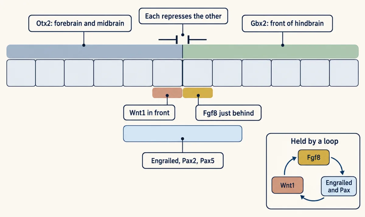
The parts of the loop work through pathways you have met elsewhere. FGF8 acts through fibroblast growth factor (FGF) receptors, which are receptor tyrosine kinases (that lesson), and through the Ras-MAPK relay (that lesson); Wnt1 is a secreted Wnt ligand (that lesson).
Design problem
Using add it, take it away and move it, predict the brain in each case, and say which kind of test each one is. (a) A mouse that loses Fgf8 early from the region. (b) A mouse in which Otx2 is forced on in the territory that would become anterior hindbrain. (c) A chick embryo with an FGF8 bead in the caudal forebrain.
Saved in this browser only.
One way to do it
(a) Without Fgf8, the loop fails: Engrailed, Wnt1 and Pax2 are not maintained, and the midbrain and cerebellum are lost. This takes it away, so it tests necessity.
(b) The boundary is drawn where Otx2 meets Gbx2. Forcing Otx2 backward pushes Gbx2 back, so the boundary and the Fgf8 band shift toward the tail, and less rhombomere 1 is left to make cerebellum, which shrinks. This is a move-it test, a gain-of-function change. Extending Otx2 is enough to shift the line in this tissue, though it does not show that Otx2 can build an organizer anywhere.
(c) The bead adds FGF8 where it is normally absent. The forebrain tissue around it, which carries Otx2, turns on Engrailed and later forms a midbrain whose order is reversed, with isthmus-like parts next to the bead. This adds it, so it tests sufficiency.
Other organizers along the tube
The isthmus is one of several. At the very front of the neural plate, the anterior neural ridge expresses Fgf8; in mouse explants, removing it blocked the forebrain gene Foxg1 (also called BF1), and FGF8 protein could induce that gene. In the diencephalon, a thin stripe called the zona limitans intrathalamica makes Sonic hedgehog and, in chick experiments, patterns the thalamus on one side and the prethalamus on the other.
Not every organizer works by adding an activating signal. In zebrafish, a small strip of cells at the front edge of the neural plate, the anterior neural boundary, is needed for normal forebrain markers. One of its products, Tlc, dampens Wnt signaling nearby so that the telencephalon can form.
Putting it together
| Experiment | Kind of test | What happened | What it supports | What it does not establish |
|---|---|---|---|---|
| Otx2 put into the En1 gene (mouse, 1999) | move it | line and Fgf8 band moved toward the tail; cerebellum smaller | the line sits where the two repressors meet | how the isthmic genes first come on |
| Gbx2 made in the back of the midbrain (mouse, 1999) | move it | line moved forward | the same, from the other side | — |
| Isthmic tissue grafted into forebrain (chick, 1991) | add it | host cells next to the graft turned on Engrailed and became midbrain | the junction instructs its neighbours | which molecule carries the message |
| FGF8 bead in the diencephalon (chick, 1996) | add it | extra midbrain, built as a mirror image; Engrailed-2 induced around the bead (Fgf8 and Wnt1 in later bead work, 1999) | FGF8 is enough to start an isthmus-like program; the mirror image fits reading distance, though the induced organizer may share the work | that the bead’s gradient alone set the order |
| Wnt1 null (mouse, 1990) | take it away | Engrailed on, then lost; most of the midbrain and much of the cerebellum gone | Wnt1 maintains the region | which steps of the loop are direct |
| Wnt1 null with En1 made from Wnt1 control elements (mouse, 1996) | take it away, then put back | early development of the region restored | a main job of Wnt1 here is to keep Engrailed on | that this is Wnt1’s only job |
| acerebellar (zebrafish Fgf8 mutant) | take it away | boundary genes on, then fade | Fgf8 maintains more than it starts | — |
| Fgf8 lost early from the region, or reduced (mouse) | take it away | after early loss, midbrain and cerebellum lacking; milder reductions spare part of both | Fgf8 is necessary | how much each part needs (timed deletions address that) |
| Both En genes, or both Pax2 and Pax5, removed (mouse) | take it away | more of the region lost than in either single mutant | the transcription factors are needed too | which gene covers for which |
Every row is consistent with a line that becomes a source and must be kept on. Only the bead row bears on distance, and no row isolates the repressor clause.
The three principles from the Hox lesson hold here, now in concrete form. Genes switched on at specific places and times: the line where Otx2 meets Gbx2. Graded signals set up the pattern: FGF8 and Wnt1 from that line. Those signals restrict transcription factors, which in turn control the genes of each region: Engrailed graded across the midbrain; and Otx2 and Gbx2, which position the source and set what each side can make of its signal.
Going deeper
Engrailed, a cousin of Hox
Engrailed was named in the fly for a defect in body segments. Its homeodomain, the DNA-binding unit it shares with the Hox proteins, is about 60 amino acids long and recognizes short DNA sequences, but the gene is not part of the Hox code. A cousin, not a sibling. The structure of the Engrailed homeodomain on DNA is among the structures for this lesson.
What makes the edge sharp
In a toy model, along a stretch of tube from x = 0 to 1, Otx2 is made at a rate a = 2(1.2 − x) and Gbx2 at b = 2(0.2 + x), and each represses the other: dO/dt = a/(1 + Gn) − O and dG/dt = b/(1 + On) − G. The exponent n, the Hill coefficient, measures how switch-like the repression is. The input alone would take 1.12 of the stretch to shift from 90 to 10 percent Otx2. At steady state, the zone where Otx2’s share, O/(O + G), falls from 90 to 10 percent narrows somewhat with n = 1 (0.82 of the stretch) and much more with switch-like repression (0.51 with n = 2, 0.17 with n = 4); stronger repression sharpens it at any n. Each steady state is reached from a start at zero. With n = 4 the system has two stable states in a narrow band at the edge (about 0.44 to 0.56), so a different start can move the switch point within that band. Adding 1.5 to Otx2’s production between 0.5 and 0.8, a stand-in for the En1-Otx2 knock-in, moved the point where Otx2’s share falls below half (n = 2) from 0.50 to 0.80: the edge follows the repressor. These are model numbers, not measurements. In the embryo the two domains overlap briefly at first.
A weaker source
What should a partial loss do? Take one steady source with the gradient used above (λ = 50 micrometres), and give three parts three thresholds. The part nearest the source forms where the signal is at least 0.6 of the full source level, the middle part where it lies between 0.25 and 0.6, and the outer part between 0.08 and 0.25. Each column gives how far that part reaches from the source.
| Source strength | Nearest part (0.6 and up) reaches | Middle part (0.25 to 0.6) reaches | Outer part (0.08 to 0.25) reaches |
|---|---|---|---|
| Full | 26 µm | 69 µm | 126 µm |
| Half | lost | 35 µm | 92 µm |
| One fifth | lost | lost | 46 µm |
Two things follow, for this single steady source with λ and the thresholds fixed. The part that needs the most signal goes first. And halving the source pulls every remaining boundary toward it by the same distance, λ × ln 2, about 35 micrometres, not by a fixed fraction. The timed Fgf8 deletions in the mouse point the same way, in time rather than level.
The chick Otx2 map
In the chick, Millet and colleagues in Alvarado-Mallart’s group reported in 1996 that the back edge of Otx2 lies at the midbrain-hindbrain junction.
Single mutants and strain background
Mice lacking En1 lose most of the colliculi and cerebellum (Wurst, Auerbach and Joyner, 1994), though on one other mouse strain most of the cerebellum is spared, apparently because En2 covers for it. Mice lacking En2 have a milder cerebellar defect. Putting the En2 coding sequence in place of En1’s rescues the En1 defect (see the anecdote below). Losing both Pax2 and Pax5 removes the posterior midbrain and cerebellum; each alone has a milder or strain-dependent effect.
Two forms of FGF8
FGF8b, the form that can induce cerebellum, binds fibroblast growth factor (FGF) receptors more strongly than FGF8a. The FGF8b structure among this lesson’s structures shows the extra receptor contact proposed for its front end.
The fish anterior neural boundary
Corinne Houart, Monte Westerfield and Stephen Wilson described the fish anterior neural boundary in 1998; moved, its cells can induce forebrain markers. In 2002 Houart, Wilson and colleagues identified Tlc as a secreted relative of the Wnt receptor Frizzled. In the fish, Wnt from behind pushes tissue toward a posterior fate, and Tlc holds it off. The same three principles apply: a place, a signal opposed by another, and transcription factors restricted by the result.
An open question: a border cells seldom cross
The FGF8 bead induces mirror-image midbrain, so the tissue reads something like distance from the source. Yet the boundary also acts as a lineage restriction that cells of the two sides seldom cross. How the same signal sets graded identity on one side, a fate choice on the other and a cell-sorting border between them is still not fully explained.
Your notes
Select any text in the lesson to highlight it or add a note to it. Your highlights and notes are saved in this browser and collected in your notebook.
This browser is not letting the site save anything (a private window or strict privacy settings), so highlights and notes will be lost when you leave the page.
How we measure it
Quail-chick grafting
A piece of neural tube is cut from a quail embryo and placed into a chosen spot in a chick embryo of similar stage. Quail cells look different from chick cells under the microscope, so you can tell whether a new structure is made of donor cells or of host cells that the graft has instructed.
Protein-soaked bead implantation
A small bead is soaked in a purified protein and pushed into embryonic tissue, where it releases the protein locally. Control beads soaked in buffer alone show what the wound and the bead do by themselves. A positive result tests sufficiency.
Gene targeting in mice
A gene is altered in embryonic stem cells, which are then used to make mice carrying the change. A knockout removes the gene, a hypomorph reduces it, and a knock-in puts another coding sequence under its control. These test necessity and position.
In situ hybridization
A labelled RNA probe binds the messenger RNA of one gene in a fixed embryo, showing where that gene is on. Comparing the domains of Otx2, Gbx2, Fgf8 and Engrailed in normal and mutant embryos shows where a boundary lies and whether it has moved.
Practice questions on this lesson (3) → MCAT-style, original, each with an explanation
Where this lesson fits
Before thisReview first
- The Organizer: Spemann and MangoldThis course · Lesson 5
Spemann's organizer is the first organizer, and this lesson sets local organizers apart from it.
- Head to Tail: Hox Genes, Retinoic Acid and RhombomeresThis course · Lesson 8
Hox genes address the hindbrain and stop near its front, which leaves the midbrain needing a different address.
- Tools for Following Cells and GenesThis course · Lesson 4
The grafts, bead implants and conditional deletions here rely on the tools for following cells and genes described there.
After thisStudy next
- Ventral to Dorsal: Sonic Hedgehog and BMP in the Neural TubeThis course · Lesson 10
Another secreted source patterns the tube by how much signal a cell receives, now across ventral to dorsal.
Suggested by an AI model from the lessons themselves, then checked. How we use AI
Finish line
Test yourself
9 questions
Practice and MCAT-style, with explained answers.
Next · Lesson 10
Ventral to Dorsal: Sonic Hedgehog and BMP in the Neural Tube
Graft a second notochord beside a chick neural tube, and the tube grows a second floor.
Continue →← Lesson 8 · Head to Tail: Hox Genes, Retinoic Acid and Rhombomeres