Developmental Neuroscience · Skim version
Seeing Neurons: Golgi, Cajal and the Neuron Doctrine
Read the full lesson → Skim the whole course →
The guiding idea
Are neurons separate cells that touch, or one fused network? The neuron doctrine says separate, and it took a new stain, a lot of embryos and the electron microscope to show it. Two men looked at the same stained brain and saw two different organs. Who was right, and how could anyone tell?
The nervous system is built of separate cells, neurons, that pass signals to one another across specialized contacts, and that is why development has to build connections between cells.
From the film: A Touch Is Not a Fusion
- A touch is not a fusion: whether two touching branches are one cell or two is decided at the membrane where they meet.
- Evidence that bears directly on that membrane, by seeing it or by testing what crosses it, can settle the question.
- A gap of about 20 nanometres at a chemical synapse sits inside the light microscope's blur of about 200 nanometres, so touching and fused branches look alike.
- In vertebrates, development has to wire separate cells to the right partners.
From the film: One Stain, Two Readings
- Golgi's black reaction fills a few cells completely, so one neuron and its branches can be traced.
- Sparse staining shows whose branch is whose, but it cannot show touch or fusion, because both partners are rarely stained and the gap is below what light can show.
- Golgi read the tangle of axon branches as a continuous net, while Cajal read the free endings he kept finding as evidence of separate cells.
- Embryo evidence and the direction of signal flow made separate cells more likely without settling the question.
From the film: The Gap, Seen
- Electron micrographs of the chemical synapses examined in the 1950s showed two membranes staying separate across a cleft, which settled the question.
- Electrical synapses join some neurons through gap junction channels, but each cell keeps its own membrane.
- A small tracer can pass gap junctions to coupled neighbours while a large dextran stays in the injected cell, so coupling is not fusion.
- A few animals really do have fused neurons, such as the squid giant axon and the comb jelly nerve net.
-
Touch or fusion: a question about 20 nanometres
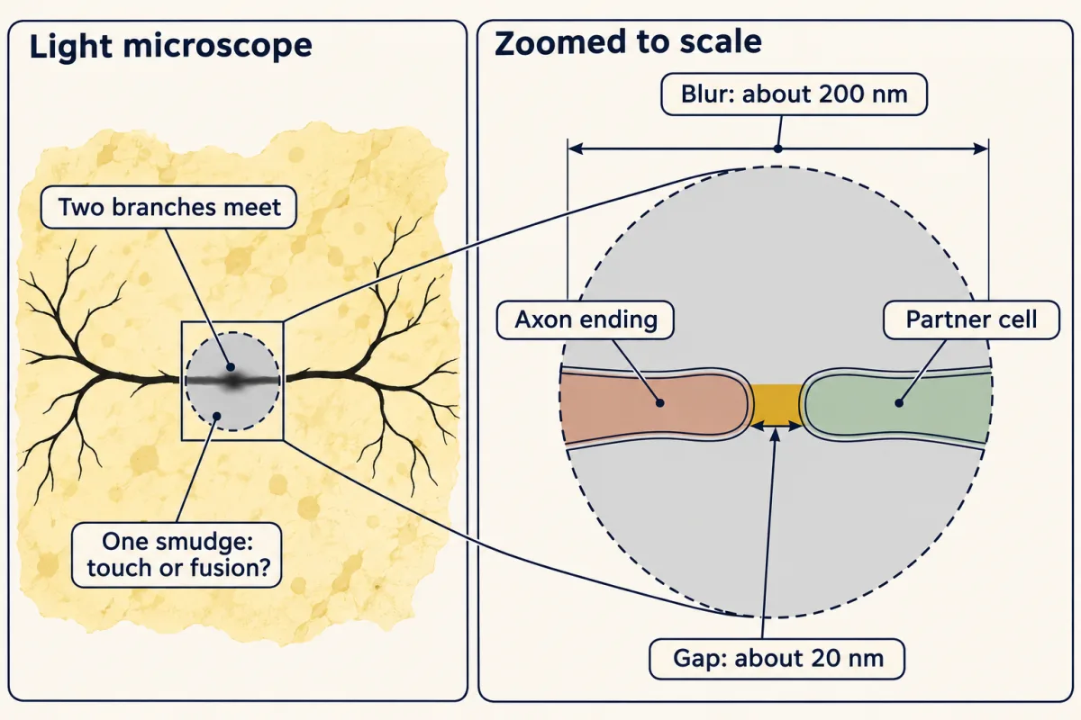
The gap at a chemical synapse, about 20 nanometres, sits inside the light microscope's blur of about 200 nanometres, so touching and fused branches give the same smudge. No light-microscope image of one contact could settle the question. AI-generated illustration -
To see one cell, stain almost none
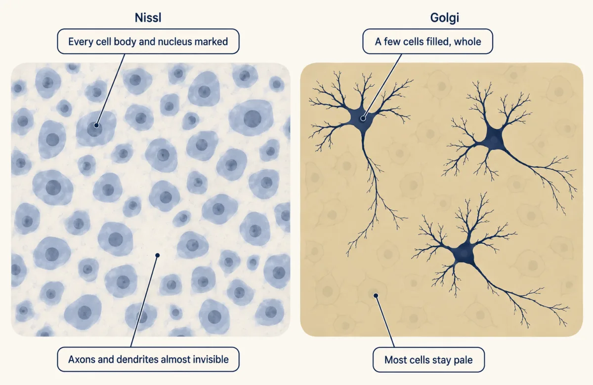
Nissl stain marks every cell body and nucleus but leaves axons and dendrites almost invisible. Golgi's black reaction fills a few cells, showing the cell body and many branches, though processes that leave the section are cut off. Why one neuron takes the stain and its neighbour does not is still unknown. AI-generated illustration -
One slide, two readings
-
Evidence from development: fibres grow out of single cells
-
The direct test: look at the membrane
-
A pore is not a fusion
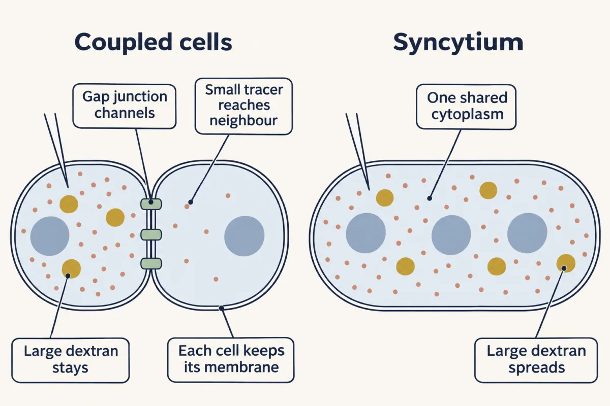
Coupled neurons each keep their own membrane: a small tracer can pass through gap junction channels to a neighbour, but a large dextran stays in the injected cell. In a true syncytium, one shared cytoplasm, the large tracer would spread too. A touch is not a fusion, and neither is a pore. AI-generated illustration -
Putting it together
-
Going deeper
Where this lesson fits
Before thisReview first
- The Wiring Problem: How a Brain Gets BuiltThis course · Lesson 1
It poses the question of how a brain gets wired, which assumes the separate cells whose evidence is weighed here.
After thisStudy next
- The Growth Cone: The Tip That Steers the AxonThis course · Lesson 3
It follows the growth cone Cajal first inferred from fixed tissue and shows how it steers an axon.
- One Axon, Many Dendrites: Neuronal PolarityThis course · Lesson 21
It explains how one axon and many dendrites arise, the polarity behind Cajal's law of dynamic polarization.
- Building the Neuromuscular Junction: Agrin, Lrp4 and MuSKThis course · Lesson 35
It shows how a specialized contact between two separate cells is built, the synapse this lesson established.
Elsewhere on the siteFilms, simulations and method films in other courses
- Counting failuresSimulation · Neuroscience
The 1950s separate-membrane synapse finding is what makes quantal chemical transmission across a gap possible.
- Blips from a Silent NerveFilm · Neuroscience
It shows the chemical relay across the cleft between two cells that the electron micrographs revealed.
- Small Space, Big ChangeFilm · Neuroscience
It shows neurons and glia as separate cells sharing a tight extracellular space, the premise of the doctrine.
Suggested by an AI model from the lessons themselves, then checked. How we use AI