Biochemistry · Introduction to Biochemistry & the Cell
Introduction to Biochemistry and Cell Biology: Compartments Create Control
Rewritten on : new text, figures and flashcards. Your notes and highlights stay, but the text around them may have moved.
Why can one missing enzyme kill a child, when her parents, with half as much, are fine? The answer is a room with no way out.
Pause when the reading reaches a film, and offer to play it. Saved on this device.
Watch the film now, or keep listening
Builds on:Vitalism: How Chemistry Captured LifeWhy these?
 ▶ Watch A Room With No Way Out · 3:05
Read the transcript of “A Room With No Way Out”
Film transcript: A Room With No Way Out
Why can one missing enzyme kill a child, when her parents, with half as much, are fine? The answer is a room with no way out. Start with a wall. A cell membrane is a double layer of lipids with an oily core. Small nonpolar molecules, like oxygen, slip straight through. Glucose and ions need doors the cell builds. Close that wall into a bag, and you have a room. The lysosome is the cell's recycling room, with digestive enzymes sealed inside, in acid. Old membrane is delivered in. The enzymes cut it into small parts, which leave through transporters. Now follow one lipid in that old membrane, GM2 ganglioside. One enzyme, HexA, snips one sugar off it, so its breakdown can go on.
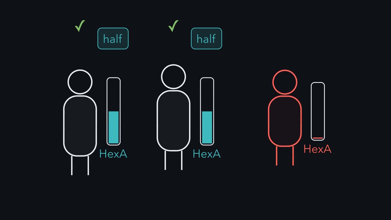
GM2's big sugar head cannot cross the lysosome's membrane. And no transporter carries it out. Nothing else in the cell does HexA's job well enough to stand in. So, in its simplest form, this room is a tap and a drain. What it holds is set by delivery in, against clearance out. Each parent has one working copy of the gene. Each makes about half the normal amount of HexA. So halve the drain. Does their lysosome fill up?
No. A normal lysosome has far more HexA than it needs. It clears GM2 many times faster than it arrives. Halve the drain, and the level settles a little higher, but stays low. In the infantile form, there is almost no working enzyme. The drain is almost shut, but delivery goes on, from before birth.
GM2 piles up, and the lysosomes swell. Yet the room stays intact, filling with something it cannot empty. Storage starts only when the drain falls below the tap. Measurements put that cut-off near 10 to 15% percent of normal, far below half. Below it, leftover enzyme slows the rise, so the illness starts later. Why are nerve cells hit hardest? Their membranes are rich in gangliosides, so delivery is high. And neurons do not divide. A dividing cell splits its stored GM2 between its daughters. A neuron cannot dilute stored GM2 that way. So a sealed room keeps whatever reaches it and is not cleared. Half a drain still keeps up, so the parents are fine.
With almost no drain, the room fills, and nerve cells are hit hardest.
From the film
- GM2 cannot cross the lysosome's membrane, so in its simplest form its level is a balance: delivery in, against clearance out by HexA.
- Tay-Sachs carriers make about half the normal HexA, which still clears GM2 faster than it arrives.
- GM2 storage starts only when HexA-dependent clearance can no longer match delivery; measurements put that cut-off near 10 to 15% of normal.
- In infantile Tay-Sachs there is almost no working HexA, so GM2 piles up from before birth in an intact lysosome.
- In Tay-Sachs, nerve cells are hit hardest: their membranes are rich in gangliosides, and stored GM2 is not diluted between daughter cells.
A single missing enzyme can kill a child before the age of five, while her parents, who each make only half the normal amount, are healthy.
The enzyme is hexosaminidase A (HexA), and the disease is Tay-Sachs. HexA’s essential job is one chemical cut in one fatty molecule. Why does losing it destroy a child’s nerve cells, when half as much is plenty? The answer is a room with no way out, and building up to it takes you through the atoms, molecules and rooms every later lesson assumes.
The one idea to carry through this lesson. A membrane turns one shared solution into separate rooms. A molecule that cannot cross a membrane on its own is changed inside a room only by the enzymes and conditions in that room, and it leaves only by a route the cell builds for it. So how much of a trapped molecule a room holds is a balance: delivery in, against breakdown and export out. When delivery outruns the room’s capacity to clear it, the molecule piles up. “Compartments create control” means exactly this: the cell decides what reaches each room, what each room contains, and what conditions it keeps.
The film above draws this balance as a tap and a drain. The rule does not say a missing enzyme is more dangerous inside a room than outside one (the last section shows why).
What a cell is made of
Strip an organism down to its atoms and 98% of them are hydrogen, oxygen and carbon. Add nitrogen, phosphorus and sulfur and you have nearly the entire chemical alphabet of biology: CHNOPS. A memory aid gives each one a job. Carbon is the backbone. Oxygen pulls shared electrons strongly toward itself (it is highly electronegative), so in O–H and C=O bonds the electrons sit closer to the oxygen and the bond is polar, slightly negative at one end and slightly positive at the other; as O₂ gas it is the final electron acceptor in aerobic respiration. Nitrogen carries information in the bases of DNA. Phosphorus brokers energy in ATP. Sulfur bridges, locking proteins into shape with disulfide bonds. (The jobs are for remembering, not exclusive: nitrogen is also in every amino acid, phosphorus in the DNA backbone and every phospholipid.)
Carbon is at the center because its four stable bonds build chains, branches and rings whose chemistry works in water, the medium every cell runs in. From these atoms cells build four classes of molecule:
- Proteins are the workers: enzymes that catalyze reactions, signals like insulin that carry messages. Which proteins a room holds is much of what makes it different from the next.
- Nucleic acids are the library. DNA is the master file, written in four letters (A, C, G, T). RNA makes the working copies that leave the shelf (A, C, G, U), and other RNAs help run the machinery that reads them. The first room, the nucleus, guards the DNA.
- Carbohydrates are the fuel and the ID tags: glucose burned for energy, sugar chains studding the cell surface as recognition labels.
- Lipids are the walls. A typical membrane lipid has a water-loving head and two water-fearing tails. In water they line up tail to tail in a double layer, a bilayer, with an oily core. That is a wall, and a wall can make a room. (Fats, another kind of lipid, are also the cell’s densest stored fuel.)
A wall makes a room
Predict. Which of these can cross a bare bilayer on its own: O₂, glucose, a K⁺ ion, a ganglioside (a lipid with a large chain of sugars on its head)? Hint: the core is oily, so ask which of these water loves.
Check your prediction
Only O₂. Small nonpolar molecules such as O₂ and CO₂ pass straight through the oily core (water crosses too, more slowly). Glucose (polar) and K⁺ (charged) each need a channel or carrier the cell builds. The ganglioside’s big sugar head cannot pass the oily core, so it stays in the layer it was put in (it flips only extremely slowly). On a bare bilayer the other three stay on the side where they start.
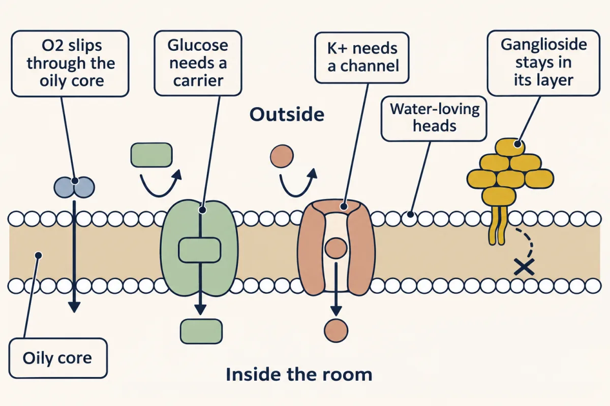
So a room can keep its own enzymes and its own conditions. Take acidity. The inside of a lysosome, the cell’s digestive room, sits near pH 4.5 to 5, against about 7.2 in the cytosol (the fluid outside the organelles). Each pH unit is a tenfold step, so the lysosome holds a few hundred times the free H⁺ concentration of the cytosol (about 300 times at pH 4.7). That acid is the second lock on the recycling room, below.
The first room: the nucleus and the central dogma
Information in a cell flows in one main direction: DNA to RNA to protein.
- Replication copies DNA so a dividing cell can hand a full set to each daughter. DNA polymerase reads one strand and builds its partner, and the copy is semi-conservative: each new double helix keeps one original strand.
- Transcription reads a stretch of DNA and writes it out as RNA (for protein-coding genes, messenger RNA, or mRNA). A cell doesn’t run every gene at once; which genes it transcribes is a large part of what makes a liver cell a liver cell and a neuron a neuron.
- Translation reads the mRNA and builds the protein, codon by codon (a codon is three bases), at the ribosome, with transfer RNA acting as the matchmaker between code and amino acid.
Try it below: send each machine to the step it runs, and notice which steps sit inside the nucleus. Each correct match shows what that step produces.
Pick a molecule, then tap the step it carries out.
Structure fits job, and one atom on the sugar makes the difference. (The sugar’s carbons are numbered 1′ to 5′, “one-prime” to “five-prime”, to tell them apart from the base’s atoms.) RNA’s ribose carries an –OH on carbon 2′, which lets its backbone break much more easily, fitting a message the cell uses and discards. DNA’s deoxyribose carries only an H there, so its backbone is far more stable, fitting long-term storage. (How long RNAs really last: Going deeper.)
In your cells, nuclear genes are transcribed in the nucleus and translated in the cytoplasm. (Mitochondria keep a small genome of their own and transcribe and translate it inside the organelle.) The message is made in one room and read in another, and the wall between them controls the flow of information. In bacteria, which have no such wall, ribosomes start reading an mRNA while it is still being made. In your cells the message can be spliced (cut and rejoined) and checked in the nucleus first, and only then exported through the pores in the nuclear membrane. That separation is one reason your cells can regulate genes in ways bacteria cannot.
Why big cells need inner walls
Prokaryotes, the bacteria and archaea, are an open floor plan: few or no internal membranes and no true nucleus. They do organize their insides (Going deeper), but most work happens in one shared space. Eukaryotes, your cells, are a building of walled offices, each a membrane-enclosed organelle with its own main jobs.
Geometry shapes both. A cell’s volume (the demand for fuel and the waste to clear) rises with the cube of its radius, while its surface (the membrane that brings fuel in and lets waste out) rises only with the square. Go from a 1 µm bacterium to a 10 µm eukaryotic cell, ten times the radius, and volume grows 1000-fold but surface only 100-fold: each square micrometre of membrane must serve ten times as much cytoplasm (1000 ÷ 100). A cell has three options: stay small, stretch out thin, or fold extra membrane inside. Drag the radius and watch the squeeze.
- Surface area, 4πr²
- —
- Volume, ⁴⁄₃πr³
- —
- SA : V = 3/r
- —
- Cytoplasm per µm² membrane, V/SA = r/3
- —
At 10 µm, SA:V is 0.30 µm⁻¹. Compared with a 1 µm bacterium: surface ×100, volume ×1000, so each µm² of membrane serves 10× as much cytoplasm.
Demand scales with volume, supply with surface. A cell can stay small, stretch out thin, or fold extra membrane inside. Inner membranes (mitochondrial cristae, the ER) give room for work done on membranes and each one encloses a room; uptake from outside still needs the plasma membrane, which is why gut cells fold it into microvilli.
Your cells fold extra membrane inside for the work done on membranes. A bacterium makes much of its ATP on its plasma membrane; a large cell makes most of its ATP on the folded inner membrane of its mitochondria, and makes secreted and membrane proteins, and lipids, at the ER. That gives it far more membrane for those jobs than its outer surface could, and each inner membrane encloses a room. Uptake from outside still depends on the plasma membrane, which is why absorbing cells such as gut cells fold their outer surface into fine projections (microvilli). (Mitochondria came from engulfed bacteria, not from folds of the cell’s own membrane.) Walk the offices:
- The plasma membrane is the outer wall, its oily core blocking most molecules and letting protein gatekeepers choose what crosses.
- The nucleus is the library, a double membrane studded with pores, holding the DNA.
- Mitochondria are the power plant, burning fuel to make ATP.
- The rough ER, ribosomes clinging to its surface, makes proteins that will be secreted or sent to membranes and lysosomes.
- The smooth ER, with no ribosomes, makes lipids and detoxifies drugs (your liver leans on it hard).
- The Golgi is the post office, modifying and addressing proteins before shipping them on.
- Lysosomes are the recycling rooms, sealed and acid (their own section below).
The sealed conveyor belt, and how we know
From rough ER to Golgi to membrane, a secreted protein travels a conveyor belt of sealed rooms, made, checked, modified and addressed without passing through the general workspace. That separation is the control. The ER interior has its own folding enzymes and conditions that favor disulfide bonds; most sugar chains on a secreted protein are added there and in the Golgi; misfolded proteins are held back, and those that cannot be fixed are pushed back out to the cytosol to be destroyed; properly folded cargo never meets that protein-destroying machinery.
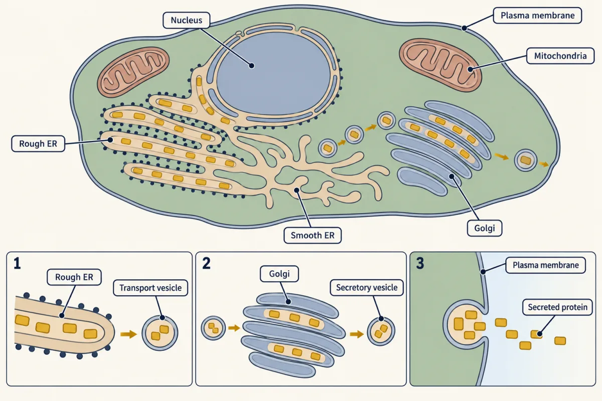
The belt had to be shown to move. George Palade and his colleagues did it with a clock, using guinea pig pancreatic cells, which export digestive enzymes. They fed the cells a brief pulse of radioactive amino acids, then chased them with ordinary ones, so a single cohort of new protein carried the label. At a series of time points they chemically fixed (preserved) a sample of cells and looked where the label was.
Predict. If proteins really travel rough ER → Golgi → secretory granules (the storage packets that wait near the cell surface), where should most of the label be at an early, a middle and a late time point?
Check your prediction
ER, then Golgi, then granules, and that is what they saw. Picture three curves of label against time, one per compartment: the rough ER curve high first, then falling; the Golgi curve rising as the ER curve falls; the granule curve rising last. When the cells were stimulated, the granules released their contents and the label appeared outside the cell. The pathway was no longer a hypothesis; it was a trip you could time.
Tagging a molecule and following it is still how cell biology makes a cell’s invisible logistics visible.
Two moves connect the rooms to the outside. In exocytosis, a vesicle fuses with the plasma membrane and dumps its cargo outside; this is how a pancreatic beta cell releases insulin into your blood. In endocytosis, the membrane folds inward to bring cargo in; this is how a cell pulls in cholesterol through its low-density lipoprotein (LDL) receptor.
The recycling room
Lysosomes hold digestive enzymes (hydrolases, which split molecules using water) that would be dangerous loose in the cell, so they sit behind two locks. The first is the membrane: the enzymes cannot get out, and their targets get in only by routes the cell builds. The second is the acid: lysosomal hydrolases work best at acid pH and poorly at the cytosol’s, so an enzyme that escapes into the cytosol is much less active there (though a large leak can still kill a cell).
Evidence for the membrane lock came from the test tube. Predict. Christian de Duve measured an acid hydrolase in a fresh fraction of liver cells, then added detergent, which dissolves membranes. What should happen to the measured activity if the enzymes sit inside sealed bags?
Check your prediction
It should rise, and it did. In a fresh fraction most of the activity was hidden, because the substrate in the test tube could not cross the bags’ membranes. Breaking the membranes released the full activity. This hidden activity, called latency, was evidence that the enzymes sit inside sealed membrane bags: lysosomes. De Duve shared the 1974 Nobel Prize with Albert Claude and Palade.
Material reaches the lysosome by delivery. Endocytosis brings in cargo and old patches of plasma membrane; autophagy wraps up worn-out parts of the cell’s interior. The enzymes break the cargo into small reusable parts, such as amino acids and sugars, which leave through transporters.
When one room lacks one enzyme
Now return to the child. GM2 ganglioside is one of those sugar-headed lipids (a glycolipid). Gangliosides sit in the outer layer of the plasma membrane, and nerve cells make a great deal of them. GM2 itself is a minor one, but it is also a step on the way down: when larger gangliosides such as GM1 are taken apart in the lysosome, they pass through GM2. Membranes are constantly renewed, so a steady stream of gangliosides reaches lysosomes with the old membrane endocytosis delivers, and enzymes there take each one apart one sugar at a time. Hexosaminidase A (HexA) makes one of those cuts: it removes the end sugar from GM2, so the next enzyme can go on.
As the film shows, GM2’s sugar head (large and charged) cannot cross the lysosome’s membrane, no transporter carries it out, and no other enzyme can stand in for HexA (Going deeper, “Partners of HexA”). So GM2 in a lysosome is a balance: delivery in, against HexA breaking it down.
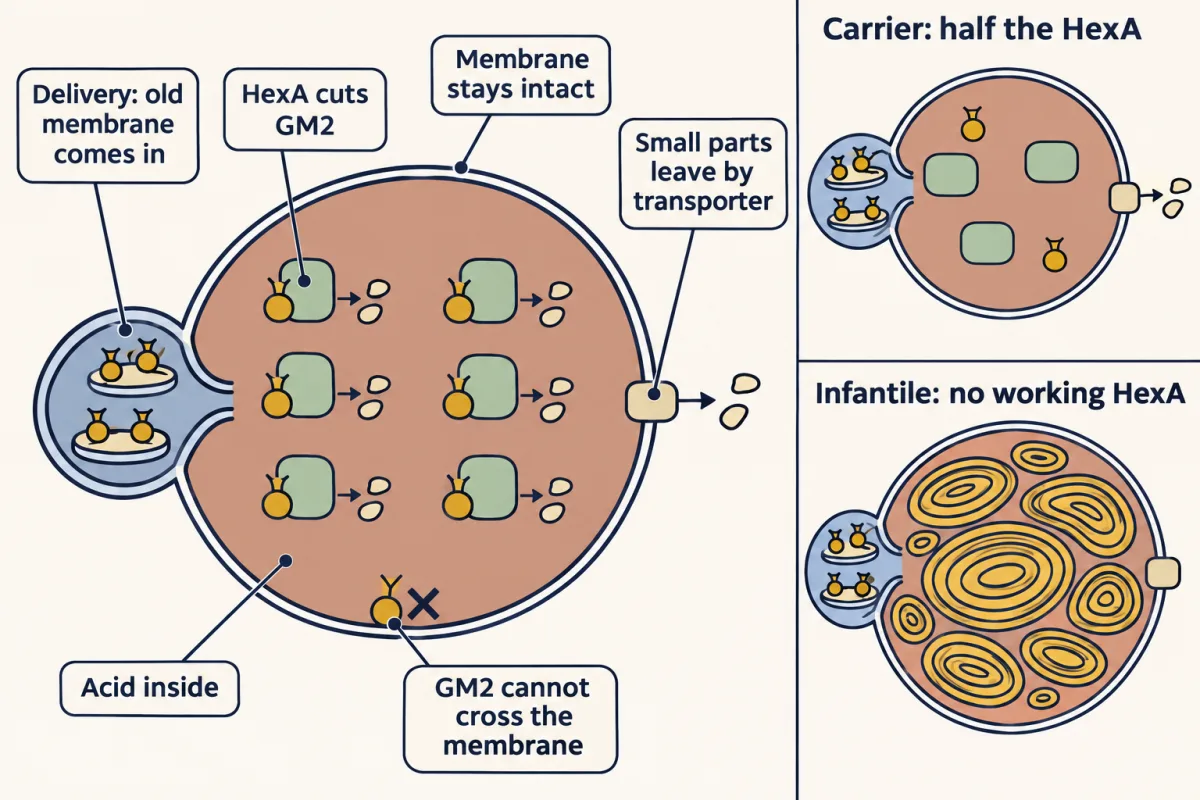
Predict. Each parent carries one working copy of the HEXA gene and makes about half the normal amount of HexA. Should their lysosomes fill with GM2?
Check your prediction
No. A normal lysosome has far more HexA than it needs: it can break down GM2 many times faster than GM2 arrives, and with half it still keeps up. Think of a drain much wider than the tap: halve it and the water sits a little higher, but never overflows. Storage starts only when the enzyme left can no longer keep up with delivery. That cut-off is far below half the normal amount, which is why Tay-Sachs is recessive: a child is affected only if both copies of the gene are broken (Going deeper, “The balance in numbers”).
The film above follows the classic infantile form, where neither copy makes enzyme that works and GM2 builds up from before birth in an intact lysosome. The lysosomes swell with layered membrane, nothing leaks out, and the neurons swell and gradually stop working. Babies develop normally for a few months, then come muscle weakness and the loss of skills they had already learned, and in this form death comes at around age four. In the rarer juvenile and adult-onset forms some enzyme activity remains, so the balance tips more slowly: the illness starts later and progresses more slowly.
Predict. A child with infantile Tay-Sachs has the same broken gene in her skin and liver cells as in her neurons. Why are the neurons hit hardest?
Check your prediction
Two reasons follow from the balance. Delivery is high in neurons, because their membranes are especially rich in gangliosides. And neurons do not divide: a dividing cell splits its stored GM2 between two daughters at every division, while a neuron keeps everything it stores, for life.
Here is what the compartment does and does not explain. Losing any enzyme with no backup can be serious, wherever it works. In phenylketonuria (PKU) the missing enzyme works in the liver cytosol, and untreated PKU also damages the brain. The difference is where the unbroken molecule ends up. Unconverted phenylalanine stays in the blood, and it comes from food and body protein, so a low-phenylalanine diet, started after newborn screening (a blood test at birth), can keep it down. GM2 is made in neurons and sealed in their lysosomes, behind the blood–brain barrier (the tight lining of the brain’s blood vessels, which keeps most large molecules out). No diet reaches it, and enzyme infused into the blood does not get there.
That is the lesson in one line: what a room holds is set by what is delivered and what it can clear, so compartments create control, and they also decide where a failure piles up and what treatment can reach it.
Going deeper
Why carbon, not silicon
Silicon sits right below carbon on the periodic table and can also make four bonds, so why not silicon? Part of the answer is bond chemistry: carbon–carbon bonds are stronger than silicon–silicon bonds, carbon forms stable double bonds, and chains of silicon atoms are attacked by water and oxygen. Silicon–oxygen bonds are much stronger still, so silicon ends up locked in silicates. Part of the answer is water: carbon dioxide is a soluble gas that moves freely through blood and air; silicon dioxide is quartz, an insoluble rock. Carbon wins because its chemistry works where life happens.
RNA lifetimes and detours from the dogma
The 2′-OH makes RNA’s backbone chemically fragile, but how long a given RNA lasts in the cell is set mostly by enzymes that destroy it on purpose, and some RNAs, such as those in ribosomes, last a long time.
The DNA → RNA → protein slogan also has detours. Retroviruses like HIV carry reverse transcriptase, an enzyme that copies RNA into DNA, and many RNA viruses copy RNA straight into RNA. Crick’s own statement of the central dogma allowed these transfers; what it forbids is information flowing out of protein back into nucleic acid or protein. Reverse transcriptase is exactly what some antiviral drugs are built to block.
Order without inner walls
Bacteria organize their insides with protein-shelled compartments and with molecules parked at set places, which gives them part of the control that membranes give your cells, so the contrast is less absolute than “open floor plan” suggests. Same planet, two designs: you are eukaryotic, while the bacteria of your microbiome, which help digest your food and make your vitamins, are prokaryotic.
The balance in numbers
In a toy model of one lysosome, GM2 arrives at a steady rate, a tenth of what normal hexosaminidase A (HexA) can clear at full speed, and the remaining HexA clears it with ordinary enzyme kinetics. The numbers are illustrative, not measurements.
| HexA left (fraction of normal) | What GM2 does |
|---|---|
| 1.0 (normal) | Settles at a low level |
| 0.5 (carrier) | Settles at about twice that level, still low |
| 0.2 | Settles higher, about nine times the normal level |
| 0.1 or less | Never settles; it keeps rising |
| 0 (infantile form) | Rises fastest |
Below the cut-off, leftover enzyme slows the rise, the logic of the later-onset forms. This is the “threshold theory” of lysosomal storage diseases (Conzelmann and Sandhoff), whose measurements put the real cut-off near 10 to 15% of normal activity. In the same toy model, add cell division and GM2 per cell levels off even with no enzyme, because each division splits the load; a neuron gets no such relief.
Partners of HexA
HexA cannot reach GM2 on its own. Inside the lysosome GM2 sits in small internal membranes, and a helper, the GM2 activator protein, lifts the lipid out so HexA can cut it. Losing the activator (the AB variant) blocks the same cut. In Sandhoff disease the broken gene is HEXB, which loses both HexA and a related enzyme, HexB. A third relative, HexS, can cut GM2 too, but far too weakly to cover the loss of HexA.
Your notes
Select any text in the lesson to highlight it or add a note to it. Your highlights and notes are saved in this browser and collected in your notebook.
This browser is not letting the site save anything (a private window or strict privacy settings), so highlights and notes will be lost when you leave the page.
How we measure it
The pulse-chase experiment
Feed cells a brief pulse of radioactive amino acids, then chase with unlabeled ones, and a single labeled cohort of protein lights up as it moves — letting you watch a pathway through the cell that no single snapshot could show.
Reading structure off function (and back)
The 2'-OH on RNA's ribose makes it chemically unstable; DNA's 2'-H makes it stable. One atom of difference helps explain why DNA suits long-term storage and RNA a short-lived message; cells also destroy messages on purpose, with enzymes. When structure and job line up this cleanly, each predicts the other.
Practice questions on this lesson (3) → MCAT-style, original, each with an explanation
Where this lesson fits
Before thisReview first
- Vitalism: How Chemistry Captured LifeThis course · Lesson 1
It shows how life's chemistry came to be treated as ordinary chemistry, the starting point this lesson assumes.
After thisStudy next
- Water, Weak Bonds, and the Generation of OrderThis course · Lesson 3
It takes the idea of water-compatible chemistry and weak bonds further to explain how molecules fold and assemble.
- Membranes: The Impermeable Sheet That Builds ItselfThis course · Lesson 12
It takes the bilayer wall idea further, showing how the sheet builds itself and which proteins choose what crosses.
- Digestion and Absorption: Turning a Meal into Molecules Your Cells Can UseThis course · Lesson 14
It uses the idea of hydrolytic enzymes cutting big molecules into small pieces that transporters carry across a membrane.
Elsewhere on the siteFilms, simulations and method films in other courses
- Forward Is the DefaultFilm · Cell Biology
It shows Palade's pulse-chase evidence for the secretory pathway that this lesson cites.
- Two Pushes, One HillFilm · Cell Biology
It shows how a membrane that cannot be crossed makes each room's contents depend on transport.
- A Loop and a One-Way TripFilm · Cell Biology
It follows cargo into the lysosome, the room where GM2 piles up here.
- A Tap, a Hole, a LevelFilm · Cell Biology
It shows how pumps balance leaks to keep a room's conditions steady, like the lysosome's.
Suggested by an AI model from the lessons themselves, then checked. How we use AI
Finish line
Test yourself
10 questions
Practice and MCAT-style, with explained answers.
Next · Lesson 3
Water, Weak Bonds, and the Generation of Order
A protein folds itself, and no instructions are needed. Water and many weak, reversible bonds make the folded state the favorable one, and burying oily groups away from water is widely considered the largest single contribution.
Continue →