Cell Biology · Skim version
Cell Shape, Migration and Cilia
Read the full lesson → Skim the whole course →
The guiding idea
Four men had sperm that could not swim, and three could not clear mucus from their lungs. How could one missing part explain both?
In a crawling cell and in a beating cilium, polymers and motors make internal motion, and constraints such as adhesion and the links between doublets help shape what visible movement it becomes.
From the film: How Internal Motion Becomes Movement
- At a lamellipodial edge, polymerization adds forward speed and rearward flow subtracts it.
- Integrin coupling can reduce backward slip in this simple crawling model.
- In motile axonemes, dynein motors slide neighbouring doublets.
- In motile axonemes, links that resist sliding, with base anchoring, turn sliding into bending.
- In these two systems, constraints and motor control shape the visible output of internal motion.
From the film: Reading a Broken Machine
- After brief trypsin treatment, sperm axonemes given ATP slid apart instead of beating.
- Afzelius's four patients had immotile sperm; three had no detectable mucociliary transport.
- Sperm tails and airway cilia share a basic axoneme, though some dynein parts differ.
- One mutation can stop cilia in one tissue and only disturb the beat in another.
- Neutrophils without a working β2-family integrin largely fail to leave the blood.
-
A crawling edge: measure the hidden motion
-
From edge advance to whole-cell crawling
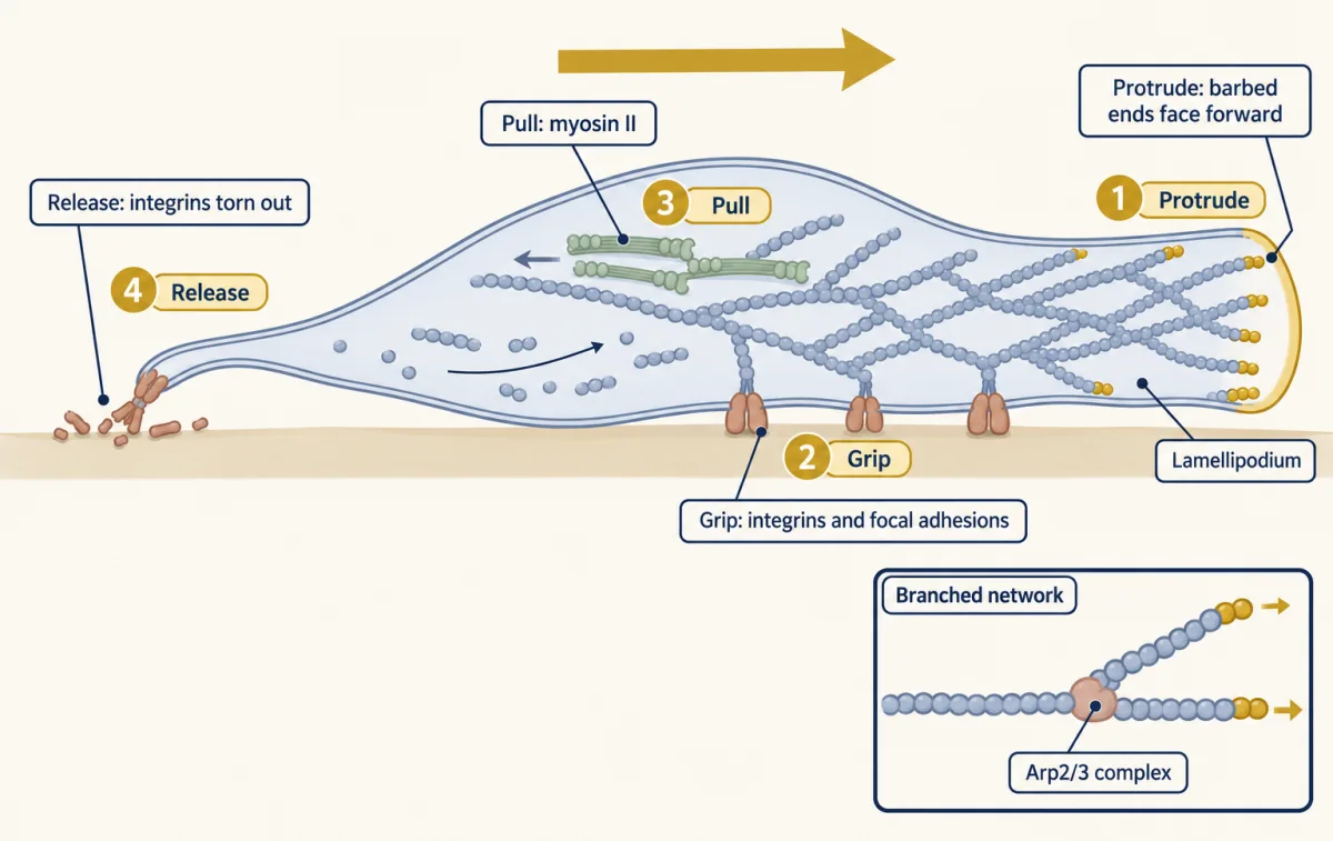
A crawling cell coordinates four concurrent processes: actin pushes the front, integrins grip, myosin II contracts the network, and the rear releases. The inset shows Arp2/3 at a branch. AI-generated illustration -
The axoneme: what the model predicts
-
From a broken part to a cellular phenotype
-
Going deeper
Where this lesson fits
Before thisReview first
- Cytoskeletal Polymers and DynamicsThis course · Lesson 14
You need actin and microtubule polymerization from there to understand protrusion and the axoneme's tubules.
- Molecular Motors and CargoThis course · Lesson 15
Dynein walking toward a minus end and myosin pulling actin are used here without re-teaching.
After thisStudy next
- Adhesion and the Extracellular MatrixThis course · Lesson 22
Integrin focal adhesions, which act as the clutch here, are developed in full there.
- Cell Identity, Cell State and DiseaseThis course · Lesson 25
It takes defects in a cellular part further, linking altered cell state to disease.
Elsewhere on the siteFilms, simulations and method films in other courses
- Optical trap (optical tweezers)Method film
Single-motor force measurements show how dynein and myosin generate the sliding and pulling used here.
Suggested by an AI model from the lessons themselves, then checked. How we use AI