Physiology · Unit 3: The heart and circulation
Capillary Exchange, Lymph and Edema
Rewritten on : new text, figures and flashcards. Your notes and highlights stay, but the text around them may have moved.
Adults born with almost no plasma albumin usually have only mild swelling. What keeps their tissues from flooding?
Pause when the reading reaches a film, and offer to play it. Saved on this device.
Watch the film now, or keep listening
Builds on:Body Water and Its CompartmentsResistance, Compliance and Local Blood FlowWhy these?
The diagram in most textbooks shows fluid leaving a capillary at its arterial end and coming back in at its venous end. When physiologists tested it, most of that fluid did not come back that way.
It was a reasonable model, one Starling himself floated: blood pressure falls along a capillary, so the outward push should win near the arterial end and lose near the venous end. In the reabsorption model as usually taught, about 24 L a day filters out and 20.4 L of it, about 85 percent, comes back in at the venous ends, leaving lymph only the remainder. In fact, in most tissues the microvessels filter a little along their whole length, and lymph carries the fluid home.
Keep one puzzle for the end. Adults born with almost no albumin in their plasma usually have only mild edema, and a few have none. (In newborns and young children the condition can be serious, with fluid retention.) Albumin supplies most of the pull that holds water in the blood, so what protects them?
Blood vessels, blood and lymph gave the first look at Starling’s balance. Two terms before the rule. Oncotic pressure is the osmotic pull of the plasma proteins, drawing water toward the side with more protein. The glycocalyx is a coat of proteoglycans and glycoproteins (sugar-covered proteins) lining every capillary, its fibres covering the openings between and through the cells.
The one idea to carry through this lesson. Water crossing a capillary wall is pushed out by the pressure difference and held back by the protein difference across the glycocalyx, and that protein difference is set by the flow itself. Filtering rinses protein from under the glycocalyx and keeps the hold-back strong. Absorbing leaves protein behind there and weakens it. So plasma protein can slow filtration but cannot reverse it for long (the no-absorption rule). In the steady state almost every microvessel filters a little along its whole length, lymph carries the fluid home, and edema is filtration outrunning lymph.
The rule covers the steady state in most systemic beds. It does not cover the moments after a sudden fall in pressure, the few beds that absorb steadily, sinusoids, or the glomerulus (its own balance). It governs water; nutrients and gases cross mainly by diffusion.
Push, hold-back and conductance
Take the three forces one at a time, with round values for resting skeletal muscle.
The push is the pressure inside the capillary, Pc, minus the pressure in the tissue, Pi. Pc sits between arterial and venous pressure, placed by the ratio of pre- to postcapillary resistance (resistance, compliance and local flow), so a rise in venous pressure moves it more than an equal rise in arterial pressure. Take Pc as 18 mmHg. Pi is slightly below atmospheric in loose tissues such as skin, intestine and lung, about -1 mmHg, though positive in some organs such as liver and kidney. A rise in Pi opposes filtration. Here the push is 18 - (-1) = 19 mmHg.
The hold-back comes from the protein difference across the wall. Plasma oncotic pressure, πp, is about 26 mmHg in adults by one review. Albumin supplies most of it, about 65 to 80 percent by different reviews, and globulins supply the rest. Some protein reaches the tissue side, and the oncotic pressure of the protein just beyond the glycocalyx, written πg and taken here as 6 mmHg, is subtracted. It is not the average for the whole tissue; the next section shows where it sits and why it is not fixed.
A protein can only pull water back across a wall it cannot easily cross. If some albumin can be carried through the wall along with the water, it pulls with only part of its strength. That fraction of its full pull is the reflection coefficient, sigma: 1 if the wall passes water and no solute, 0 if it hinders the solute no more than water. For albumin it is high, about 0.8 to 0.96 in the single vessels and tissues measured, varying with the tissue; take 0.9. Here the hold-back is 0.9 x (26 - 6) = 18 mmHg.
The conductance is the hydraulic conductivity, Lp, times the wall area, S; for whole tissues their product is the filtration coefficient, Kf. It sets how much water flow each mmHg of net pressure buys. Push minus hold-back: 19 - 18 = +1 mmHg, outward.
In words Flow across the wall equals how freely the wall passes water, times the push of pressure minus the hold-back of protein.
- Jv“J v”
- volume flow across the wall, positive outward (filtration)cm³/s for one vessel; mL/min for a tissue
- Lp“L p”
- hydraulic conductivity: water flow per unit area per unit pressurecm per s per cmH2O (1 mmHg = 1.36 cmH2O)
- S“S”
- wall area; for a whole tissue Lp S is Kf, in mL per min per mmHg per 100 gcm²
- Pc“P c”
- mean capillary hydrostatic pressuremmHg
- Pi“P i”
- interstitial hydrostatic pressuremmHg
- sigma“sigma”
- reflection coefficient for plasma proteinno unit
- πp“pi sub p”
- plasma oncotic pressuremmHg
- πg“pi sub g”
- oncotic pressure just beneath the glycocalyx (next section)mmHg
Sanity check If sigma is 0, protein holds nothing back and the net is just the push. If the push equals sigma times the protein difference, the net is zero and no fluid moves.
Try one number
Arterial end of a muscle capillary, with πg held at 6 mmHg for now (the next section shows it is lower here): Pc = 32, Pi = -1, πp = 26, πg = 6 (all mmHg), sigma = 0.9.
Push 33 mmHg, hold-back 18 mmHg, net +15 mmHg outward. Multiply by Lp S to get the flow.
Need a refresher? subscripts and Greek letters · units and prefixes
Why sigma differs from bed to bed
Capillary walls differ in how their endothelial cells join (epithelia), and sigma follows the wall.
- Continuous capillaries (skeletal muscle, skin, lung; the tightest kind in the brain) have an unbroken lining. Cells meet at narrow intercellular clefts and, outside the brain, carry many caveolae, small membrane pits and vesicles (endocytosis). They hold back most plasma protein.
- Fenestrated capillaries (intestinal villi, renal peritubular capillaries, endocrine glands) add fenestrae, pores through the thinnest parts of the cell, here covered by a thin diaphragm. They pass more water and small solute.
- Sinusoidal capillaries (liver, spleen, bone marrow) have wide openings, large open fenestrae in the liver and gaps between cells elsewhere, and an incomplete or absent basement membrane. They hold back even large molecules little.
Oxygen, carbon dioxide and other lipid-soluble molecules diffuse through the endothelial membranes over the whole surface (membrane transport). Outside the brain, small water-soluble solutes such as glucose, ions and urea diffuse through clefts and fenestrae; brain capillaries close this route, and glucose crosses them on transporters. For them, diffusion moves far more across the wall than filtering water carries. In the two-pore model, many small pores, thought to be the spaces between glycocalyx fibres over clefts and fenestrae, pass water and small solutes, and rare large pores pass plasma proteins and other macromolecules. What the large pores are is not settled.
Why the venous end does not reabsorb
Now do the textbook picture’s arithmetic. Pressure falls from about 32 mmHg where blood enters a capillary to about 12 mmHg where it leaves (round textbook end values; because resistance places the mean nearer the venous end, the resting mean used above is 18 mmHg, not the 22 mmHg midpoint of 32 and 12). Hold everything else at the values above, so the hold-back is 18 mmHg everywhere.
| Point along the capillary | Pc (mmHg) | Push, Pc - Pi | Net, push minus 18 |
|---|---|---|---|
| Arterial end | 32 | 33 | +15, filters |
| 24 | 25 | +7, filters | |
| 20 | 21 | +3, filters | |
| 16 | 17 | -1, absorbs | |
| Venous end | 12 | 13 | -5, absorbs |
This is the familiar diagram. It predicts that a venule held at low pressure should keep absorbing for as long as you watch it.
Predict. The table assumed that the tissue-side protein stays at 6 mmHg wherever water is moving in. But the wall holds protein back. What happens to the protein on the tissue side when water is drawn into the blood across that wall?
Check your prediction
It is left behind. Water crosses the sieve into the plasma, but most of the protein cannot, so the protein just outside the sieve gets more concentrated. As it rises, the protein difference across the wall shrinks, the hold-back weakens, and absorption slows to a stop. The table’s venous rows assumed a hold-back that the absorption itself destroys.
That is the revised Starling principle in one move: the hold-back is set by the flow. The sieve is the glycocalyx, lying over the mouths of the clefts between endothelial cells like a coffee filter over a narrow channel. Just outside the filter, inside the cleft, is a thin film of fluid, the subglycocalyx space. Its oncotic pressure is πg, not the average protein of the whole interstitium.
- When the vessel filters, nearly protein-free fluid streams out along the cleft, rinsing the film. Protein reaching the tissue by large pores must diffuse back against this stream, so πg stays below average interstitial oncotic pressure and the hold-back stays strong. Faster filtration rinses harder.
- When the vessel absorbs, the stream reverses. Water leaves the film for the plasma and protein is left behind, so πg climbs until the hold-back falls to match the push. Then absorption stops.
- If water stopped moving, protein would diffuse out until the difference vanished. So there is no true Starling equilibrium.
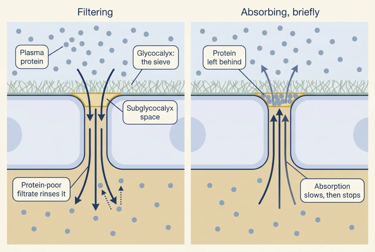
Now let πg follow the flow and redo the capillary. The steady-state numbers come from a simple sieving model tuned so that resting muscle (push 19 mmHg) settles at πg = 6 mmHg; they are illustrative, not measurements.
| Pc (mmHg) | Net with πg fixed at 6 | πg at steady state | Net at steady state |
|---|---|---|---|
| 32 | +15 | about 2.6 | about +12 |
| 24 | +7 | about 3 | about +4 |
| 20 | +3 | about 4.5 | about +1.6 |
| 16 | -1 | about 7.8 | about +0.6 |
| 12 | -5 | about 11.9 | about +0.3 |
Every row filters. Near the arterial end, fast filtration rinses πg toward its floor. Near the venous end, where the push is weaker than the plasma proteins’ full pull, πg rises until only a trickle of filtration is left, and the trickle never becomes absorption.
Plot steady filtration against capillary pressure and you get the hockey stick. Above a bend where the push nears sigma x πp (0.9 x 26, about 23 mmHg; Pc about 22 mmHg here, just below plasma oncotic pressure), filtration rises steeply, close to a straight line. Below it the filtrate is too slow to wash protein away, so the curve flattens toward zero but stays positive. The old diagram is the straight line continued below zero.
Two refinements. Absorption is real but temporary. After a sudden fall in capillary pressure, as after blood loss, a vessel absorbs until protein builds up under the glycocalyx, then filters slightly again. That is autotransfusion (reflexes in blood pressure regulation; Part C below). And steady absorption needs a second stream that keeps the tissue side rinsed with protein-poor fluid. That happens in the intestinal mucosa (absorption and the liver) and around renal peritubular capillaries, where an epithelium delivers nearly protein-free fluid, and in lymph nodes, where incoming lymph, lower in protein than plasma, does the rinsing. Everywhere else, absorption switches itself off, and lymph is the way home.
How we know. Levick concluded in 1991 that sustained downstream reabsorption is difficult to uphold, and Woodcock and Michel’s 2021 review states that the arterial-end, venous-end model has no reproducible experimental evidence behind it. In single frog mesenteric capillaries perfused through a micropipette (Michel and Phillips, 1987), a vessel held at low pressure absorbed only briefly; in the steady state, fluid movement fell to near zero or slight filtration. When enzymes strip part of the glycocalyx, the same vessel becomes leakier to water and albumin. Both fit the glycocalyx being the sieve. Whether the glycocalyx or the junctions beneath it are the main barrier to protein is still argued, and nearly all of this evidence comes from animal vessels.
Where the fluid goes: lymph
Predict. A resting muscle filters a little all along its capillaries, day and night. Where must that fluid go, and what else must the same route carry?
Check your prediction
Into lymph vessels, and through them back to the blood. They must also carry the plasma protein that leaked through the large pores, or protein would build up in the tissue and the hold-back would fail.
Filtrate enters blind-ended initial lymphatics, whose overlapping cells meet at discontinuous button junctions, with flap-like openings between the buttons acting as the primary valves. Collecting lymphatics have secondary valves, and each segment between two of them is a lymphangion, a contractile pump. Lymph moves by this intrinsic pumping plus extrinsic squeezing from muscle and breathing. Lymphatics running inside skeletal muscle are reported to have little or no smooth muscle, so muscle contraction probably drives flow there.
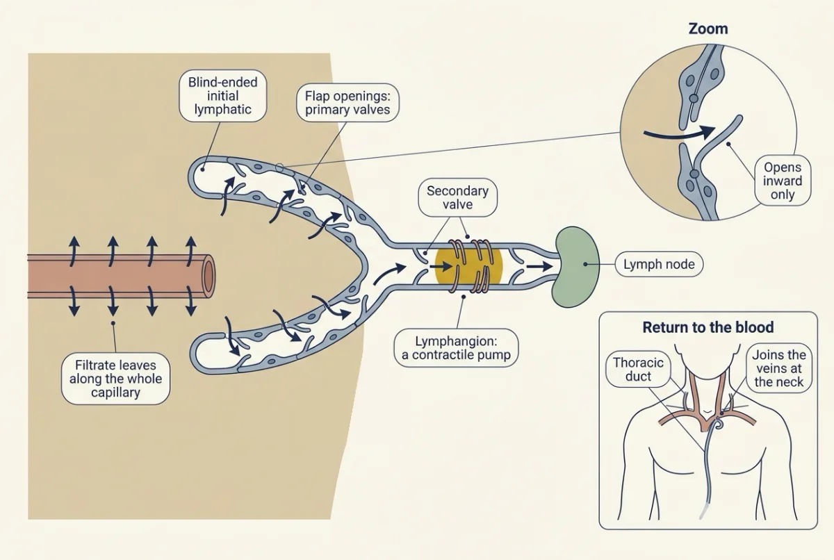
Recent reviews put the fluid filtered from an adult’s blood capillaries at about 8 L a day and lymph transport at 8 to 12 L a day. Roughly half of it is thought to be absorbed back into blood within lymph nodes, an estimate resting mainly on animal studies (lymphoid organs and immune traffic). The rest enters the veins by the thoracic duct, at the junction of the left subclavian and left internal jugular veins, and by the right lymphatic duct on the right.
Lymph also carries fat. Long-chain fats leave intestinal cells as chylomicrons, fat-carrying lipoprotein particles, enter the gut’s lymph vessels, the lacteals, and reach the blood through the thoracic duct, bypassing the liver at first. Medium-chain fatty acids go mostly by the portal vein.
Worked problem: one muscle, three states
Made-up round numbers for resting adult human skeletal muscle, steady state unless stated: Kf = 0.005 mL per min per mmHg per 100 g (illustrative, of the order estimated in human limbs), 10 kg of muscle (100 units of 100 g), Pc = 18 mmHg, Pi = -1 mmHg, πp = 26 mmHg, πg = 6 mmHg, sigma = 0.9. Assume one lumped Pc and constant Kf and sigma.
A, rest. Push 19 mmHg, hold-back 18 mmHg, net +1 mmHg, as above. Jv = 0.005 x 1 x 100 = 0.5 mL per min, or 720 mL per day, returned as lymph.
B, venous pressure rises so Pc = 28 mmHg (as in heart failure; cardiac output and venous return). At first net = 29 - 18 = 11 mmHg and Jv = 5.5 mL per min. Over hours, in this example, Pi rises to +2 mmHg and the faster filtrate rinses πg down to 3 mmHg: net = (28 - 2) - 0.9 x (26 - 3) = 5.3 mmHg, and Jv = 2.65 mL per min, about half the first value.
Predict. Lymph from this muscle can carry at most 1.5 mL per min. Does the muscle swell?
Check your prediction
Yes. Filtration of 2.65 mL per min beats a lymph ceiling of 1.5 mL per min by 1.15 mL per min, about 1.7 L a day: edema. The rise in Pi and the fall in πg roughly halved the filtration, but not enough.
C, blood loss lowers Pc to 10 mmHg. Net = 11 - 18 = -7 mmHg, absorption of 3.5 mL per min at first. It stops when 0.9 x (26 - πg) = 11, at πg = 13.8 mmHg, and then the vessel goes back to filtering slightly, on the shallow arm of the hockey stick.
Set Pc to 12 mmHg in each mode: just after the fall the vessel absorbs; in the steady state protein collects under the glycocalyx and a small filtration returns. Then lower albumin to 2 g per dL and block the lymph to see which route makes edema.
- πg beneath the glycocalyx
- 6.0 mmHg
- Net pressure
- +1.00 mmHg, outward
- Steady filtration Jv
- 0.50 mL per min · 0.72 L per day
- Lymph flow
- 0.50 mL per min · 0.72 L per day
- Accumulation in the tissue
- 0.00 mL per min · 0.00 L per day
In this sketch: adult human skeletal muscle, one lumped Pc. All numbers are made up for the sketch, as in the lesson’s worked problem: Kf = 0.005 mL per min per mmHg per 100 g (illustrative) for 10 kg of muscle, so Jv = 0.5 × net; Pi = −1 mmHg unless you set it; πp = 2 + 6 × albumin (26 mmHg at 4 g per dL), a linear simplification; lymph capacity of 1.5 mL per min is the lesson’s example, and the blocked capacity of 0.3 mL per min is invented here. In the steady state πg is computed from a simple sieving model tuned so Part A settles at 6 mmHg (illustrative, the same model as the lesson’s table); just after a sudden change it is still 6 mmHg. Values of σ other than 0.9 and the two lymph states are sketch settings. Lymph capacity does not rise and Pi does not rise on its own as fluid collects, so accumulation here is an upper estimate.
Rule to keep: plasma protein can slow filtration but not reverse it in the steady state: the hold-back is set by the flow. Edema forms when that filtration outruns lymph.
Four ways to make edema, and what holds it back
Edema is interstitial fluid accumulation when filtration exceeds lymph drainage. Each cause moves mainly one term of the equation, or the lymph.
| Route | What changes | Example |
|---|---|---|
| Raised capillary pressure | The push rises | Heart failure (Part B) |
| Low plasma protein | πp falls, so the hold-back falls | Hypoalbuminemia, as in congenital analbuminemia; a contributor in liver disease and nephrotic syndrome, where renal sodium retention also drives swelling |
| Leaky microvessels | sigma falls, so the hold-back falls and protein escapes | Inflammation: with histamine in rat cremaster, the leak is at venules, between endothelial cells |
| Blocked lymph | Drainage fails, and protein may build up in the tissue and shrink the hold-back | Lymphedema, including hereditary forms |
The inflammation row comes from Guido Majno and George Palade’s electron microscopy of the rat cremaster in 1961 (innate immunity). In lymphedema, chronic impaired drainage, the swelling includes fat deposition; one hereditary form is tied to a receptor on lymphatic endothelium.
Three safety factors limit swelling: Pi rises as fluid collects, washout (protein rinsed away by faster filtration) lowers tissue and subglycocalyx protein, and lymph flow rises. Part B used the first two.
Design problem
Raised venous pressure and a histamine leak can both raise lymph flow severalfold. What in the lymph tells them apart?
An anaesthetized dog has a cannula in a prenodal lymphatic draining the hind paw, so lymph reports that tissue’s capillaries before a node alters it. Plasma protein is 60 g per L. You can raise limb venous pressure, infuse histamine into the femoral artery, or lower plasma protein by plasmapheresis. Predict lymph flow, the lymph-to-plasma protein ratio (L/P) and protein flux for each, and interpret these invented results: baseline 0.05 mL per min, L/P 0.5; raised venous pressure 0.25 mL per min, L/P 0.25; histamine 0.25 mL per min, L/P 0.7.
Saved in this browser only.
One way to do it
- Protein flux = lymph flow x lymph protein. Baseline 0.05 x 30 mg per mL = 1.5 mg per min; venous pressure 0.25 x 15 = 3.75 mg per min; histamine 0.25 x 42 = 10.5 mg per min, seven times baseline.
- Pressure. Water flow rises more than protein flow, so L/P falls (sieving, the washout safety factor).
- Permeability. Histamine opens venular gaps and sigma falls, so L/P stays high or rises and protein flux climbs. Histamine also dilates arterioles and raises Pc, so the rise in lymph flow alone does not separate the two causes; the high L/P is what points to permeability.
- Low plasma protein. Lymph flow rises while lymph protein falls with plasma protein, so L/P may change little (a simplification).
- Controls. Reach a new steady state at each step, keep the cannula prenodal, compare the other limb, allow for anaesthesia, measure Pi if you can. A dog’s paw does not stand for every tissue or for people.
Back to the opening: life without albumin
Start with hypoalbuminemia, low plasma albumin. It contributes to edema in liver disease and nephrotic syndrome, where renal sodium retention also plays a large part. Suppose albumin falls from 4 to 2 g per dL and πp from 26 to 14 mmHg, a round illustrative value, not a clinical conversion (oncotic pressure rises more steeply than linearly with protein concentration). With πg fixed at 6 mmHg, Part A’s net rises from 1 to 19 - 0.9 x 8 = 11.8 mmHg, enough for severe edema if nothing else adjusted. Washout lowers πg: at 2 mmHg, net = 19 - 0.9 x 12 = 8.2 mmHg. Lower, but still large.
| Quantity | Direction after plasma protein falls |
|---|---|
| Plasma oncotic pressure, πp | Down |
| Filtration, Jv | Up, most at first |
| Lymph flow | Up |
| Interstitial protein | Down |
| Interstitial pressure, Pi | Up |
So low protein is not harmless: filtration does rise. What holds the line is the three safety factors above, acting together, and in congenital analbuminemia, where albumin is almost absent from birth, globulins, which may partly stand in for albumin. That may be why edema in congenital analbuminemia is common but usually mild, and a few people have none.
By the same rule, raising plasma oncotic pressure can slow steady filtration but not reverse it.
Going deeper
The main line above does not depend on these details.
Who wrote the equation
Starling argued in 1896, from perfused hind limbs of anaesthetized dogs, that capillary pressure and plasma oncotic pressure set fluid exchange. Staverman defined the reflection coefficient, and Kedem and Katchalsky gave the flow equation that uses it. Putting the subglycocalyx oncotic pressure into it came later, from Michel’s work on the revised principle. The hockey stick, the absence of steady absorption, transient absorption and the steadily absorbing beds are drawn from reviews by Levick and Michel (2010) and Michel, Woodcock and Curry (2020).
Tissue pressure
The push used above takes Pi as about -1 mmHg, slightly below atmospheric, and that round value rests on few human measurements: Noddeland found a mean Pi of about -1.4 mmHg in subcutaneous tissue at heart level in 10 people.
Routes across the wall
In rat tissues a thin diaphragm across each fenestra is labelled by the protein PV-1 (Stan, Kubitza and Palade, 1999). Glomerular fenestrae have no diaphragms (glomerular filtration). Bruns and Palade (1968) found intravenous ferritin inside endothelial vesicles of rat diaphragm-muscle capillaries, not in junctions, and concluded that vesicles are the large pores; a 2021 review lists rare channels or possibly vesicles.
Testing the sieve
If the glycocalyx is the sieve, stripping part of it should raise Lp and lower sigma in the same vessel. Adamson (1990) found that a minute of pronase, a broad protease, raised Lp of single perfused frog mesenteric capillaries from 2.0 to 4.9 x 10⁻⁷ cm per s per cmH2O with clefts unchanged. Betteridge, Arkill and colleagues (2017) cannulated single mesenteric microvessels in anaesthetized rats and measured Lp by Landis-Michel occlusion (the slope of filtration against pressure), the albumin reflection coefficient, and glycocalyx depth. Lp and sigma were measured in each vessel before and after neuraminidase, which removes sialic acids (n = 7 pairs): Lp rose 1.9-fold, from 1.39 to 2.68 x 10⁻⁷ cm per s per cmH2O, and sigma for albumin fell from 0.96 to 0.87. Depth by electron microscopy was 78 nm in vehicle-treated vessels and 40 nm in neuraminidase-treated vessels, a comparison between different vessels.
That ties sialic acids to water and albumin permeability in rat mesentery. It does not show the effect in human vessels or disease, neuraminidase may act beyond the glycocalyx, and depth estimates in one rat study ranged from 0.17 to 3.02 micrometres with label and method. Dull and Hahn (2022) argue that junctions, not the glycocalyx, limit protein permeability; the debate is open.
Where an inflammatory leak happens
Majno and Palade injected histamine or serotonin under the scrotal skin of rats, carried a colloidal mercuric sulfide tracer in the blood, and fixed the cremaster muscle 1 to 12 minutes later for electron microscopy. If leak occurred where exchange normally happens, capillaries should open. Instead, openings about 0.1 to 0.8 micrometres wide appeared between endothelial cells in vessels of 7 to 8 micrometres or larger, venular-sized, while capillary-sized vessels (3 to 5 micrometres) looked intact, and the tracer passed the gaps between endothelial cells but was held back by the intact basement membrane. That located this leak in venules, between cells, and ruled out capillaries as its site in this tissue. It did not show how the gaps form, that every organ and mediator behaves this way, or anything about baseline protein leak.
More on lymphatics
In isolated rat mesenteric lymphatics and thoracic ducts, imposed flow lowered contraction frequency and amplitude (Gashev, Davis and Zawieja, 2002). In mice, endothelial deletion of Nrp1 and Flt1 raised VEGF-A signalling through VEGFR2, zippered lacteal junctions, blocked chylomicron uptake and gave resistance to diet-induced obesity (Zhang and colleagues, 2018). The hereditary form behind the blocked-lymph row of the edema table has been traced to the same lymphatic endothelium: in one family, Milroy disease, a congenital hereditary lymphedema, cosegregated with a mutation in VEGFR3 (Irrthum and colleagues, 2000).
Nephrotic protein loss
Low plasma albumin is only part of the story in nephrotic edema: the underfill hypothesis (low oncotic pressure drains the plasma) is debated, and renal sodium retention also drives the swelling.
Your notes
Select any text in the lesson to highlight it or add a note to it. Your highlights and notes are saved in this browser and collected in your notebook.
This browser is not letting the site save anything (a private window or strict privacy settings), so highlights and notes will be lost when you leave the page.
How we measure it
Landis-Michel micro-occlusion of single perfused microvessels
Cannulate one capillary or venule of a thin tissue such as frog or rat mesentery, perfuse it at a set pressure, block it briefly and follow marker red cells to get the filtration rate per unit area. The slope against pressure gives hydraulic conductivity (Lp); the pressure intercept gives the effective oncotic pressure (sigma times the oncotic difference).
Tracer electron microscopy
Put a particle or protein that electron microscopy can see (colloidal mercuric sulfide, carbon, ferritin) into the blood, fix the tissue at set times, and find where the tracer sits: in vesicles, in junctions, or in gaps between cells. It shows a route at the moment of fixation, not the rate of transport.
Venous-occlusion plethysmography
Inflate a cuff around a human limb to raise venous pressure in steps and record limb volume. After a fast rise as the veins fill, the slow continuing rise is fluid filtering into the tissue, and its slope against pressure estimates the limb's filtration coefficient.
Lymphatic cannulation
Cannulate a lymph vessel draining one tissue, before it reaches a node, and collect lymph in an anaesthetized animal. Lymph flow and the lymph-to-plasma protein ratio report that tissue's filtration and its capillaries' sieving of protein.
Practice questions on this lesson (4) → MCAT-style, original, each with an explanation
Where this lesson fits
Before thisReview first
- Body Water and Its CompartmentsThis course · Lesson 1
You need the split of body water between blood plasma and tissue fluid to follow where filtered fluid goes.
- Resistance, Compliance and Local Blood FlowThis course · Lesson 8
Capillary pressure is placed by the ratio of pre- to postcapillary resistance, which that lesson explains.
After thisStudy next
- Regulating Arterial PressureThis course · Lesson 10
Autotransfusion after blood loss uses the transient absorption described here to refill the blood volume.
- Lymphoid Organs and Immune TrafficThis course · Lesson 25
It follows lymph into the nodes, where part of the fluid is absorbed back into blood.
- Absorption, the Liver and Enterohepatic CirculationThis course · Lesson 24
It covers the gut's lacteals carrying fat as lymph and the intestinal capillaries that absorb steadily.
Elsewhere on the siteFilms, simulations and method films in other courses
- Starling forces along a capillarySimulation · Organismal Biology
Starling forces along a capillary shows the same push and hold-back balance in a simpler first treatment.
Suggested by an AI model from the lessons themselves, then checked. How we use AI
Finish line
Test yourself
11 questions
Practice and MCAT-style, with explained answers.
Next · Lesson 10
Regulating Arterial Pressure
A reflex pushes back on every change in arterial pressure within seconds. Cut its nerves in a dog, and what happens to the daily average?
Continue →