Cell Biology · Skim version
Epithelia and Tissue Organization
Read the full lesson → Skim the whole course →
The guiding idea
The gut lining is one cell thick. How does it keep pulling glucose into the blood, even when the gut holds less than the blood does?
An epithelium is a sheet of cells with two different faces, joined by junctions that seal, grip and connect neighbours.
From the film: Two Faces and a Seal
- In the gut lining, each cell has an apical face toward the gut and a basolateral face toward its neighbours and the blood.
- In these cells, sodium flowing in through SGLT1, the apical turnstile, pays for glucose to come in, even uphill.
- Glucose leaves by a passive exit door on the basolateral face, where the sodium pump throws sodium back out.
- Under the usual gut gradients, apical SGLT1 and basolateral GLUT2 bias glucose flow toward the blood.
- The seal between the cells does not choose the direction; it decides what can slip between them, and so what can leak back.
From the film: The Seal and the Sides
- The tight junction is a selective gate near the apical edge, not a watertight plug.
- Claudin proteins build the branching strands of the tight junction.
- TEER measures how freely ions cross the whole sheet, not which ion or route, or whether larger molecules leak.
- Polarity proteins and targeted traffic help keep apical and basolateral faces distinct.
- Other junctions grip neighbours, connect their cytosols or anchor cells to the basement membrane.
-
How far uphill, and at what cost
-
The seal: what passes between the cells
-
Measuring the seal: TEER
-
Is the seal also a fence?
-
How each cell knows its top from its bottom
-
Holding the sheet together: the rest of the stack
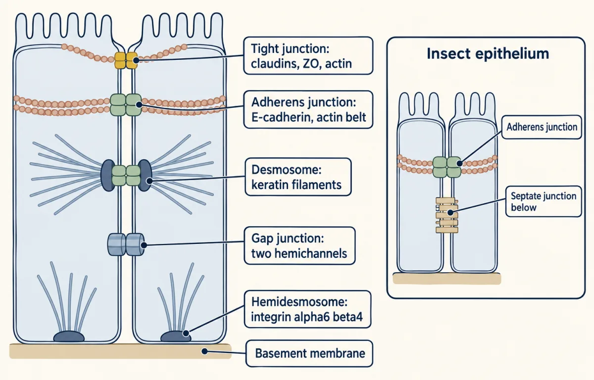
In a vertebrate epithelium the tight junction is the most apical, with the adherens junction just below it, then desmosomes and gap junctions along the lateral membrane, and hemidesmosomes at the base. Many insect epithelia seal with septate junctions that lie below the adherens junction, so the vertebrate order is not the rule for all animals. AI-generated illustration -
Plants: walls and plasmodesmata
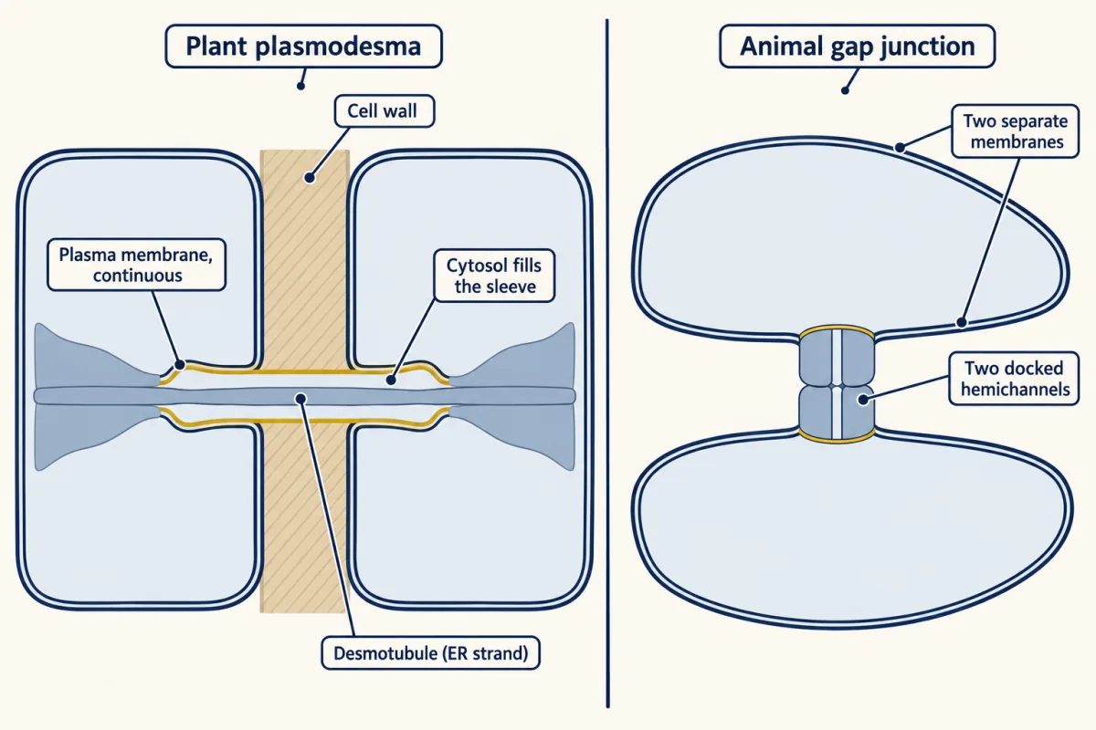
A plasmodesma is a channel through the cell wall lined by plasma membrane that is continuous from one cell to the next, with the desmotubule, a strand of endoplasmic reticulum, down its middle. A gap junction is a protein pore joining two separate membranes, so a plasmodesma makes the two cells' membranes one. AI-generated illustration -
Going deeper
Where this lesson fits
Before thisReview first
- Transport Across MembranesThis course · Lesson 5
You need electrochemical gradients and coupled transport from there to work out how far sodium can push glucose uphill.
- Adhesion and the Extracellular MatrixThis course · Lesson 22
It introduces cadherins, desmosomes and the basement membrane that this lesson stacks into the sheet.
- Vesicle Budding, Targeting and FusionThis course · Lesson 11
Polarized delivery of carriers to the apical or basolateral face here builds on targeted vesicle traffic.
After thisStudy next
- Stem Cells and Tissue RenewalThis course · Lesson 24
It uses the gut lining as its example of a sheet that renews while keeping its faces and seals.
- Cell Identity, Cell State and DiseaseThis course · Lesson 25
It builds on the idea of tissue organization, asking what goes wrong in sheets of cells with altered state.
Elsewhere on the siteFilms, simulations and method films in other courses
- Why Order Matters in a GutFilm · Organismal Biology
It shows the gut as a one-way tube where absorption happens across the lining.
- Downhill for FreeFilm · Biology
It shows how molecules cross a membrane downhill for free, as glucose does through the passive exit.
- Which Way Does the Water Go?Film · Organismal Biology
It shows water crossing epithelia toward higher osmolality, the same water movement the seal governs.
- The bile acid budgetSimulation · Physiology
It follows what the gut lining absorbs and how that is budgeted.
Suggested by an AI model from the lessons themselves, then checked. How we use AI