Developmental Neuroscience · Skim version
The Death Machinery in Neurons
Read the full lesson → Skim the whole course →
The guiding idea
Stop a starving neuron from making new proteins, and it lives longer. Why?
The killers of a developing neuron are already built, and a survival signal keeps brakes on them: one in the worm's core pathway, two in the NGF-dependent sympathetic neuron.
From the film: A Neuron Must Work to Die
- A young sympathetic neuron deprived of NGF does not just run down, because it runs a program that needs new RNA and protein.
- Stopping protein synthesis delays the death of deprived neurons.
- Apoptosis shrinks the cell and condenses its chromatin, while necrosis spills the contents and inflames the tissue.
From the film: Cells Marked with an X
- In the worm, the same named cells die in every animal, so a death can be screened for as a phenotype.
- Loss of ced-3 or ced-4 saves doomed cells, while loss of ced-9 kills cells that should have lived.
- If removing the killer cancels the effect of removing the brake, the brake works through the killer.
From the film: The Same Parts in Us
- Each worm death protein has a mammalian counterpart: caspases, Bcl-2 family proteins and Apaf-1.
- Bax is a gate in the outer mitochondrial membrane that the worm has no clear counterpart for.
- The match is imperfect: Apaf-1 needs cytochrome c, and the worm brake holds CED-4 directly.
From the film: The First Brake: The Mitochondrial Gate
- Brake 1 is a gate at the mitochondrion, and NGF loss opens it through new gene expression.
- Loss of NGF raises c-Jun and BH3-only proteins such as Bim, which free Bax and let cytochrome c escape.
- Without Bax the gate stays shut and the neuron survives, though as a shrunken cell.
From the film: The Second Brake: Competence to Die
- Cytochrome c injected into neurons kept in NGF does not kill them, so the gate is not the only brake.
- Brake 2 is XIAP on the caspases, and deprivation lifts it without any new protein.
- A neuron dies by apoptosis only when the gate is open or skipped and the caspase brake is off.
From the film: Take the Machinery Away
- Deleting caspase-3 or caspase-9 changes how these neurons die, not whether they die.
- In Bax-dependent populations, caspases decide how and Bax decides whether.
- Neurons saved by Bax deletion are not normal, and NGF is also needed for axons to grow into many targets.
-
Two ways to die
-
Cells marked with an X
-
The same parts in us
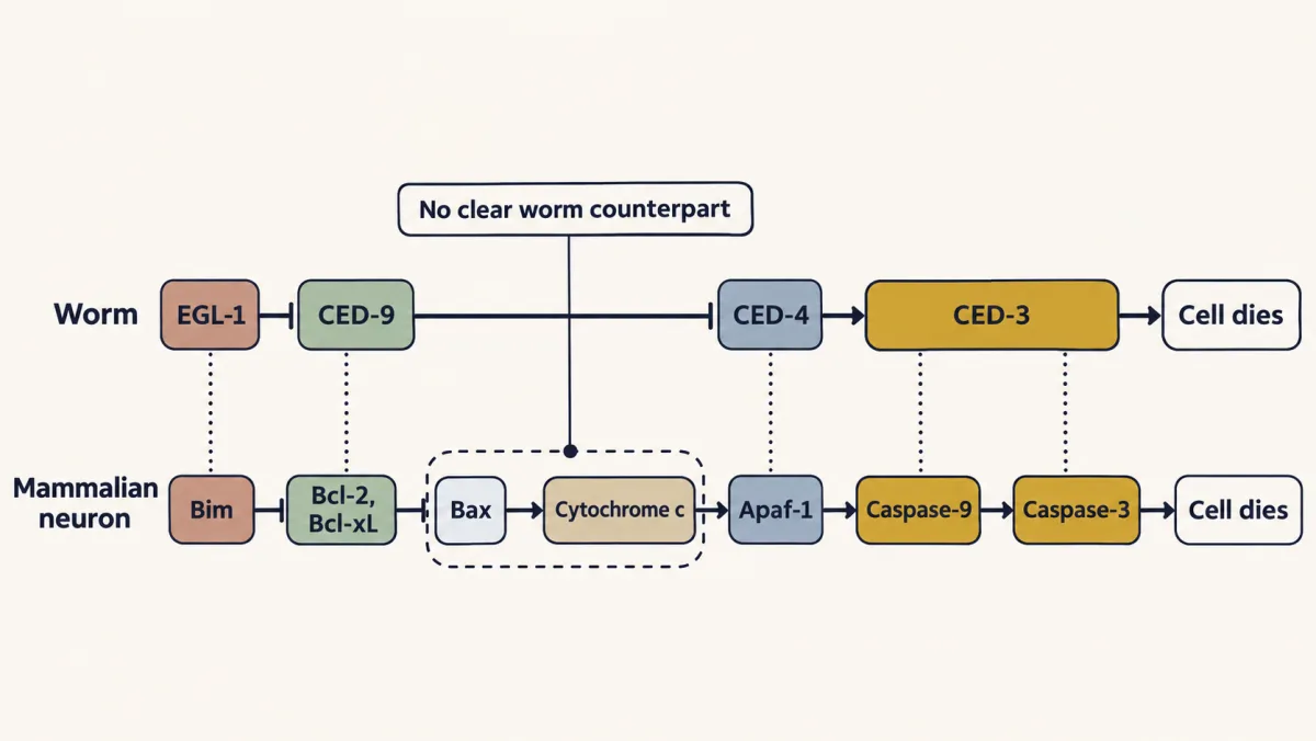
The worm's death genes have mammalian counterparts: EGL-1 and BH3-only proteins such as Bim release the brake, CED-9 and Bcl-2 are the brake, CED-4 and Apaf-1 gather the killer, and CED-3 and the caspases do the killing. The mammalian gate, Bax and the release of cytochrome c, has no clear worm counterpart. AI-generated illustration -
The first brake: from a lost signal to cytochrome c
-
The second brake: competence to die
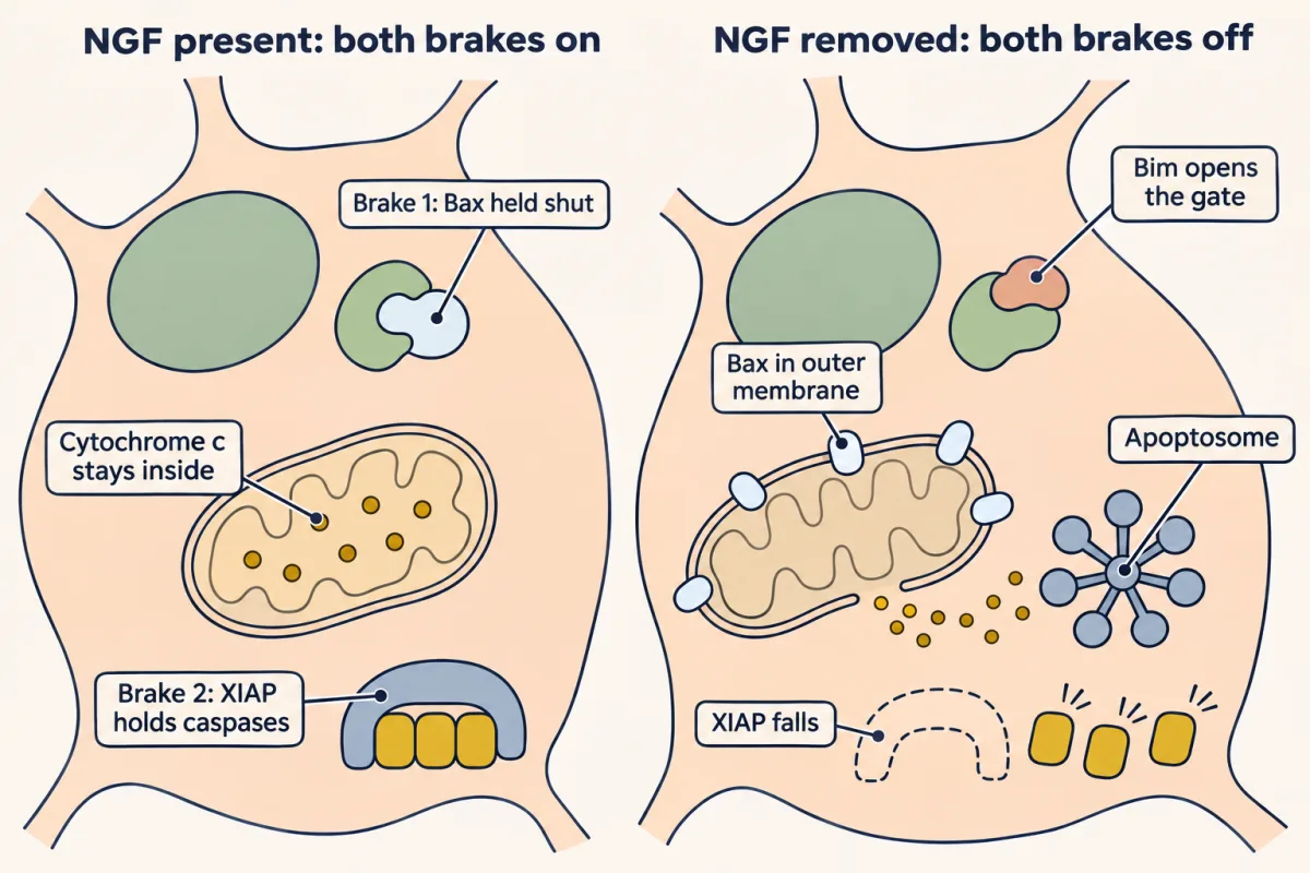
A young NGF-dependent sympathetic neuron has two brakes on apoptosis. Brake 1 is the mitochondrial gate, where protective BCL-2 proteins keep Bax shut until Bim opens it; brake 2 is XIAP on the caspases, which falls during deprivation. The neuron dies only when both are off. AI-generated illustration -
Cleaning up: the corpse sends a signal
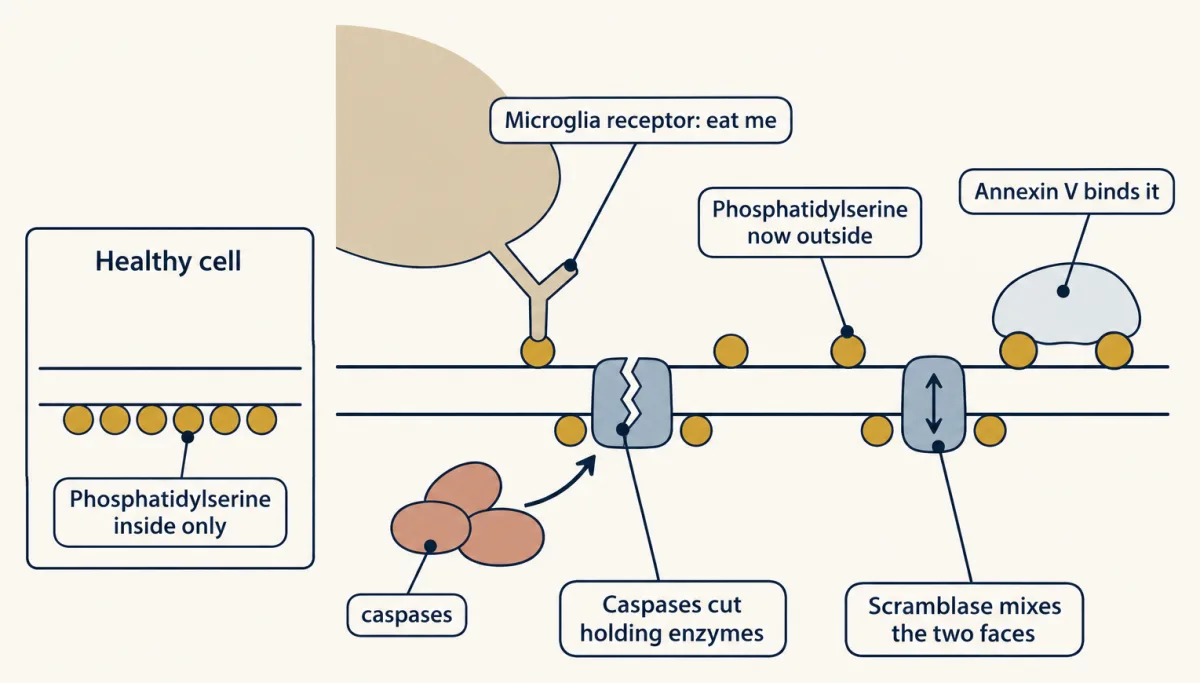
A healthy cell keeps phosphatidylserine on the inner face of its membrane. In the cells where this has been studied, caspases cut the holding enzymes and switch on a scramblase, so phosphatidylserine appears outside as an "eat me" signal that phagocyte receptors, and the lab label annexin V, recognise. AI-generated illustration -
Take the machinery away
-
Putting it together
-
Going deeper
Where this lesson fits
Before thisReview first
- Too Many Neurons: Naturally Occurring Cell DeathThis course · Lesson 28
It shows that neurons are overproduced and die during development, which is the death this lesson explains mechanistically.
- The Neurotrophic Hypothesis TestedThis course · Lesson 29
You need the idea that target-derived NGF keeps neurons alive, since this lesson asks what happens when it is withdrawn.
- Neurotrophins, Trk Receptors and p75This course · Lesson 30
NGF acts through TrkA and Akt to hold back death, which is the survival signal the two brakes depend on.
After thisStudy next
- Competition: How Neurons Win a TargetThis course · Lesson 32
It takes the two-brake survival rule further to ask which neurons win limited NGF and which are lost.
- Pruning Without Dying: Axons That Cut Themselves BackThis course · Lesson 34
It builds on local caspase use in axons to show how a branch is removed while the neuron lives.
- Microglia and Complement Prune SynapsesThis course · Lesson 41
It follows the clearance step, where microglia remove dying cells and synapses.
Elsewhere on the siteFilms, simulations and method films in other courses
- Sealed or TornFilm · Cell Biology
It shows apoptosis as caspases taking the cell apart while its membrane stays sealed, then clearance of the corpse.
- The Safeguard Comes Before the CutFilm · Cell Signaling
It covers BCL-2 family control of cytochrome c release and the caspase cascade in signaling terms.
- Reading annexin V and PISimulation · Cell Biology
It lets you read annexin V and propidium iodide to tell apoptotic from necrotic cells.
Suggested by an AI model from the lessons themselves, then checked. How we use AI