Physiology · Unit 8: Defense, skin and temperature
Lymphoid Organs and Immune Traffic
Rewritten on : new text, figures and flashcards. Your notes and highlights stay, but the text around them may have moved.
Drain a rat's thoracic duct for a few days and its lymph runs short of lymphocytes. Where were they coming from?
Pause when the reading reaches a film, and offer to play it. Saved on this device.
Watch the film now, or keep listening
Builds on:Capillary Exchange, Lymph and EdemaBlood Cells, Hematocrit and ClottingInnate Immunity: Recognize and RespondWhy these?
 ▶ Watch Cells Go Around, Exit Sets the Stay · 2:43
Read the transcript of “Cells Go Around, Exit Sets the Stay”
Film transcript: Cells Go Around, Exit Sets the Stay
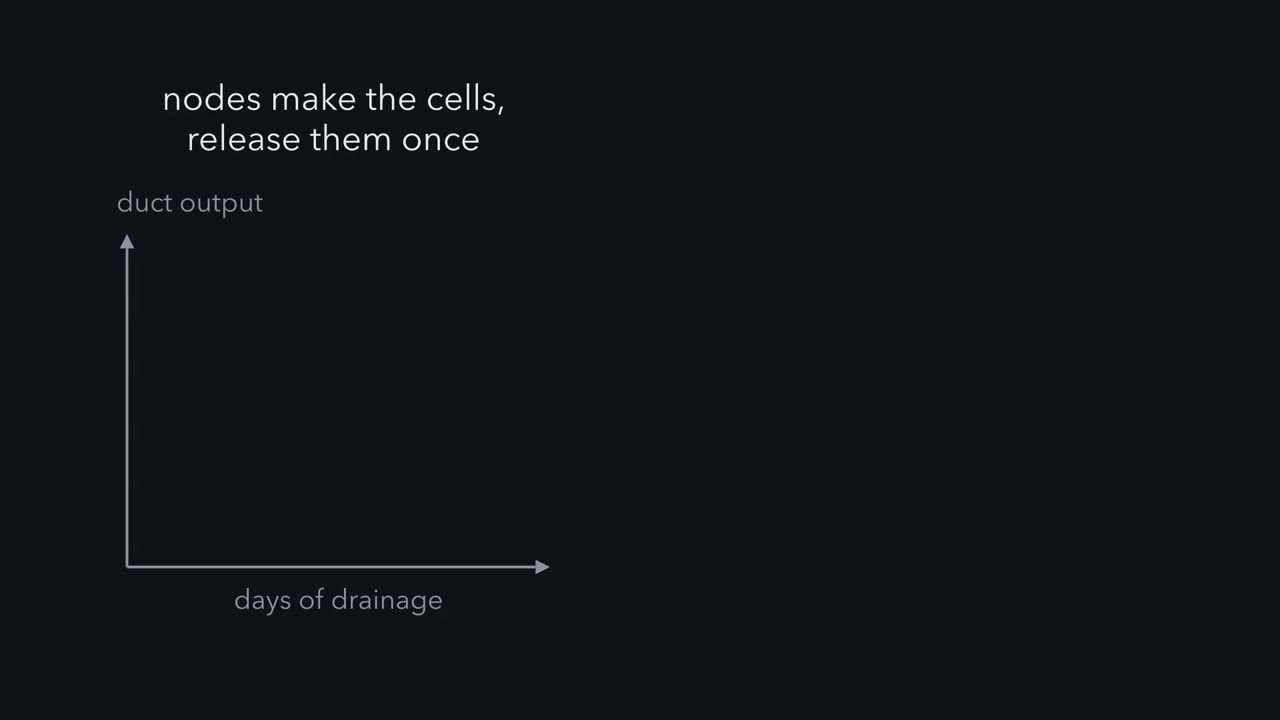
Drain the thoracic duct of a rat for a few days, and the lymph runs short of lymphocytes. The usual view was that lymph nodes made those cells. If so, and they were released once, output should hold roughly steady. If the same cells go around a loop, removing them should empty it. Gowans left a cannula, a fine tube, in the thoracic duct of rats and collected lymph for days. Output fell progressively. When he returned the collected cells by vein, output recovered, and the returned cells turned up again in duct lymph. The cells were going around: from blood, through lymphoid tissue, into lymph, and back to blood.
Labelled lymphocytes injected by vein into rats moved quickly into nodes, across the walls of high endothelial venules. A node has two entrances, afferent lymph and these venules, and one exit, the efferent lymphatic. In large animals at rest, more than 90 percent of lymphocytes in efferent lymph came in through the venules. L-selectin tethers the cell, and it rolls. A chemokine signal switches the integrin LFA-1, which grips ICAM-1 and ICAM-2. The cell stops, then crosses. Getting out needs a receptor. The lipid sphingosine 1-phosphate is high in blood and lymph, and low inside lymphoid tissue. Lymphocytes carry a receptor for it, S1P1. In mice whose blood-forming cells lacked it, mature T cells could not leave the thymus. Deficient T and B cells transferred into normal mice could not leave lymphoid organs. So the need lies in the lymphocyte itself.
Exit sets how long a cell stays. The mean residence time is 1 divided by the exit rate constant. In a fitted rat model, the spleen's exit rate constant is 0.007 per minute. That gives about 2.4 hours. The subcutaneous nodes together hold a cell about 10 hours, and blood about 26 seconds per pass. At steady state, relative to blood, the spleen holds 8 times as many cells, the subcutaneous nodes about 15. This keeps only those three; lung, liver and cell loss are left out. A node that drains a site of immune challenge changes its traffic. In mouse studies, its lymphocytes express CD69, which removes S1P1. Entry from blood rises, and for a time exit slows.
With entry fixed and the exit rate constant halved, residence time doubles, and so does the number held. The shutdown is transient. Keep this: at steady state, cells held equal entry times residence.
Drain the thoracic duct of a rat for a few days and the lymph flowing out of it runs short of lymphocytes.
If lymph nodes were making those cells, drainage should not have used them up. Most were not made there; they were passing through. A naive lymphocyte (one that has not yet met the antigen it recognises) spends its life on a loop: blood, lymph node, lymph, blood again. It never settles in one place, so how many lymphocytes a node holds is a question about traffic.
The one idea to carry through this lesson. Every secondary lymphoid organ is like a bathtub with the tap and the drain both running. At steady state (entry equals exit, so the count holds still), the number of lymphocytes an organ holds = the number entering per hour × the average time each one stays. The stay is set by the way out, not the way in: if cells leave through a single exit step at a fixed rate (for slow exits, the same small fraction of those present per minute), the average stay is 1 / that rate. Entry has its own molecular gate, and so does exit. Open or shut either gate and the cells move.
Where the rule holds. Naive lymphocytes at steady state, with no cell division or death; it is false in the first hours after something changes. Holding an organ’s entry fixed while changing it assumes it is a small part of the pool, such as one node among hundreds (last section).
One real number: in a fitted rat model, about 0.7% of the lymphocytes in the spleen leave per minute, so the average cell stays 1 / 0.007 per min = 143 minutes, about 2.4 hours.
Lymphocytes go around a loop, not out of a factory
Lymphocytes are made in the primary lymphoid organs. In humans and mice, B cells develop in the bone marrow from blood-forming stem cells (stem cells), and T cells mature in the thymus from marrow-derived precursors. The secondary lymphoid organs (lymph nodes, the spleen, and mucosal tissue such as the tonsils and the Peyer’s patches of the gut wall) host mature lymphocytes and are where those cells meet antigen. In the 1950s the usual view was that the lymphocytes in lymph were newly made in the nodes upstream. James Gowans tested it in rats fitted with a chronic cannula in the thoracic duct, the large lymph vessel that empties into the veins at the base of the neck (capillary exchange and lymph covers how lymph forms and moves). The cannula let him collect lymph for days.
If nodes made the cells and released them once, duct output should have held roughly steady while production continued. If the same cells were going around, removing them should empty the loop. Output fell progressively.
Predict. Gowans then returned the collected cells into a vein. What should happen to duct output if the nodes are a factory? What if the cells go around a loop?
Check your prediction
A factory would not care: cells put into blood have no way back into its production line, so output would stay low. A loop would refill: cells put into blood re-enter the nodes and come out in lymph again. Output recovered, and the returned cells were later found in duct lymph.
The cells go around, and the nodes are not the main source of duct lymphocytes in the rat. This is rat data, from a duct that samples only part of the pool (Going deeper, “What Gowans’s experiment did not show”).
Gowans’s 1964 papers asked where the cells leave the blood. Radioactively labelled lymphocytes injected by vein into rats moved quickly into lymph nodes across the walls of high endothelial venules (HEVs), small venules lined by plump, almost cuboidal endothelial cells instead of flat ones. Gowans concluded that HEVs are the site of large-scale migration of lymphocytes from blood into the secondary organs.
A node samples lymph and blood; the spleen samples blood alone
Why tour at all? Only a tiny fraction of T cells, of the order of one naive T cell in a hundred thousand to a million, can recognise any one antigen, so the antigen has to be brought to a place they all pass through. Dendritic cells carry it there: they pick up antigen in skin or gut and travel in afferent lymph (lymph flowing into the node) to its T-cell zone, a trip that needs the chemokine receptor CCR7 (in CCR7-deficient mice, activated skin dendritic cells fail to reach the nodes). So the lymphocytes touring the node have something to find.
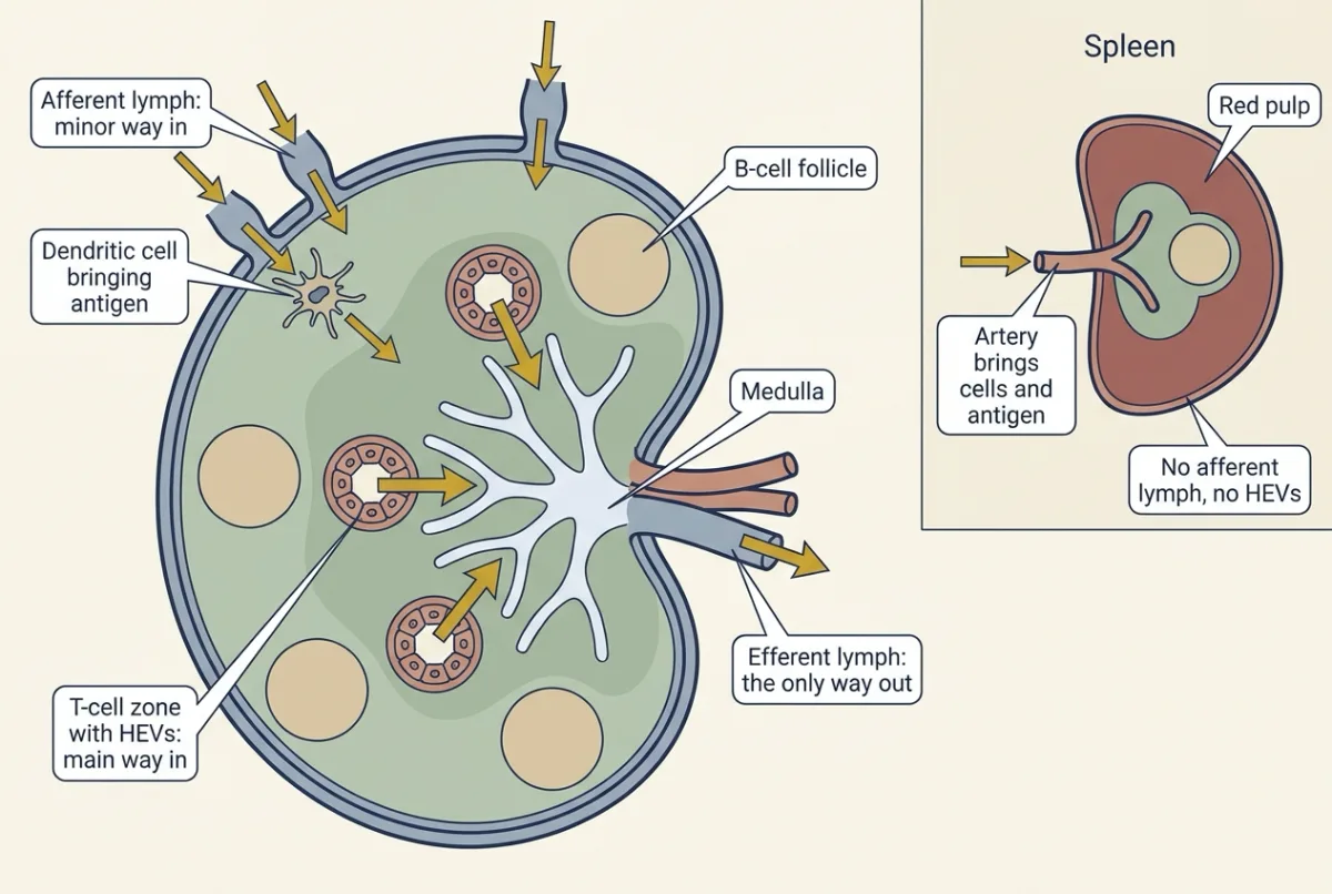
A lymph node is a bean-shaped filter on a lymph vessel. Afferent lymphatics enter its convex surface and empty into the subcapsular sinus, a space under the capsule. Inside lie B-cell follicles in the outer cortex, a T-cell zone (the paracortex) where the high endothelial venules (HEVs) sit, and medullary cords and sinuses. One efferent lymphatic, sometimes two, leaves at the hilum, beside the artery and vein.
Think of the node as a hotel with two entrances, afferent lymph from tissue and HEVs from blood, and one exit, the efferent lymphatic. The blood entrance is much the busier. At rest, afferent lymph has only about 5 to 10 percent of the cell concentration of efferent lymph, and cannulation studies in large animals found that more than 90 percent of the lymphocytes in efferent lymph of resting nodes had entered through HEVs.
The hotel also carries the guiding idea: 100 check-ins a night, each staying 3 nights, fills 300 rooms.
The spleen, the largest secondary lymphoid organ, has no afferent lymphatics and no HEVs, so it samples blood, not lymph. Arterial blood brings both antigen and lymphocytes. Its white pulp holds T-cell and B-cell zones around arterioles, and its red pulp removes old red cells (blood cells and hemostasis).
Getting in on chemokines, getting out on S1P
The way in: three steps at the high endothelial venule
In a peripheral lymph node:
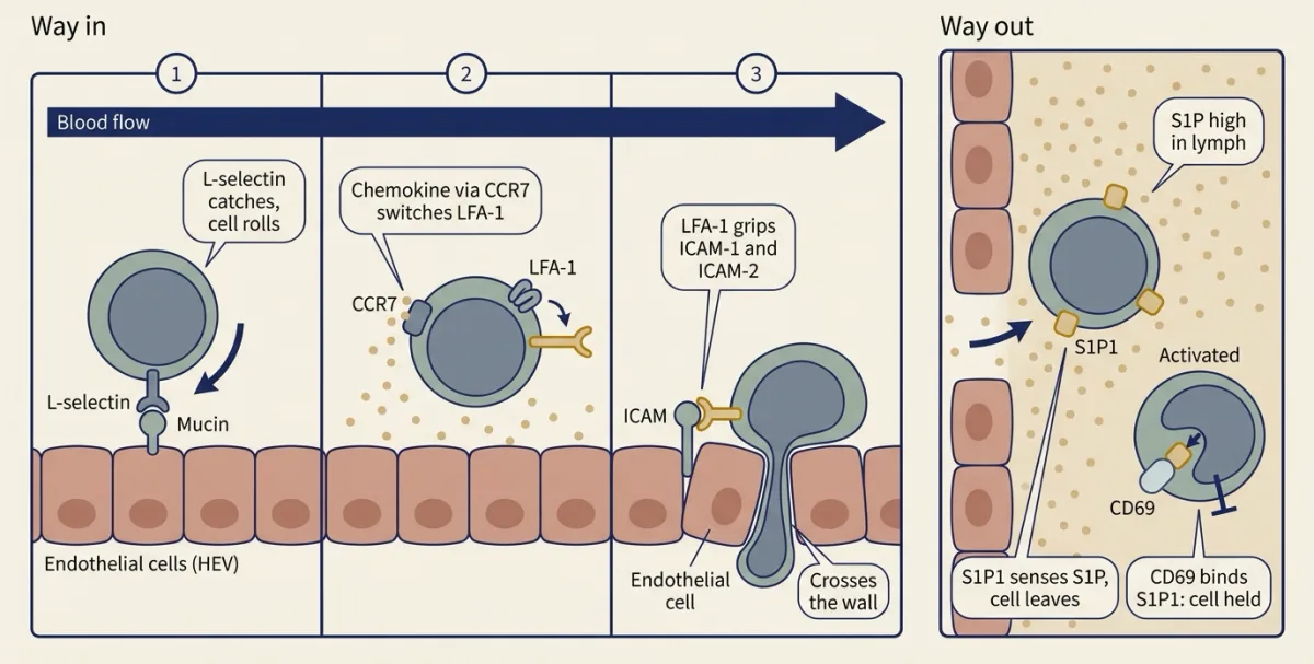
- Tether and roll. L-selectin (CD62L) on the lymphocyte binds sulfated mucin-like glycoproteins on the HEV, which tethers a fast-moving cell and sets it rolling. L-selectin needs no signal before it binds, though its grip is stable only above a minimum shear from flowing blood.
- Switch on the integrin. The integrin LFA-1 stays in a low-affinity shape until a chemokine (CCL21, CCL19 or CXCL13) signals through its receptor, CCR7 for CCL21 and CCL19. In mice the HEVs make CCL21 themselves; human HEVs carry it but do not make it.
- Arrest and cross. The signal changes LFA-1’s shape so it binds ICAM-1 and ICAM-2 on the endothelium, and the cell stops, then crosses the wall.
Predict. L-selectin is already sticky. Why would a lymphocyte need a second adhesion molecule that only grips after a chemokine signal?
Check your prediction
L-selectin catches and lets go, so the cell rolls but cannot stop. Requiring a chemokine signal before the firm grip means the cell commits to stopping only where that signal is presented, on the HEVs of a lymphoid organ.
The way out: S1P1 reads the S1P in lymph
Exit was a later question. The lipid sphingosine 1-phosphate (S1P) is high in blood and lymph and low inside lymphoid tissue, and lymphocytes carry a G protein-coupled receptor for it, S1P1. In 2004 Mehrdad Matloubian, Charles Lo, Guy Cinamon and colleagues in Jason Cyster’s laboratory asked whether lymphocytes need it to leave. Mice lacking S1P1 in every cell do not survive embryonic development, so they gave irradiated mice blood-forming cells from S1P1-deficient fetal liver. If S1P1 is needed for exit, cells should enter lymphoid organs and pile up there.
They did. Mature T cells could not leave the thymus, so no T cells reached the periphery. S1P1-deficient T and B cells transferred into normal mice could not leave lymphoid organs, so the requirement lies in the lymphocyte, not its surroundings. This is mouse work: the study gave no human values, and did not show how the S1P gradient is kept or that every subset depends on S1P1 equally.
The exit gate can also be closed from inside. The same study found that S1P1 falls when lymphocytes are activated, which goes with retention. CD69, a surface protein that appears early after activation, binds S1P1 and opposes its function, and the pair is internalized and degraded, so T cells expressing CD69 stay in the node.
Why a cell’s stay is set by the way out
With both gates in hand, ask how long a lymphocyte stays, starting with the spleen. A lymphocyte there has no scheduled departure. At each moment it either happens to reach a place where it can sense the sphingosine 1-phosphate (S1P) of blood and leave, or it does not. So the fitted model treats leaving as a matter of chance: the same fraction of the cells present leaves each minute, not the same number: the rat spleen’s 0.7% per minute holds whether it contains a thousand cells or a billion. A process like that is called first-order, and the 0.007 per min is its exit rate constant. (A lymph node adds a delay before exit becomes possible, so its fit needs two such steps in series; see Going deeper.)
That one property fixes the average stay. If 10% left each hour, the average cell would stay about 10 hours; at 0.007 per min it stays the opening’s 143 minutes. The entry rate is not in that calculation: how fast cells arrive has no effect on how long any one of them stays.
At steady state, as many cells leave per hour as arrive, so the two rules combine into the guiding idea:
In words The number held equals the number entering per unit time, times the mean stay, and the mean stay is one over the exit rate constant.
- N“N”
- the number of lymphocytes the organ holds at steady statecells
- J“J”
- the number of cells entering the organ per unit time (the entry flux)cells per h
- e“e”
- the exit rate constant: the fraction of the cells present that leaves per unit time, for an organ with a single first-order exit stepper h
- 1 / e“one over e”
- the mean stay (mean residence time) of one cellh
Sanity check Double J and N doubles, but each cell stays as long as before. Halve e and the mean stay doubles. Entry sets how many; exit sets how long. This is Little's law, a general rule for queues, and it needs a steady state.
Try one number
Made-up entry, to keep the arithmetic clean: 1,000 naive CD4 T cells and 1,000 naive CD8 T cells enter one mouse node per hour.
In mice, naive CD4 T cells cross a node in about 12.2 h and naive CD8 T cells in about 21.2 h.
CD4: 1,000 per h × 12.2 h = 12,200 cells. CD8: 1,000 per h × 21.2 h = 21,200 cells.
The node holds 21.2 / 12.2 = 1.74 times as many CD8 as CD4 cells, and entry did not differ at all: the ratio is the stay and nothing else. What makes CD8 cells linger is still an open question.
Need a refresher? rates and “per” · units and prefixes
Predict. In the rat model, the spleen receives about twice as many lymphocytes per minute from blood as the subcutaneous nodes (those under the skin) do. Yet at steady state the spleen holds only about half as many. How can that be?
Check your prediction
Look at the other side of the rule. A cell stays about 2.4 hours in the spleen but about 10 hours in the subcutaneous nodes, about four times longer. Twice the entry times a quarter of the stay gives about half the holding. Traffic alone does not tell you what an organ holds.
Predict. A drug strips S1P1 from the surface of a patient’s lymphocytes. Entry through the high endothelial venules (HEVs) is untouched. What happens to the number of lymphocytes in the patient’s blood, and to the size of their lymph nodes?
Check your prediction
The exit gate is shut, so the stay in lymphoid tissue lengthens, and the extra cells held there come out of blood: the blood lymphocyte count falls steeply. The nodes gain only a little, because blood was a small part of the pool to begin with. Fingolimod, a multiple sclerosis drug that removes S1P1 from lymphocytes, lowers blood lymphocyte counts this way: the cells are held in lymphoid tissue, not killed.
- Spleen stay = 1 / exit rate
- Node stay = 2 / exit rate (two parts)
- Steady-state ratio blood : spleen : nodes
- Steady-state shares, blood · spleen · nodes
- Shares of the cohort at 24 h, blood · spleen · nodes
- Nodes, relative to defaults: stay × entry flux = held
- Spleen, relative to defaults: stay × entry flux = held
In this sketch: three compartments only, blood, spleen and subcutaneous nodes (two parts in series), with first-order entry and exit, steady state, no cell death or division, and labelled cells behaving like the rest of the pool; the nodes here are the lumped rat compartment. The default rates are the lesson’s fitted values for a rat (Ganusov and Auerbach, 2014; per min): spleen entry 0.056 and exit 0.007, node entry 0.026 and exit 0.0034 for each part. The slider ranges, the factor of 0.1 for lymphocytes lacking S1P1 and the doubling of node entry are made up for the sketch. The curves over 24 h are computed in this sketch from those first-order rules; the lesson gives no time course. Lung, liver, loss and other organs are omitted, so the blood share is a teaching value, not a measurement. Because lung and liver are left out, cells leave blood in about 12 min here at the default entry rates, 1 / (0.056 + 0.026) per min, not the lesson’s 26 s per pass.
Rule to keep: mean residence time = 1 / exit rate constant, and the number held at steady state = entry rate × residence time (Little’s law), so a longer stay or a faster entry gives a compartment a larger share relative to blood.
In the sandbox, the sliders start at the fitted values; watch where a cohort starting in blood settles. Then halve the node exit rate: each cell stays twice as long, but because every node slowed at once, blood drains, entry falls, and the nodes hold about 1.23 times as many, not twice.
A draining node holds on to its visitors
A node that drains a site of immune challenge changes its traffic. Migrating dendritic cells arrive in afferent lymph and settle in the T-cell zone. More lymphocytes enter from blood through the high endothelial venules (HEVs), and for a time fewer leave: in mouse studies, lymphocytes in the node express CD69, which removes S1P1 from their surface. The node enlarges and holds more lymphocytes than its partner on the other side of the body.
Predict. A draining node comes to hold twice as many lymphocytes as before. Using the guiding idea, give two different ways that could happen.
Check your prediction
Double the entry (each cell staying as long as before), or halve the exit rate constant (the same entry, each cell staying twice as long), or any mix of the two. The rule cannot tell you which happened; only measuring entry and exit separately can. How much of the enlargement comes from more entry and how much from less exit is still debated. A model fitted to rat data (Ganusov and Auerbach, 2014) put it down mainly to increased entry.
When halving exit doubles the holding. The second answer kept entry fixed. That is fair for one draining node out of hundreds: slowing its exit barely changes the blood count, so entry keeps its old rate and halving exit doubles both the stay and the number held. It is not fair if every node slows at once, as with fingolimod. Then blood is drained, entry into each node falls with it, and the holding rises by much less than the stay (Going deeper, “When every node slows at once”). The film’s line about entry fixed and exit halved is this one-node case.
If entry also rises, the node holds more again. The longer stay gives the rare cells that recognise the antigen more time to find it. The shutdown is transient, and exit resumes afterwards.
Design problem (extension)
You want to measure how long naive T cells stay in a mouse lymph node and test whether S1P1 sets that time. Design an adoptive-transfer experiment, say what each control rules out, and interpret these made-up results: at the start, one node holds 10,000 labelled normal T cells and 10,000 labelled S1P1-deficient T cells; 6, 12 and 24 h later it holds 6,100, 3,700 and 1,400 normal cells, and 9,800, 9,500 and 9,100 S1P1-deficient cells.
One way to do it
Take naive T cells from normal mice and from mice in which S1P1 can be deleted in T cells after they have left the thymus (for example with an inducible Cre); fetal liver chimeras will not do, because their mature T cells never reach the periphery. Give each population a different congenic surface marker, and both a photoconvertible fluorescent protein that turns from green to red under violet light. Inject them together by vein into one normal host, so differences cannot come from the host. Prepare both donor populations the same way, and swap the two markers between repeats so the labels themselves are not the cause.
A day later, expose one peripheral node to violet light under anaesthesia. Transferred cells inside it turn red; cells arriving later stay green, so new arrivals do not blur the cohort. Harvest that node from separate groups of mice at 0, 6, 12 and 24 h and count red cells of each type by flow cytometry.
Controls: equal time-zero counts show both populations start from the same cohort, so later differences reflect exit or death, and checking the viability of each population in the node helps tell them apart; illuminating cells and checking their viability tests whether the light harms them; counting red cells in blood and in an unexposed node shows where leavers go.
Interpretation: for first-order exit, cells remaining in the node = N0 x exp(-k t). For normal cells, ln(10,000 / 3,700) = 0.99 over 12 h, so k is about 0.083 per h and mean residence about 12 h; the 6 h and 24 h points fit (10,000 x exp(-0.5) = 6,070; 10,000 x exp(-2) = 1,350). For S1P1-deficient cells, ln(10,000 / 9,100) = 0.094 over 24 h, about 0.004 per h, or roughly 250 h: exit is nearly abolished, and the small loss could be cell death. Limits: one species and one node type, a few red cells may re-enter from blood, and the result shows S1P1 is needed for exit, not that it alone sets the normal residence time.
Going deeper
The main line above does not depend on these details.
Miller’s controls
How the thymus’s role was found, by Miller’s neonatal thymectomy in mice and by Good’s laboratory in rabbits, belongs to adaptive immunity. Two of Miller’s controls carry the logic: sham-operated mice had the same surgery without losing the thymus, and adult mice thymectomized, irradiated and given marrow could not recover immune function, unlike sham-operated controls given the same irradiation and marrow (Cross, Leuchars and Miller, 1964).
What Gowans’s experiment did not show
It did not show which lymphocytes recirculate, how fast each kind does, or what molecules let them cross a vessel wall, and it gave no human values. In the rat model below, the duct is assumed to collect lymph from the mesenteric nodes and about half of the output of the subcutaneous nodes.
How the entry molecules were found
Thome had described the shape of high endothelial venules (HEVs) in 1898; for decades their role went unrecognised, until Gowans’s 1964 work. Stamper and Woodruff in 1976 laid rat and mouse lymphocytes over frozen sections of lymph node and found that they stuck to HEVs and not to other vessels. In 1983 Gallatin, Weissman and Butcher described MEL-14, an antibody to a mouse lymphocyte molecule that appeared to be required for homing to nodes; the molecule is L-selectin. In 1994 mice lacking L-selectin had a severe reduction in lymphocytes in their peripheral nodes.
Other routes in
HEVs in the Peyer’s patches of the gut also use a second pair: the integrin alpha4beta7 on the lymphocyte binds MAdCAM-1 on the venule. The HEVs depend on lymph: tying off a node’s afferent lymphatics makes them lose their specialised features. L-selectin is shed from the cell surface on activation. The figure of more than 90 percent entry through HEVs is for resting peripheral nodes; afferent lymph contributes more during inflammation.
How antigen gets into a node
Antigen arrives in afferent lymph, free or carried by cells. Small soluble molecules travel from the subcapsular sinus toward the HEVs through conduits, fine tubes of reticular fibres wrapped in fibroblastic reticular cells; in mice, resident dendritic cells in the T-cell zone pick up soluble antigen this way before migrating dendritic cells arrive from the tissue. Steinman and Cohn described and named dendritic cells in 1973, in mouse spleen, lymph node and Peyer’s patch, where they were only 0.1 to 1.6 percent of nucleated cells.
The spleen has its own route. Intravital imaging in mouse spleen showed T cells leaving arterial blood into the red pulp, latching onto perivascular stromal cells without needing CCR7, then moving into the T-cell zones, which does need CCR7.
The rat model in full
This is where the film’s figures come from (about 26 seconds per pass in blood; the spleen holding eight times as many cells as blood and the subcutaneous nodes about fifteen times), and the sandbox’s starting values.
Ganusov and Auerbach (2014) fitted a compartment model to data from chromium-51-labelled thoracic duct lymphocytes injected into rats. It assumes first-order entry and exit, steady state, no cell death or division, and that labelled cells behave like the rest of the pool.
(a) Spleen: entry from blood 0.056 per min, exit 0.007 per min. Mean residence = 1 / 0.007 per min = 142.9 min, about 143 min or 2.4 h. The fitted table gives 144 min (115 to 173).
(b) In a made-up cohort of 1,000,000 labelled cells in blood, the initial rate of entry into the spleen is 0.056 per min × 1,000,000 = 56,000 cells per min.
(c) Time in blood. The model’s entry rates from blood are 1.83, 0.41, 0.056, 0.026, 0.0106 and 0.0053 per min; the two largest are into the vessels of lung and liver, from which cells return quickly. The sum is 2.338 per min, so the mean time in blood per pass is 1 / 2.338 per min = 0.428 min = 25.7 s, about 26 s, as the model reports. Of cells leaving blood, 0.056 / 2.338 = 2.4 percent go to the spleen and 0.026 / 2.338 = 1.1 percent to the subcutaneous nodes.
(d) Where the pool sits. Keep only blood (B), spleen (S) and subcutaneous nodes (N). At steady state entry equals exit for each, so S / B = 0.056 / 0.007 = 8.0. The model treats the subcutaneous nodes as two sub-compartments in series, each with exit 0.0034 per min. This is a mathematical device for the delay before cells leave, not two regions of a node, and it gives a peaked spread of stays rather than the simple exponential of a one-step compartment. N1 / B = 0.026 / 0.0034 = 7.65, the second holds the same, so N / B = 15.3. With two exit steps in series, node residence = 2 / 0.0034 per min = 588 min, about 9.8 h (table: 593 min), not 1 / 0.0034. Blood’s share is 1 / (1 + 8.0 + 15.3) = 1 / 24.3 = 4.1 percent.
Checks: 2.338 × 0.428 = 1.00; 0.024 × 2.338 = 0.056; 24.3 × 0.041 = 1.00. The blood share is a teaching value, since lung, liver, other organs and cell loss are left out.
When every node slows at once
Take the three-compartment version in (d) and halve the exit rate constant of all the subcutaneous nodes together. The pool is closed, so blood’s share falls from 4.12% to 2.53% (× 0.61). Entry into the nodes is proportional to the cells in blood, so it falls by the same factor, and the nodes’ share rises from 62.95% to 77.27%: × 1.23, not × 2. Cut exit tenfold from both spleen and nodes, the direction a drug like fingolimod pushes, and in this model blood’s share falls about tenfold, from 4.12% to 0.43%, while the nodes’ share rises only from 62.95% to 65.38%: the drop in the blood count is the whole-pool effect, not a single-node one. The tenfold cut is made up; fingolimod itself lowers the blood lymphocyte count by about 70 percent, and spares cells that do not need CCR7 to re-enter nodes.
Your notes
Select any text in the lesson to highlight it or add a note to it. Your highlights and notes are saved in this browser and collected in your notebook.
This browser is not letting the site save anything (a private window or strict privacy settings), so highlights and notes will be lost when you leave the page.
How we measure it
Thoracic duct cannulation
A fine tube is placed in the thoracic duct of an anaesthetized animal and left in place, so lymph can be collected for hours or days. Counting the cells in it gives lymphocyte output per hour; in rats the duct drains the mesenteric nodes but only part of the subcutaneous node output (a fitted model assumes about half), so it samples part of the pool, not all of it.
Adoptive transfer of labelled lymphocytes
Lymphocytes are collected, tagged with a radioactive label such as chromium-51 or a fluorescent dye, and injected by vein. Measuring the label in blood, lymph and each organ over time gives entry and exit rates, and fitting a compartment model turns them into residence times.
Knockout mice and haematopoietic chimeras
A gene is deleted in every cell (L-selectin, CCR7) or only in blood-forming cells, by transplanting mutant fetal liver or marrow into irradiated hosts (S1P1). Comparing the two designs, or transferring mutant cells into normal mice, tells you whether a molecule is needed in the lymphocyte or in its surroundings.
Frozen-section adhesion assay
Lymphocytes are laid over thin frozen sections of lymph node, washed, and counted where they stick. Stamper and Woodruff found that they stick selectively to high endothelial venules, which made homing a measurable binding event that antibodies could block.
Practice questions on this lesson (3) → MCAT-style, original, each with an explanation
Where this lesson fits
Before thisReview first
- Capillary Exchange, Lymph and EdemaThis course · Lesson 9
You need how lymph forms and flows back to the veins to follow the thoracic duct and the loop.
- Blood Cells, Hematocrit and ClottingThis course · Lesson 11
Lymphocytes are blood cells, and this lesson counts them in blood, nodes and spleen.
- Innate Immunity: Recognize and RespondOrganismal Biology · Starting college
It introduces the dendritic cells and immune basics that carry antigen to the node.
Elsewhere on the siteFilms, simulations and method films in other courses
- Receptors First, Antigen ChoosesFilm · Organismal Biology
It shows how naive lymphocytes with receptors made before meeting antigen are what the node's tour is sampling.
- Lineage tracingMethod film
Following the descendants of marked cells is the logic behind tracking lymphocytes around the loop.
Suggested by an AI model from the lessons themselves, then checked. How we use AI
Finish line
Test yourself
8 questions
Practice and MCAT-style, with explained answers.
Next · Lesson 26
Skin, Sweat and Human Thermoregulation
A gram of sweat that evaporates carries off about 2.4 kJ of heat. A gram that drips carries off almost none.
Continue →← Lesson 24 · Absorption, the Liver and Enterohepatic Circulation