Sensory Systems · Skim version
Meissner and Pacinian Corpuscles: Flutter and Vibration
Read the full lesson → Skim the whole course →
The guiding idea
Slide a fingertip across cloth and Meissner corpuscles report the movement; hold a buzzing phone and Pacinian corpuscles report the hum. Each is a nerve ending wrapped in cells that help it feel. How do the two split the work of sensing change?
A corpuscle reports change, not level, and the brevity of its response can arise in three places: the capsule, the terminal's channels and the spike generator.
From the film: Reporters of Change
- Meissner and Pacinian corpuscles report change, not level: their afferents fire when the skin starts to move, stops or keeps moving, and go quiet while a press is held.
- Meissner afferents serve low-frequency flutter and Pacinian afferents high-frequency vibration, and the best frequency differs between humans and mice.
- The brevity of the response can arise in three places: the capsule, the terminal's channels and the spike generator.
From the film: Peel the Onion
- Brevity can arise in the capsule, the terminal's channels or the spike generator, and the classical account put the filtering in the capsule.
- In cat corpuscles with most layers peeled, the generator potential decayed more slowly but the impulses did not persist, so brevity had two stages.
- In the duck preparation, the terminal stayed fast without the outer core, so fast adaptation does not need the onion there.
From the film: The Cells That Help It Feel
- In mice, force opens Piezo2 in the sensory axon, but the wrapping cells also feel force and contribute to detection; how they normally do so is not yet known.
- Switching off Pacinian wrapping cells in mice meant a vibration had to be about five times larger to be detected.
- Mice without Meissner corpuscles needed about twice the push to detect a touch yet still detected stronger pushes, so these corpuscles help set sensitivity.
-
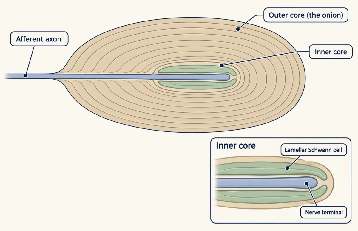
A Pacinian corpuscle has an outer core of many concentric layers, the onion, around an inner core where lamellar Schwann cells hug the nerve terminal. AI-generated illustration -
The typical response: a burst at each end
-
Where force becomes current: Piezo2 in the axon
-
Why the response is brief: capsule, terminal and spike
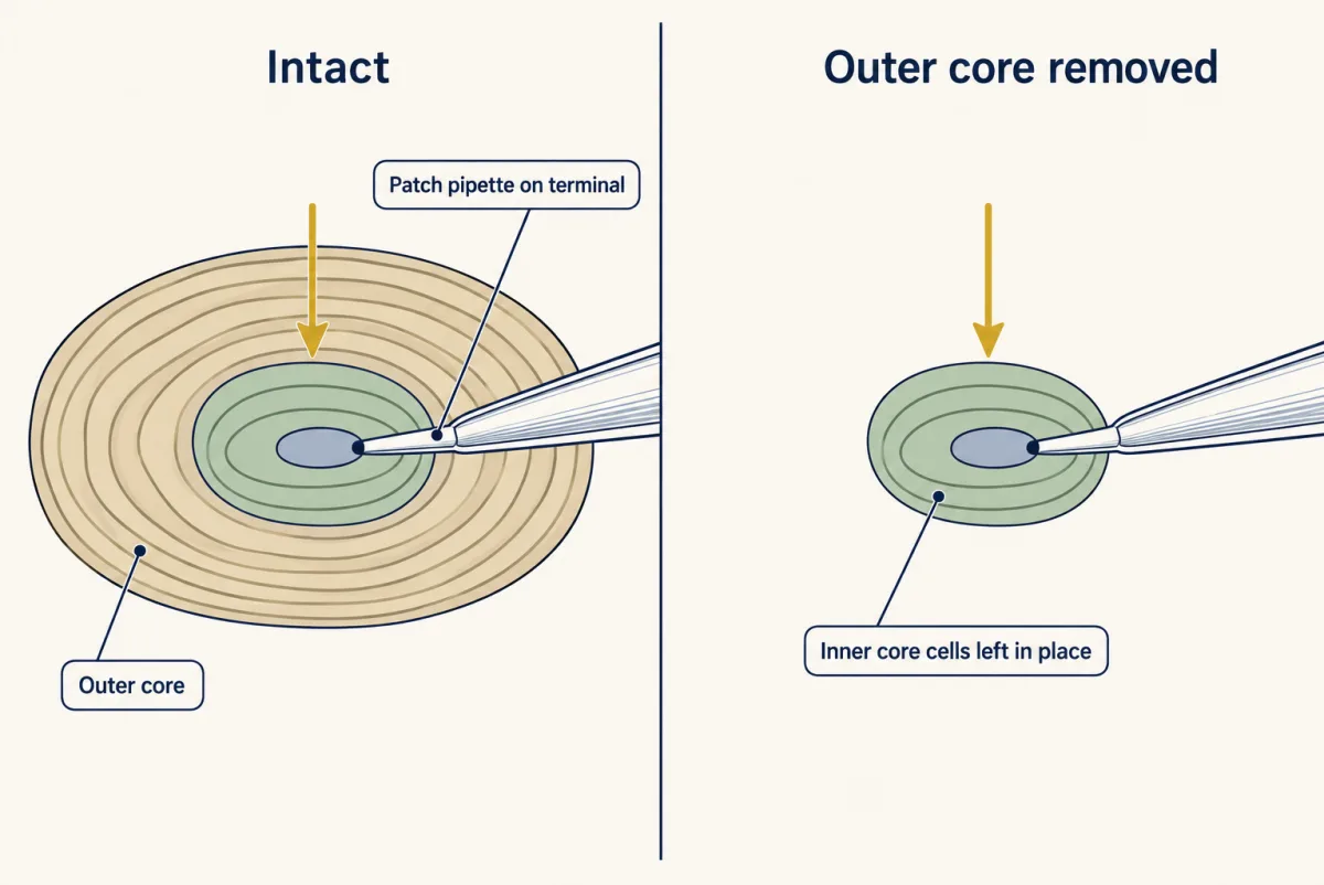
Ziolkowski and colleagues patch-clamped the terminal inside duck Herbst corpuscles, with and without the outer core. Removing it left the rapid kinetics of the transduction current intact in this ex vivo embryo preparation. AI-generated illustration -
What is left once the onion is gone: the lamellar cells
-
Two afferents in one Meissner corpuscle
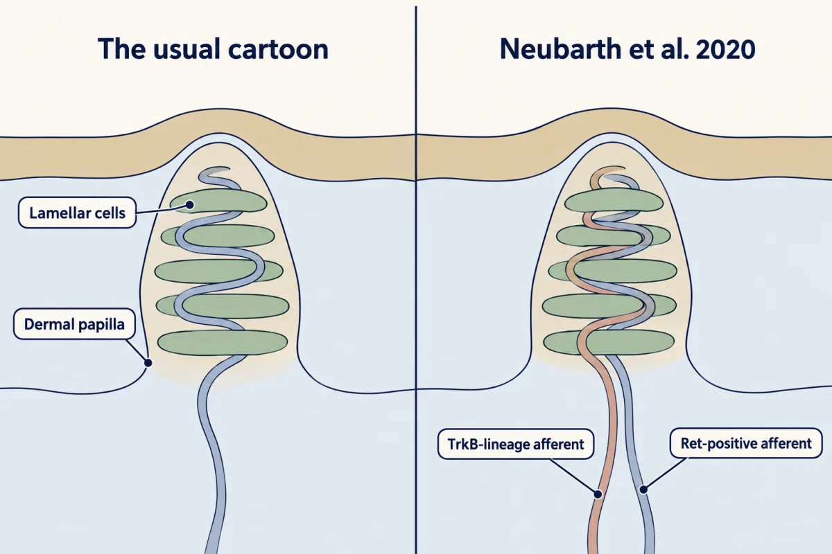
Most Meissner corpuscles in adult mouse paw pads held one Ret-labelled and one TrkB-lineage Aβ afferent, intermingled among lamellar cells. Both are developmental genetic labels. Neubarth and colleagues (2020) distinguished two afferent types; several endings per corpuscle had been described before. AI-generated illustration -
Vibration in the brain and in the wild
-
Going deeper
Where this lesson fits
Before thisReview first
- Piezo Channels and the Physics of ForceThis course · Lesson 2
You need Piezo2 as the force-gated channel and the proof a transducer claim requires, which this lesson takes as settled.
- The Low-Threshold Mechanoreceptors: One Touch, Several SignalsThis course · Lesson 4
This lesson builds on the class table that places Meissner and Pacinian afferents among the touch neurons.
- Five Ways to Turn a Stimulus into a CurrentThis course · Lesson 1
The split between generator potential and spike adaptation here rests on how a stimulus becomes a current.
After thisStudy next
- Where Touch Is First Processed: Dorsal Horn and Dorsal Column NucleiThis course · Lesson 8
It follows these rapidly adapting afferent signals into the spinal cord and brainstem.
- Hairy Skin, Stroking and Affective TouchThis course · Lesson 7
It applies the same afferent-response ideas to other touch fibres, comparing them with the corpuscle afferents.
- Touch Over-reactivity in Autism ModelsThis course · Lesson 9
It tests how changes in these touch afferents alter behaviour in mice.
Elsewhere on the siteFilms, simulations and method films in other courses
- The Spike and Its PuzzlesFilm · Neuroscience
It shows spike generation, one of the three places where brevity can arise.
- What a Spike MeansFilm · Neuroscience
It shows that receptor spikes carry stimulus change as rate, the basis for flutter and vibration coding.
Suggested by an AI model from the lessons themselves, then checked. How we use AI