Sensory Systems · Skim version
Balance: Two Codes for Every Head Movement
Read the full lesson → Skim the whole course →
The guiding idea
People with severe loss in both balance organs can see the world bounce as they walk. Yet in monkeys, head-motion neurons go largely quiet for a turn the animal makes itself, which may keep the head-steadying reflex from fighting it. How does one nerve report motion fast and precisely, yet let the brain set your own turns aside?
The vestibular nerve reports each head movement on two overlapping channels, whoever caused it: a steady, precise firing rate and a higher-gain, well-timed signal.
From the film: Report It Twice
- The vestibular nerve reports each head movement on two overlapping channels, whoever caused it.
- In the canals, regular fibres follow how fast the head is turning in a steady rate, while irregular fibres add higher gain, more response to how quickly the motion changes, and well-timed spikes.
- In the head-posture neurons tested, your own turn is largely cancelled when neck feedback matches the prediction, which may help the head reflex spare the turns you intend.
From the film: A Synapse That Does Not Wait
- Type I hair cells release glutamate from vesicles, as type II cells do, though sustained release in mature mouse tissue was about tenfold smaller.
- In mouse tissue in a dish, type I cells lacking otoferlin or a needed calcium channel could still make the calyx fire.
- Cleft potassium and cleft potential can move the calyx without vesicles; the animal recordings are measurements, while the model's order of events is a prediction.
From the film: Pass On the Surprise
- In the neurons tested in monkeys, the response to a head turn is cancelled only when the turn is self-made and neck feedback matches the motor command's prediction.
- This is not the ear switching off: canal afferents kept firing, so any cancelling happens in the brain.
- A vestibular implant supplies a motion-modulated rotation signal without the nerve's natural two channels or an otolith signal, and in a small cohort hearing in the implanted ear fell in all but one.
-
Two channels for one head movement
-
Where each channel comes from
-
A synapse that need not wait for vesicles
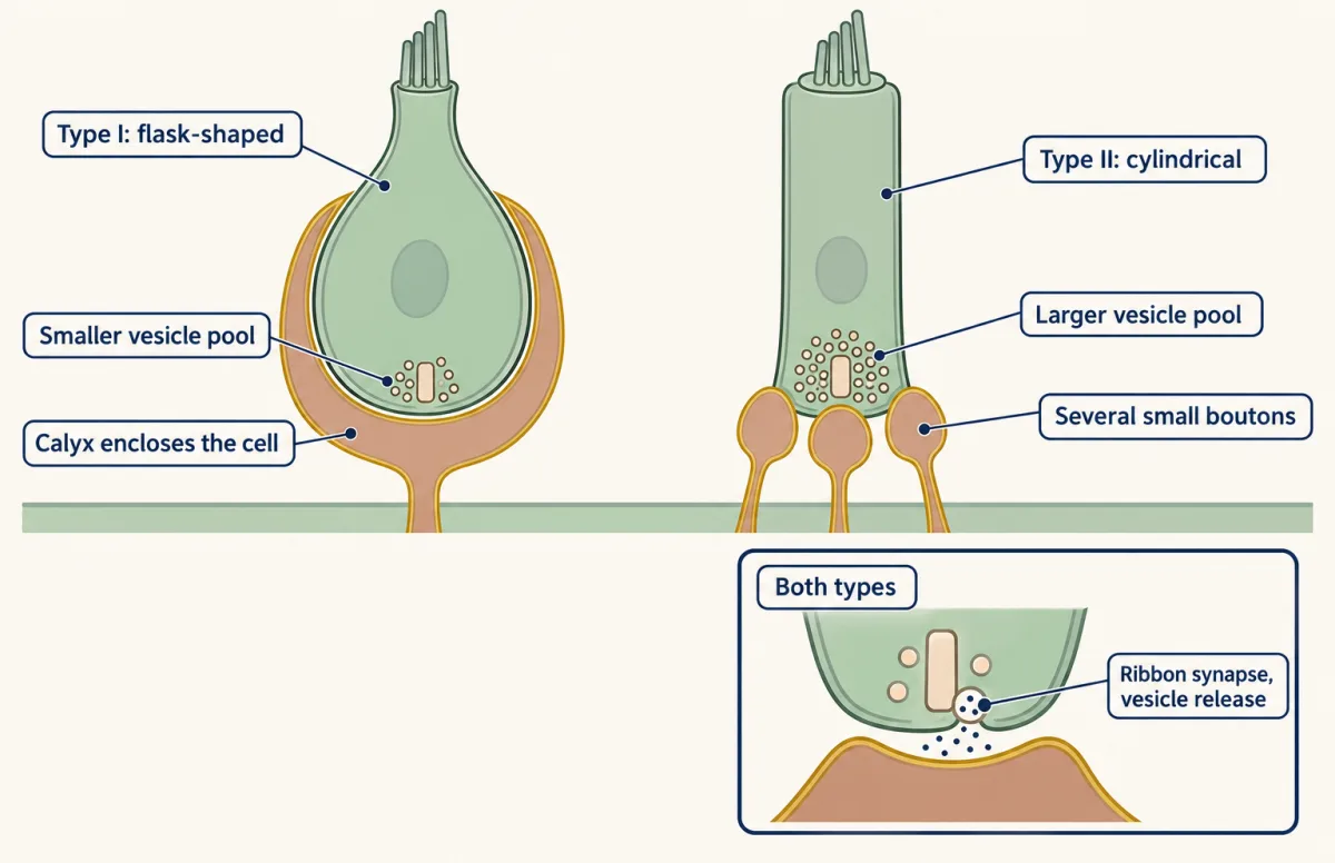
A type I vestibular hair cell is flask-shaped and its afferent calyx almost completely encloses the basolateral membrane; a type II cell is cylindrical and contacts several small afferent boutons. Both release glutamate from vesicles at ribbon synapses. In the mature mouse utricle studied, sustained calcium-evoked exocytosis was about tenfold smaller in type I cells. The large transient capacitance change in type I cells reflects potassium-channel gating, not vesicle fusion; the vesicle clusters shown are schematic. AI-generated illustration 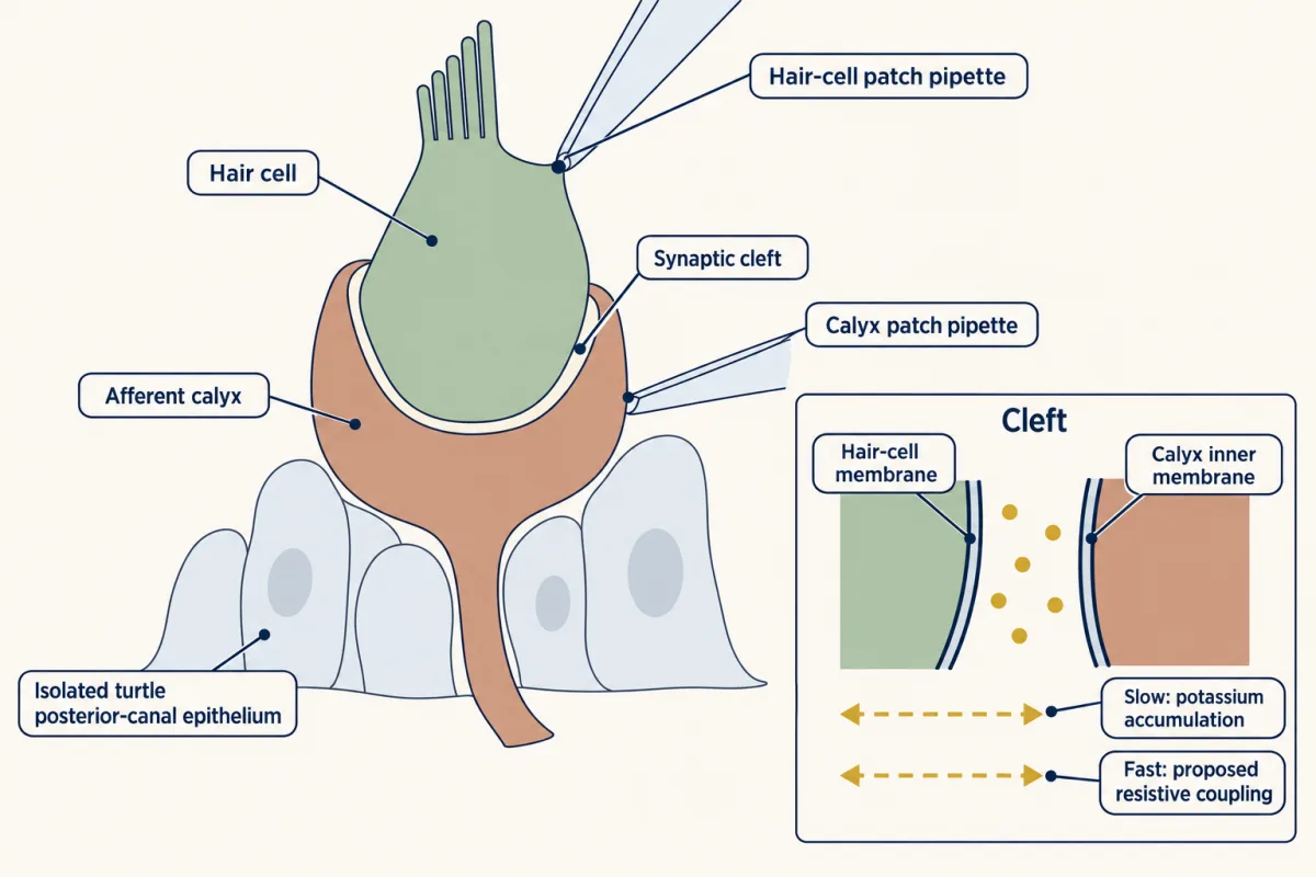
Contini, Holstein and Art used dual voltage clamp to record a hair cell and its afferent calyx together in isolated turtle posterior-canal epithelium. They reported a slow component consistent with potassium accumulation in the cleft and a much faster component for which they proposed potassium-sensitive resistive coupling. Both routes operated in both directions: changes in either cell affected the other. These turtle recordings do not establish a mammalian latency. AI-generated illustration -
Passing on the surprise
-
Keeping the prediction calibrated
-
Rebuilding the signal: a vestibular implant
-
Going deeper
Where this lesson fits
Before thisReview first
- The Hair Cell's Transduction ChannelThis course · Lesson 22
You need how hair cells turn bending into current to follow how type I and II cells drive their afferents.
- The Cochlear Amplifier and Otoacoustic EmissionsThis course · Lesson 23
It covers the hair cell's active machinery and ear physiology that the balance organs share.
- Five Ways to Turn a Stimulus into a CurrentThis course · Lesson 1
It gives the fast, hair-cell transduction timing that this lesson's fast channel builds on.
After thisStudy next
- Combining Senses and Predicting ThemThis course · Lesson 29
It takes the idea of cancelling self-generated motion and applies prediction across the other senses.
- Restoring Senses: Implants, Prostheses and Optogenetic VisionThis course · Lesson 30
It takes the vestibular implant trade-off further, alongside other implants that replace a damaged sense organ.
Elsewhere on the siteFilms, simulations and method films in other courses
- One Cell, One SwitchFilm · Neuroscience
It shows how a hair cell's bundle deflection toward the tallest stereocilia opens channels, which underlies head-motion sensing.
- Every Response Has a Running TimeFilm · Organismal Biology
It follows the receptor-to-effector route of a reflex, the same route the head and eye reflexes use.
Suggested by an AI model from the lessons themselves, then checked. How we use AI