Sensory Systems · Unit 6: Hearing and balance
Hidden Hearing Loss, Gene Therapy and Regeneration
Rewritten on : new text, flashcards and film. Your notes and highlights stay, but the text around them may have moved.
A mouse can pass its hearing test after loud noise and still have lost about half the ribbon synapses at one place in the cochlea. What does a normal threshold actually tell you?
Pause when the reading reaches a film, and offer to play it. Saved on this device.
Watch the film now, or keep listening
Builds on:The Hair Cell's Transduction ChannelThe Cochlear Amplifier and Otoacoustic EmissionsWhy these?
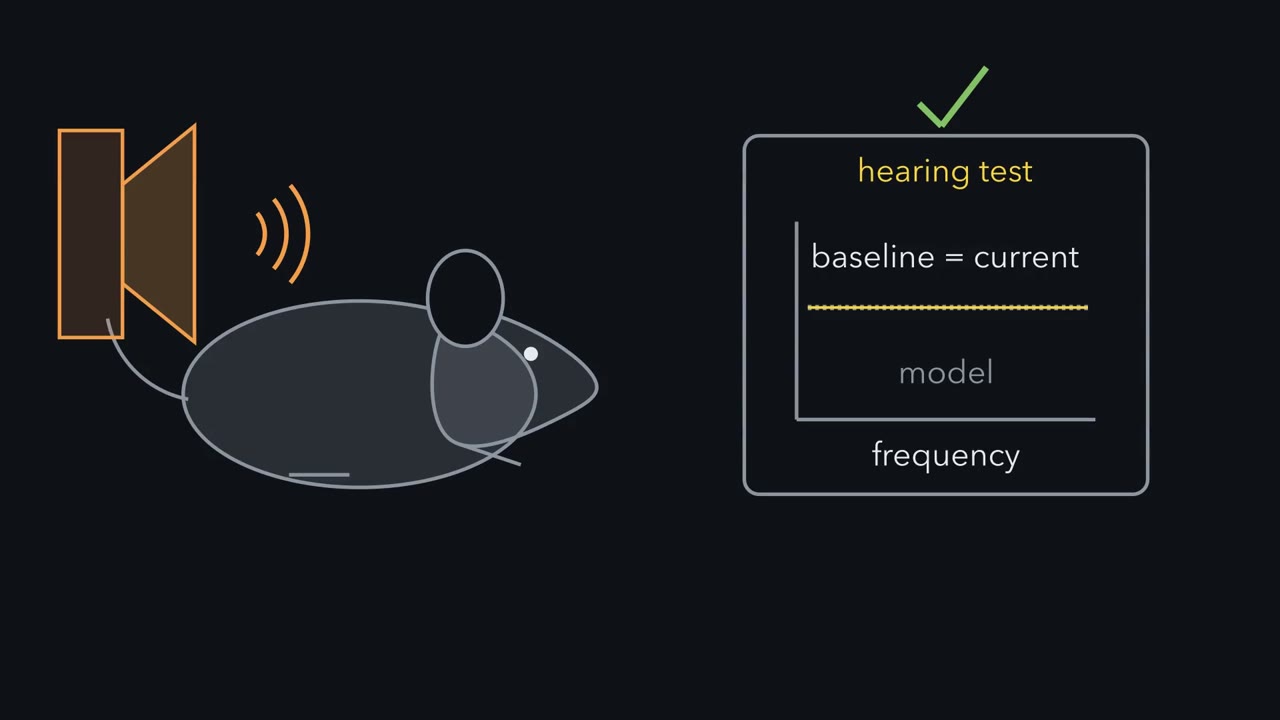 ▶ Watch A Threshold Is Not a Count · 2:59
Read the transcript of “A Threshold Is Not a Count”
Film transcript: A Threshold Is Not a Count
A mouse passes its hearing test after loud noise. Can its cochlea still be damaged? A threshold is the quietest sound that evokes a response. That is what a hearing test measures. Inner hair cells pass sound to nerve fibres at ribbon synapses. The ribbon holds a ready supply of vesicles. Kujawa and Liberman exposed male mice of one lab strain, sixteen weeks old. The loud noise lasted two hours. Thresholds rose after the noise, then returned to their pre-exposure values by two weeks. The hair cells remained intact. Eight weeks later, what would you predict for the number of ribbon synapses?
About half were gone at the 32 kHz place. Nerve terminals had been lost early. Losing these synapses while the hair cells survive is called cochlear synaptopathy. Why would losing half the synapses leave the threshold where it was? Nerve fibres at one place differ in sensitivity. Some respond to very quiet sounds, others need louder sound. A threshold asks when the response to a quiet sound first stands out from background firing. At the quietest levels, only the most sensitive fibres do that. Here is a small model of sixteen fibres. It is illustrative and passive, not fitted to mouse data. Remove half the fibres at random, or remove the most sensitive half.
Which moves the threshold? Losing a random half moved the threshold by about 2 dB. A hearing test moves in 5 dB steps. Losing the most sensitive half moved it by about 8 dB. That is more than one 5 dB step. In this model, the sound-driven response to a loud sound halved in every case. So here is the one idea. Hearing runs through a chain: hair cell, ribbon synapse, nerve fibre. A threshold depends mostly on the most sensitive links. It does not count the links. In these mice, a cochlea lost many synapses and still passed a hearing test. And a repair has to put back what is missing: a molecule in a surviving cell, or the cell itself. In people, temporal bones show nerve fibre loss after death.
No one can count synapses in a living ear. Living ears give only indirect signals, and the evidence from them is mixed. A normal threshold is good news about sensitivity. It is not a census of synapses.
From the film
- In these mice, a cochlea lost many synapses and still passed a hearing test.
- A threshold depends mostly on the most sensitive fibres and does not count them.
- A normal threshold is good news about sensitivity, but it is not a census of synapses.
A mouse can pass its hearing test after loud noise and still have lost about half the ribbon synapses at one place in its cochlea. What does a normal threshold actually tell you?
Three terms first.
- A threshold is the quietest sound that evokes a response. The response can be behavioural, or electrical, recorded from the nerve and brainstem.
- An audiogram plots behavioural threshold against frequency. It is the standard hearing test.
- A ribbon synapse is the contact where an inner hair cell passes sound to auditory nerve fibres. The cell releases transmitter continuously, graded with its receptor potential (its voltage change in response to sound). The ribbon, an electron-dense structure at the presynaptic active zone, helps by holding a ready supply of vesicles.
Hearing and balance covered transduction, the amplifier and the ribbon synapse in one paragraph. Here we start with what noise does to the synapse, then ask which molecule makes it fire and how that molecule can be put back. For quantal release and calcium sensors in general, see synaptic transmission and vesicle traffic.
A hearing test passed by a damaged cochlea
For decades, a threshold that came back to normal after noise was read as recovery. Kujawa and Liberman (2009) tested that assumption. They exposed male CBA/CaJ mice in vivo, at 16 weeks of age, to 8 to 16 kHz octave-band noise at 100 dB SPL for two hours. Response thresholds rose after the noise and returned to their pre-exposure values by two weeks, and the hair cells remained intact.
Predict. By the usual reading, this ear has recovered. Eight weeks after the noise, what would you predict for the number of ribbon synapses at the 32 kHz place along the cochlea?
Check your prediction
About half were gone. Afferent nerve terminals were lost acutely, right after the noise. At eight weeks the ribbon count in the 32 kHz region was about 50 percent below control, and synaptic loss extended through the basal half of the cochlea. The cochlear nerve degenerated later, over about one to two years.
Losing the synapses between hair cells and nerve fibres while the hair cells survive is called cochlear synaptopathy. Later you will meet a second kind, congenital and molecular rather than caused by noise, in otoferlin deafness.
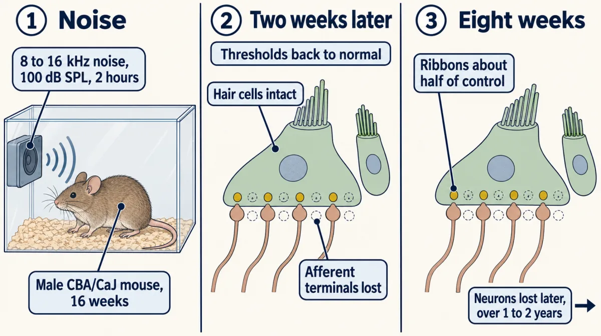
This is one mouse strain, sex, age and exposure. It shows that recovered sensitivity can conceal injury, not how common that is in people or what it does to their hearing.
Why a threshold can miss half the synapses
Why would losing half the synapses at one place leave the threshold where it was? A threshold asks when the response to a quiet sound first stands out from background firing. The nerve fibres at one cochlear place differ in sensitivity. Some respond to very quiet sounds and fire a lot even in silence (a high spontaneous rate). Others need louder sound and fire little at rest (a low spontaneous rate). So the quietest sounds are detected through the most sensitive fibres.
A small population model tests the idea; it is illustrative and passive, not fitted to mouse data. It has 16 fibres at one place, on a relative level scale: 10 sensitive fibres whose rate curves sit at 0 to 10 dB, with a spontaneous rate of 60 spikes a second, and 6 less sensitive fibres whose curves sit at 15 to 40 dB, with 2 spikes a second. (These positions are rate-curve parameters, not measured fibre thresholds.) Each fibre’s sound-driven firing grows over about 20 dB to the same ceiling, and spike counts are random. The population’s threshold is the quietest level at which the summed sound-driven count stands out from the spontaneous background by a fixed criterion (the full specification is in Going deeper). That is why spontaneous firing matters: it is the noise the signal has to beat.
Predict. Remove half the fibres in three ways: at random, the less sensitive half, or the most sensitive half. Which removal moves the threshold most? And what happens to the summed response to a loud, 80 dB sound?
Check your prediction
Averaged over 400 random populations:
| Which half of the fibres is removed | Threshold shift | Sound-driven response at 80 dB, relative to intact |
|---|---|---|
| none (intact) | 0 dB | 1.00 |
| a random half | about +2 dB | 0.50 |
| the less sensitive half | about 0 dB | 0.50 |
| the most sensitive half | about +8 dB | 0.50 |
A typical hearing test moves in 5 dB steps. Losing a random half or the less sensitive half moves the threshold by less than one step. Losing the most sensitive half moves it by more than one step, so a test could pick it up. The loud-sound response halves in every case, because at 80 dB every fibre is near the same ceiling.
In the model, then, the threshold stays near normal while most of the sensitive fibres survive, and moves if they are the ones lost. So which fibres noise removes matters. In guinea pigs it took mainly the less sensitive fibres (Furman and colleagues, 2013); in noise-exposed mice it took both kinds (Suthakar and Liberman, 2021). Neither is a selective loss of the most sensitive fibres, the case that moves the threshold most.
The loud-sound response is different: it falls with every lost fibre. The auditory brainstem response (ABR) is a series of electrical waves recorded from the scalp after a sound, and its earliest peak, wave I, comes from the auditory nerve. That is the reasoning behind proposing wave I amplitude as a sign of synapse loss. The model counts firing and does not compute wave I, which also depends on how synchronously the fibres fire.
The one idea to carry through this lesson. Hearing runs through a chain: hair cell, ribbon synapse, nerve fibre. A threshold asks when the response to a quiet sound first stands out from background firing. That depends mostly on the most sensitive fibres, and it does not count synapses. So a cochlea can lose many synapses and still pass a hearing test. And a repair has to put back the link that is actually missing: a molecule in a cell that survives, or the cell itself when it is gone.
The idea covers three cases. Noise synaptopathy is the first. In severe otoferlin deafness, the inner hair cells survive but a molecule their synapses need for release does not work, so putting back the gene can work, even if some synapses have already been lost. When the hair cell itself is gone, replacing its gene cannot help. A new hair cell has to be made, for example by giving surviving supporting cells genes that push them toward a hair-cell fate, and then wired to a fibre.
Finding the lesion in people
In a human ear you can count nerve fibres only after death, in the temporal bone, the part of the skull that holds the cochlea. These counts show nerve fibre loss directly, though they do not count synapses. Wu and colleagues (2021) compared 52 cochleas from people with a noise history with age-matched controls. Nerve fibre loss was substantial at every cochlear frequency and worse with noise. Noise history also made outer hair cell loss worse at 2 kHz and above, but not inner hair cell loss or strial atrophy. Among people with matched thresholds, the neural loss tracked poor word discrimination.
In a living ear no one can count synapses, so studies look for a proxy: an electrical signal that should shrink when fibres are lost. Electrocochleography, recorded near the cochlea, gives two signals.
- The summating potential (SP) is mainly pre-neural. It comes largely from the hair cells, with some neural contribution.
- The action potential (AP) here is the compound action potential of the auditory nerve, the summed, synchronized firing of many fibres at once.
Predict. If noise removes nerve fibres but leaves hair cells intact, what should happen to SP, to AP, and to their ratio, SP/AP?
Check your prediction
If SP stays about the same and the lost fibres were contributing to AP, AP gets smaller and the ratio rises. That is the logic behind the proxy, and it says what to check in a result: a higher ratio should come from a smaller AP.
Two studies used proxies like these, with different comparisons, and came out differently.
| Liberman and colleagues (2016) | Guest and colleagues (2018) | |
|---|---|---|
| Who | 34 young adults, mean age about 24 to 25, grouped by self-reported noise exposure and use of hearing protection | Adults aged 18 to 40 with normal audiograms from 0.25 to 8 kHz: 16 with verified difficulty understanding speech in noise and 16 closely matched controls |
| Proxy | SP/AP ratio | ABR markers, and the EFR (envelope-following response, a measure of neural phase locking to a sound’s slow amplitude changes) |
| Result | Higher SP/AP in the higher-risk group, which the authors said was consistent with selective neural loss | No association between the impairment and either marker of synaptopathy; the authors allowed that these measures might not be sensitive enough |
| Main limit | The ratio rose mainly because SP was larger, with no significant fall in AP, so it is not specific to neural loss. The same group also had raised thresholds at 10 to 16 kHz despite normal conventional audiograms, itself a cochlear difference, so synapse loss cannot be separated from it | Groups were matched on thresholds up to 14 kHz, a stronger comparison, but the proxies’ sensitivity to synapse loss in people is unknown |
Go back to the prediction. In the first study the ratio rose, but not for the reason the proxy assumes. In the second, the match was better, but a negative result from a test of unknown sensitivity cannot rule the lesion out. A listening complaint, a proposed lesion and a validated test are three different things, and a good study has to establish each one.
The link that has to fire: what makes the synapse release
Read the transcript of “The Link That Must Fire, and How to Put It Back”
Film transcript: The Link That Must Fire, and How to Put It Back
Which molecule turns the calcium signal into release at this synapse? When an inner hair cell depolarizes, calcium enters. Something must turn that signal into vesicle fusion. The ribbon is not that something. It keeps a larger resident pool of vesicles. Mutations in the OTOF gene cause deafness, and otoferlin carries calcium-binding C2 domains. Roux and colleagues recorded from inner hair cells of mice lacking otoferlin. A rise in membrane capacitance tracks how much vesicle membrane fused. So it measures release. If otoferlin acts after calcium enters, at fusion itself, what should the recording show?
Calcium current and ribbon morphogenesis were preserved at the age recorded. Release was almost abolished. They proposed that otoferlin may be the major calcium sensor for fusion. The word may mattered. A knockout shows a protein is needed. It does not show the protein is the sensor. Michalski and colleagues made a sharper test. A change in the C2C domain reduced calcium binding. The calcium sensitivity of release fell, which fits a sensing role, without making otoferlin the only sensor. Now the repair. In severe otoferlin deafness, the inner hair cells are there to treat, but their synapses release almost nothing. The outer hair cells still amplify, at least early in life.
The ear's own faint sounds, otoacoustic emissions, show it. But the gene is too large for its vector. A conventional adeno-associated virus carries about five kilobases. The otoferlin coding sequence is about six kilobases, before a promoter is added. So Al-Moyed and colleagues split it between two vectors. A cell receiving both can rebuild the full gene. If each vector independently reaches seventy percent of hair cells, what fraction can make the protein?
About half, because a cell needs both halves to rebuild the gene. The real mice are a separate question. Full-length otoferlin appeared in up to half their inner hair cells. Fast release came back in those cells. Vesicle replenishment reached only part of wild-type values. Treated mice had click thresholds of 40 to 60 dB SPL. That is partial rescue, not normal hearing. Replacing a missing link can work because the cells that need it are still alive.
From the film
- A knockout shows that a protein is needed, and it does not by itself show that the protein is the sensor.
- In severe otoferlin deafness the inner hair cells are present, but their synapses release almost nothing.
- Replacing a missing link can work because the cells that need it are still alive.
If repair means putting back what is missing, which molecule makes that link fire? When an inner hair cell depolarizes, calcium enters, and something must turn that calcium signal into vesicle fusion. The ribbon is not that something; it holds the vesicle supply.
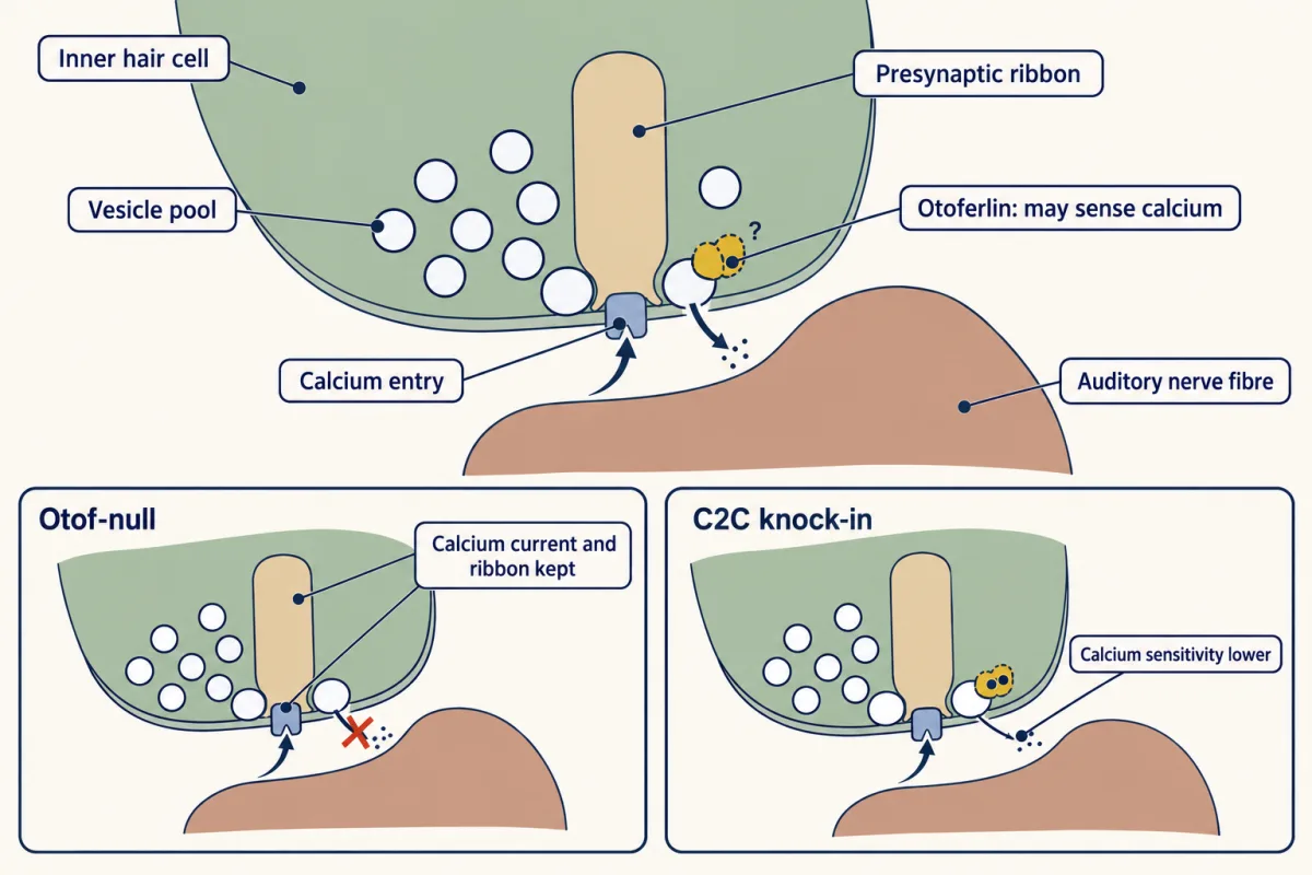
The candidate came from human genetics. Mutations in the gene OTOF cause a form of deafness, and its product is otoferlin, a protein with several C2 domains, modules that often bind calcium. Roux and colleagues (2006) recorded from inner hair cells of Otof-null mice by whole-cell patch clamp. The rise in membrane capacitance tracks how much vesicle membrane fuses, so it measures release.
Predict. Suppose otoferlin were needed for calcium entry, or for forming ribbons. What would you see? Now suppose it acts after calcium enters, at fusion itself. What would you see then?
Check your prediction
If otoferlin were needed for calcium entry, the recorded calcium current would fall. If it were needed specifically to form ribbons, imaging would show defective or missing ribbons (other defects in synapse assembly could leave ribbons in place). If it acts at fusion, calcium current and ribbons would look normal and the capacitance rise would be missing. Roux and colleagues found the last: at the age recorded, calcium current and ribbon morphogenesis were preserved, but exocytosis was almost abolished. That places the defect downstream of calcium entry, without naming the exact step. They proposed that otoferlin may be the major calcium sensor for fusion.
The word may mattered. A knockout shows that a protein is needed, not that it is the sensor, because a protein could be needed to dock or prime vesicles without sensing anything. Michalski and colleagues (2017) made a sharper test. In a knock-in mouse, a change in the C2C domain reduced calcium binding. The protein was present, so its absence could not explain any change, and in inner hair cells the calcium sensitivity of both fast and sustained exocytosis dropped. That fits a calcium-sensing role, and it does not show that otoferlin is the only sensor.
Putting the link back: a gene too big for its vector
Loss of otoferlin function causes a recessive deafness, the second kind of synaptopathy. Otoacoustic emissions (OAEs) are faint sounds the ear itself makes when the outer hair cells amplify. In otoferlin deafness they are present, at least early in life, while the auditory brainstem response is absent or grossly abnormal.
That pattern makes the gene a good target, and it is why the approved indication asks for preserved outer hair cell function.
There is a size problem. A conventional adeno-associated virus (AAV) vector carries about 4.7 to 5 kb. The otoferlin coding sequence is about 6 kb, before a promoter is even added, so it cannot ride in one vector. Al-Moyed and colleagues (2019) split the cDNA between two AAV2/6 vectors. Each carries part of the gene, and a cell that receives both can join the parts back into the full-length cassette.
Predict. In a made-up case, each vector independently reaches 70 percent of inner hair cells. What fraction of cells can make full-length otoferlin?
Check your prediction
A cell needs both, so the expected fraction is 0.7 × 0.7 = 0.49, about half. The chance of receiving at least one vector is 1 − 0.3 × 0.3 = 0.91, but a cell with only one has only part of the gene. Real transduction need not be independent, so this only shows why coverage is capped.
- Expected with both (p1 × p2)
- 49.0 of 100
- Observed in this drawn field
- 0 of 100
- Reported maximum
- up to 50 percent
In this sketch: independent delivery, every cell with both halves counted as reconstituted, made-up reach values. The reference line is from Otof-null mice injected at P6 to P7 with dual AAV2/6 (Al-Moyed and colleagues, 2019).
This sketch does not say: that the product is a prediction (real transduction is not independent); that a cell with both vectors works normally (vesicle replenishment reached only 35 to 50 percent of wild type in transduced cells); that coverage equals hearing (click ABR thresholds in treated mice were 40 to 60 dB); or anything about children or speech.
A cell can rebuild full-length otoferlin only if it received both halves, so, in this sketch, the fraction with full-length otoferlin is limited by how many cells receive both, not by either vector alone.
The mouse result was a partial rescue. Al-Moyed and colleagues injected neonatal Otof-null mice through the round window, a membrane at the base of the cochlea. Full-length otoferlin appeared in up to 50 percent of inner hair cells. That is of the same order as the made-up figure, which is not an estimate for this experiment. In transduced cells, fast exocytosis was restored, but vesicle replenishment reached only 35 to 50 percent of wild-type values. Click-evoked ABR thresholds in treated mice were 40 to 60 dB SPL, in mice that were deaf without treatment: better, but not normal hearing.
At postnatal days 26 to 29, Otof-null inner hair cells also had about 40 percent fewer ribbons than wild-type cells, and delivery at days 6 to 7 did not restore the count. (The release recordings were made earlier, at days 14 to 18.) That suggests otoferlin matters for keeping or maturing synapses as well as for release, and that timing may limit rescue (the study did not test timing). Receiving both vectors is one limit on coverage; gene reassembly, expression level and the synapse loss already present also held this rescue back.
Reading an early-phase trial without inflating it
Read the transcript of “Reading the Trial, and When the Cell Is Gone”
Film transcript: Reading the Trial, and When the Cell Is Gone
Nine of twelve children met a trial's endpoint. Does that mean most of their hearing came back? The DB-OTO trial treated twelve children with OTOF-related profound deafness. Nine met the week 24 endpoint: an average of 70 dB HL or better. Three of twelve reached average normal sensitivity. So nine of twelve is a share of children meeting an endpoint. It is not a share of hearing. In a separate report by Wang and colleagues, one child's baseline was above the test ceiling of 95 dB. At week 26 the threshold was 58 dB. What does that tell you about the improvement?
Only that it was more than 37 dB. The true baseline was never measured. A floor on the improvement is not the improvement, and it is not a percentage of hearing. Now the harder case. When the hair cell itself is gone, putting back its gene cannot help. A new hair cell has to be made, perhaps from a surviving supporting cell. Birds replace damaged hair cells. In adult quail, counts recovered to within five percent of the original. The adult mammalian cochlea does not do this. One mammalian exception is vestibular, in the guinea-pig utricle.
In newborn mice, the factor ATOH1 pushes nonsensory cells of the cochlea, chiefly in the greater epithelial ridge, toward hair cells. Would the same factor work just as well a week later? By day eight, the factor alone gave very few new hair cells. Adding two more factors gave many more.
Hair-cell genes also became less accessible with age. That makes chromatin a candidate barrier. In mature mice, some reprogrammed cells grew rudimentary bundles. Nerve fibres were reported to contact them. Whether those contacts are working synapses is the unsettled step. Replacing a link in a surviving cell is being done in children. Rebuilding the cell is not.
From the film
- Nine of twelve is a share of children meeting an endpoint, not a share of hearing.
- When the hair cell itself is gone, a new hair cell has to be made, and the adult mammalian cochlea does not do this.
- In newborn mice, reprogramming works less well a week later, so age is a barrier.
The human trials use otoferlin gene replacement. It regenerates no hair cells and treats no other form of deafness.
The DB-OTO trial (New England Journal of Medicine) treated twelve children with OTOF-related profound deafness in an open-label single-group design. Nine of twelve met the week-24 primary endpoint, an average of 70 dB HL or better, and three of twelve reached average normal sensitivity. (dB HL is measured against average normal hearing, so 0 dB HL is average normal; the mouse studies use dB SPL, measured against a fixed sound pressure.) It recorded 67 adverse events. The United States Food and Drug Administration is reported to have granted accelerated approval on 23 April 2026, with a larger analysis in which 16 of 20 efficacy-evaluable participants met the endpoint.
In the other small, single-arm reports from several centres, most or all of the treated children heard better, by margins that varied. Thresholds and sound localization improved, and the earliest report, from Lv and colleagues, had no dose-limiting toxicity or serious adverse event. Later reports followed some participants for up to about two and a half years, which is evidence of persistence over those years and not proof of lifelong durability.
Worked problem: reading a censored number
In the Wang trial (Nature Medicine, 2024), patient 1 had baseline average ABR thresholds above 95 dB in each ear and 58 dB at week 26. A student writes: “the treatment improved hearing by exactly 37 dB, so it restored 39 percent of hearing.”
Step 1, the bound. Let the true baseline be B, with B above 95. The improvement is B minus 58, which exceeds 95 minus 58, or 37 dB. So 37 dB is a strict lower bound, because the baseline was above the test ceiling and was not measured.
Step 2, the percentage. The student’s 39 percent is 37 divided by 95. It divides a bound by the ceiling and calls it a fraction of hearing. These censored values do not identify a percentage of hearing restored.
Step 3, a different kind of fraction. In DB-OTO, 9 of 12 is 75 percent: the share of children meeting a stated endpoint (70 dB HL or better), not 75 percent of normal hearing. The later label analysis of 16 of 20 (80 percent) is a different analysis set and must not be pooled with it.
When the cell itself is gone: regeneration
When noise, drugs or age kill the hair cells, there is no cell to put a gene back into, and a new hair cell has to come from somewhere. Some animals simply make new ones. Corwin and Cotanche (Science, 1988) reported replacement of damaged hair cells in the chicken cochlea after acoustic trauma. In a separate Science paper that year, Ryals and Rubel found that in adult Coturnix quail, hair-cell counts recovered to within 5 percent of the original count. Fish regrow hair cells too, in the zebrafish lateral line, a surface mechanosensory organ built of neuromasts rather than a cochlea.
The adult mammalian cochlea does not do this. Mammals do have one exception, and it is vestibular: after gentamicin damage, hair cells reappeared in guinea-pig utricles (Forge and colleagues, 1993).
In mice the question became which barriers stop supporting cells from converting into hair cells. Iyer and colleagues (eLife, 2022) used Cre-inducible mice to express the transcription factor ATOH1 alone, or with GFI1 and POU4F3, in cochlear nonsensory cells, starting at postnatal day 1 or day 8.
Predict. If the barrier to conversion were only the absence of the right transcription factors, the same factors should work at any age. Would you expect day 1 and day 8 to behave alike?
Check your prediction
They did not. At day 1, every ATOH1-containing combination tested gave cells with hair-cell morphology and rudimentary mechanotransduction, measured partly by rapid uptake of the dye FM1-43. At day 8, the three-factor combination gave about 50 to 60 new hair cells per 200 micrometres, counted at day 15, against fewer than 5 for ATOH1 alone or with GFI1. Joint RNA and chromatin profiling of untreated mice showed hair-cell gene loci becoming less accessible between the two ages, so closing chromatin, and hair-cell-to-supporting-cell signalling including Notch signaling, are candidate barriers.
McGovern and colleagues (PNAS Nexus, 2024) pushed the age further. Using lineage tracing in mature mouse organ of Corti, they found that three factors could reprogram some supporting cells into hair-cell-like cells with rudimentary stereociliary bundles, more of them when the native hair cells had been killed around the time of induction. The new cells were reported to be innervated and to carry ribbon-synapse proteins, but functional transmission through those contacts and restored hearing were not demonstrated.
Back to the one idea: a cell with a rudimentary bundle is not a restored hearing organ, because the chain has one more link. The unsettled step is whether such cells form working synapses with spiral ganglion neurons, which would be tested by recording from them and by measuring auditory responses in deafened mature animals. Replacing a molecule in a cell that survives is now being done in children; rebuilding the cell itself is still being tested in animals. For the biology of regeneration in general, see injury and regeneration and stem cells; for the devices that substitute for what is lost, see sensory prostheses.
Design problem
Design a study that could show whether noise-induced synapse loss explains trouble understanding speech in noise in people with normal audiograms. Then say what the published work can and cannot support.
Saved in this browser only.
One way to do it
No living human synapse count exists, so the design must name its physiological proxy in advance (for example ABR wave I amplitude or an envelope-following measure) and set the sample size from that proxy’s repeatability and variability and a prespecified effect size, not from hope, while stating plainly that its sensitivity to human synapse loss is unknown. Match groups on extended high-frequency thresholds through 14 kHz, as Guest and colleagues did, so a raw cochlear threshold difference cannot masquerade as synapse loss. Measure noise exposure prospectively, because recalled history is biased. Fix the speech-in-noise test and its endpoint before looking at the proxy.
What the published work supports: the mouse noise experiment supplies the lesion and its timing, but not its prevalence in people, a diagnostic criterion, or proof that it causes a listening complaint. The otoferlin rescue in mice was partial and neonatal and shows normal speech perception in no one. The trial reports cannot be pooled, and none supports lifelong benefit. The reprogramming work shows nerve contacts on new hair-cell-like cells in mature mouse cochleas, not yet a working synapse or restored hearing.
Going deeper
Which fibres does noise take?
Single-fibre recordings in guinea pig (Furman and colleagues, 2013) have been read as showing that noise preferentially removes low-spontaneous-rate, high-threshold fibres. In noise-exposed mice, Suthakar and Liberman (2021) found that both low- and high-rate fibres were lost, which they judged not consistent with a selective loss of the low-rate group. This also bears on the proxies: wave I is weighted toward fibres that fire in synchrony at sound onset, so if the lost fibres contribute little to that onset, wave I could under-report the loss.
The fibre model in full
Each fibre has a level parameter t (uniform over 0 to 10 dB for the 10 sensitive fibres, 15 to 40 dB for the other 6) and a spontaneous rate b (60 or 2 spikes a second). Its driven rate is r(L) = 200 / (1 + exp(−0.22 (L − t − 10))) spikes a second, so even at L = t it fires about 20 spikes a second above background; there is no hard onset. Counts are independent Poisson counts over T = 0.05 s. With S = T × Σr and B = T × Σb, the threshold is the first level on a 0.1 dB grid where S / √(B + S) ≥ 1. The three removals (a random 8, the 8 with the highest t, the 8 with the lowest t; random seed 1) give +2.0, −0.1 and +8.4 dB. At 80 dB every fibre is near the 200 spikes a second ceiling, so removing 8 of 16 halves the expected driven response: (8 × 200) / (16 × 200) = 0.50.
What the ribbon itself contributes
Becker and colleagues (2018) tested what the ribbon adds by deleting its main protein, RIBEYE, in mouse. The ribbon supported transmission by keeping a larger resident pool of vesicles, so it tunes the supply, but inner hair cell synapses still formed without it.
The two dual-vector designs
Al-Moyed and colleagues tested two designs that rebuild the cassette by different routes, a trans-splicing design and a hybrid design, both injected in vivo.
The early gene therapy reports, one by one
Lv and colleagues (Lancet, 2024) reported a single-centre, single-arm study of six children with biallelic OTOF variants, treated in one ear with an AAV1-hOTOF vector and followed for 26 weeks. Five showed hearing recovery, with average ABR threshold reductions of 40 to 57 dB among the five. Wang and colleagues (Nature Medicine, 2024) reported an interim analysis of five children, aged 1.2 to 11.0 years, treated in both ears. All five improved, and all five localized sound better, with 13 or 26 weeks of observation and 36 adverse events, none dose-limiting or serious. Qi and colleagues (Advanced Science, 2024) reported two children, one treated in one ear and one in both, and a click-ABR threshold of 30 dB in the treated ear of the five-year-old one month after injection, with frequency-specific thresholds still raised at that point and improving over three months. (A separate Qi trial of ten participants appeared in Nature Medicine in 2025, and the 2026 Nature report of 42 participants is the one that followed some for up to 2.5 years.)
What the label asks for
The reported accelerated approval covers patients with severe-to-profound or profound sensorineural hearing loss, any frequency above 90 dB HL, with confirmed biallelic OTOF variants, preserved outer hair cell function and no prior implant in the treated ear. Regulatory status changes.
The C2C knock-in and the reprogramming methods
In the Michalski knock-in, two alanines (Ala515 and Ala517) replaced residues in the C2C domain. Inner hair cells were recorded ex vivo at postnatal days 14 to 18, with patch clamp and calcium uncaging.
Iyer’s joint profiling was single-nucleus RNA and ATAC sequencing (ATAC reads which stretches of chromatin are open). McGovern and colleagues traced lineage with Lfng-CreERT2, inducing at three weeks and collecting at four to six weeks.
Regeneration in birds and fish, in detail
Ryals and Rubel found thymidine incorporation over hair-cell and supporting-cell nuclei within ten days of the trauma in adult quail. In the zebrafish lateral line, Pinto-Teixeira and colleagues (2015) found regeneration after repeated severe injury and in adult animals, with hair cells restored within 72 hours in animals aged three months, one year and two years.
Your notes
Select any text in the lesson to highlight it or add a note to it. Your highlights and notes are saved in this browser and collected in your notebook.
This browser is not letting the site save anything (a private window or strict privacy settings), so highlights and notes will be lost when you leave the page.
How we measure it
Whole-cell patch clamp with membrane capacitance
A recording pipette on one inner hair cell measures calcium current while the cell is depolarized, and the rise in membrane capacitance tracks how much vesicle membrane fused. A short pulse probes fast release and a longer one probes replenishment, which is how the knockout, the knock-in and the gene replacement were compared.
Auditory brainstem response and electrocochleography
Scalp or ear-canal electrodes record the synchronized firing of the auditory nerve and brainstem to sound (ABR); in electrocochleography the summating potential, SP, is mainly pre-neural and comes largely from hair cells but has some neural contribution, and the action potential, AP, reflects synchronized auditory-nerve firing. The threshold is the quietest sound that evokes a response, and suprathreshold wave I amplitude and the SP/AP ratio are used as proxies for nerve integrity.
Dual-AAV gene delivery
A coding sequence too large for one vector is split across two, and the halves are joined inside a cell that receives both. Only cotransduced cells express the full-length protein, which is one cap on how many cells can be rescued.
Joint single-nucleus RNA and ATAC sequencing
The same nuclei are read for gene expression and for chromatin accessibility, so a cell's identity can be compared with how open its hair-cell genes are at different ages.
Practice questions on this lesson (3) → MCAT-style, original, each with an explanation
Where this lesson fits
Before thisReview first
- The Hair Cell's Transduction ChannelThis course · Lesson 22
You need the hair cell's transduction step from there, since the chain here starts with that cell and ends at the synapse.
- The Cochlear Amplifier and Otoacoustic EmissionsThis course · Lesson 23
Outer hair cell amplification explains why otoacoustic emissions stay present in otoferlin deafness while the brainstem response is absent.
After thisStudy next
- Restoring Senses: Implants, Prostheses and Optogenetic VisionThis course · Lesson 30
Cochlear implants bypass lost hair cells and talk to the nerve, the alternative when gene replacement cannot work.
Elsewhere on the siteFilms, simulations and method films in other courses
- Counting failuresSimulation · Neuroscience
It shows how calcium entry triggers vesicle release at a synapse, the step that otoferlin loss removes.
- One Cell, One SwitchFilm · Neuroscience
It shows how hair cells turn sound into signals sent to the nerve, the chain that fails here.
Suggested by an AI model from the lessons themselves, then checked. How we use AI
Finish line
Test yourself
8 questions
Practice and MCAT-style, with explained answers.
Next · Lesson 25
Balance: Two Codes for Every Head Movement
People with severe loss in both balance organs can see the world bounce as they walk. Yet in monkeys, head-motion neurons go largely quiet for a turn the animal makes itself, which may keep the head-steadying reflex from fighting it. How does one nerve report motion fast and precisely, yet let the brain set your own turns aside?
Continue →← Lesson 23 · The Cochlear Amplifier and Otoacoustic Emissions