Developmental Neuroscience · Skim version
Topographic Maps: Sperry's Eye and Eph Gradients
Read the full lesson → Skim the whole course →
The guiding idea
Turn a newt's eye upside down, let the nerve regrow, and it turns its head down toward a lure held above it. Why does it not learn?
Neighbouring cells in the retina connect to neighbouring cells in the brain, so the eye's image is laid out in order on its target.
From the film: A Rank Against a Slope
- An axon's place in the map is a rank set against a slope, not an address in its own receptor level alone.
- In chick and mouse, the retina uses gradients and neighbours together to place its axons.
- More receptor, less ligand: the axon with the most EphA ends where ephrin-A is lowest.
From the film: Reading the Slope
- In the chick stripe assay, temporal retinal axons avoided posterior tectal membrane, while nasal axons grew on both kinds.
- Heating the posterior membrane removed the avoidance, which pointed to a heat-sensitive repellent at the back of the tectum.
- In the toy model, two opposite slopes, EphA in the retina and ephrin-A in the target, put axons in order.
- Where an axon stops means where it keeps its branches, its termination zone.
From the film: Two Maps from One Retina
- In mice with extra EphA3 in a scattered half of ganglion cells, two orderly maps formed, so an axon's place depends on the other axons and not on its own receptor level alone.
- The amount of ephrin-A also matters: with less ephrin-A, mapping was less accurate.
- The ligand slope sets the scale, and comparison among axons decides who goes where on it.
- Slopes help lay out the coarse map, and correlated retinal activity helps sharpen it.
-
The map, and three ways to build it
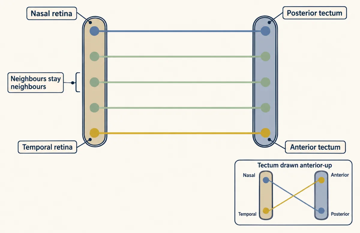
Retinal axons map in order onto the optic tectum: temporal retina ends in the anterior tectum and nasal retina in the posterior. The inset redraws the tectum with its anterior end up, so the same map appears crossed; both drawings show one map. AI-generated illustration -
Sperry’s newts: does the wiring follow the eye or the world?
-
Is the cue in the target? The stripe assay
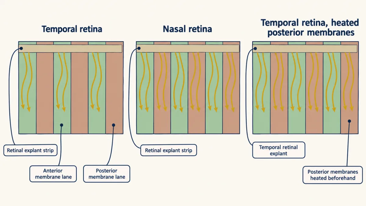
In the chick striped-carpet assay, temporal retinal axons avoided posterior tectal membrane lanes, whereas nasal axons grew on both membrane types. Heating posterior membrane preparations before the assay abolished temporal axons’ avoidance, consistent with a heat-sensitive posterior repellent. Lane and axon numbers are illustrative. AI-generated illustration -
The molecules: two opposite slopes
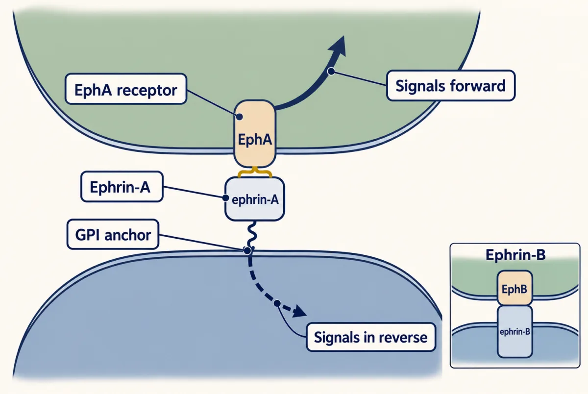
Eph receptors and ephrins sit on cell surfaces, so signalling needs contact and can run both ways. Ephrin-As hang on the outer surface by a GPI anchor, while ephrin-Bs cross the membrane. AI-generated illustration -
Is the slope needed? Ephrin knockouts
-
Absolute or relative? Two maps from one retina
-
Where slopes stop being enough
-
Going deeper
Where this lesson fits
Before thisReview first
- The Growth Cone: The Tip That Steers the AxonThis course · Lesson 3
You need how growth cones sense and respond to nearby cues to follow how axons read ephrin slopes here.
- Pioneers, Guideposts and the Logic of Guidance CuesThis course · Lesson 22
The logic of guidance cues, including repulsion, is assumed when ephrin-A is treated as a repellent.
After thisStudy next
- Activity Before Experience: Spontaneous Retinal WavesThis course · Lesson 38
Correlated retinal waves sharpen the coarse ephrin map into fine termination zones, as this lesson ends by saying.
- Choosing a Partner: Layers, Self-Avoidance and SmellThis course · Lesson 27
It extends target choice to layers and partners, where axons must find the right address after coarse mapping.
- Ocular Dominance and the Critical PeriodThis course · Lesson 39
It shows how activity from the two eyes refines retinal projections after chemical cues set the coarse map.
Suggested by an AI model from the lessons themselves, then checked. How we use AI