Biochemistry · Supplement · Nucleotide Metabolism
Nucleotide Metabolism: Build It, Recycle It, or Pay in Uric Acid
Rewritten on : new text, figures and flashcards. Your notes and highlights stay, but the text around them may have moved.
Nucleotides are costly to build — Lesch-Nyhan lives where recycling breaks, and gout and allopurinol live at the uric-acid end.
Pause when the reading reaches a film, and offer to play it. Saved on this device.
Watch the film now, or keep listening
Builds on:The Pentose Phosphate Pathway: NADPH and Five-Carbon SugarsMetabolism: Basic Concepts and DesignProtein Turnover and Amino Acid Catabolism: Tag, Recognize, Destroy, RecycleWhy these?
 ▶ Watch Too Costly to Throw Away · 2:58
Read the transcript of “Too Costly to Throw Away”
Film transcript: Too Costly to Throw Away
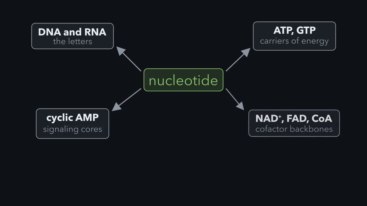
A nucleotide is costly for a cell to build. Nucleotides are the letters of DNA and RNA, and the carriers of energy, ATP and GTP. They form the cores of signaling molecules like cyclic A M P. They also form the backbones of cofactors such as NAD +, FAD, and coenzyme A. Building their rings from scratch costs a small fortune in ATP. So the cell recycles much of what it has. There are two routes to purine nucleotide, and the difference is mostly about cost. De novo synthesis builds the purine ring atom by atom, directly onto an activated ribose called P R P P. It runs about ten enzymatic steps and spends roughly six ATP to make the first finished purine nucleotide, I M P. Both A M P and G M P branch from I M P.
Salvage takes a free base that already exists, such as hypoxanthine, guanine, or adenine, and reattaches it to P R P P in a single step. The enzyme H G P R T salvages hypoxanthine and guanine. A P R T salvages adenine. Recycling is so much cheaper that many cells recover a large share of their free purines. Salvage is a major source in many tissues. Now imagine the recycling is gone. In Lesch-Nyhan syndrome, an X-linked defect, H G P R T is missing. Free bases that should have been reclaimed are dumped into the catabolic pathway and turned into uric acid. The P R P P that salvage would have used piles up and pushes de novo synthesis even higher. That makes still more purines to degrade.
The result is severe hyperuricemia alongside profound neurological disease. When a purine is finally retired, it is taken apart down a short tail. A M P and adenosine funnel through inosine to hypoxanthine. Then the enzyme xanthine oxidase oxidizes hypoxanthine to xanthine, and xanthine to uric acid. Guanine joins the route one step later, at xanthine. Most mammals carry uricase, which converts uric acid into highly soluble allantoin. Humans and the other apes lost a working uricase during evolution. For us, uric acid is the end product, and it is only sparingly soluble. That missing enzyme makes us prone to gout, along with how well we excrete urate.
Once serum urate climbs past roughly 6.8 milligrams per deciliter, it can crystallize as monosodium urate. A flare is the inflammation those needle-sharp crystals trigger. One fix is to make less of it. Allopurinol, a hypoxanthine look-alike, inhibits xanthine oxidase, so urate production falls and the crystals slowly dissolve. Gertrude Elion and George Hitchings designed it by reasoning about the enzyme.
Lose one enzyme that recycles purines, and the body builds purines far faster than normal and makes several times more uric acid.
That is Lesch-Nyhan syndrome; a classic early sign is orange, sand-like urate crystals in a baby’s diaper. A broken recycling enzyme ought to leave the cell short of purines. Why does it make a flood instead, and why does the flood leave as uric acid?
About the film above: it covers purines only; its “roughly six ATP” counts making the phosphoribosyl pyrophosphate (PRPP) (in ATP equivalents, explained below); and uricase, which it says converts uric acid to allantoin, does only the first of three steps.
Nucleotides are the letters of DNA and RNA, the energy carriers ATP and GTP and the core of cyclic AMP, and NAD⁺, FAD and coenzyme A each contain one. Their rings cost a small fortune in ATP, so the cell recycles much of what it has.
Three words first: base, nucleoside, nucleotide
A purine base is a two-ring molecule on its own (a pyrimidine base has one ring). Add a ribose and it is a nucleoside; add a phosphate to that ribose and it is a nucleotide.
| Base | + ribose = nucleoside | + phosphate = nucleotide |
|---|---|---|
| hypoxanthine | inosine | IMP |
| guanine | guanosine | GMP |
| adenine | adenosine | AMP |
IMP is the first purine nucleotide the cell builds, and the parent of both AMP and GMP.
Two doors in, one way out
Nucleotides do not last: RNA is made and broken down all day, and nucleotides are turned over constantly, so a cell is always releasing free bases.
The one idea to carry through this lesson. A cell’s purine nucleotides are a pool on a loop, with two doors in (build and recycle) and one way out. Salvage puts a large share of freed bases straight back (how large depends on the tissue); whatever it misses leaves the loop, almost all of it as uric acid. De novo synthesis refills the pool through a gate that phosphoribosyl pyrophosphate (PRPP) pushes open and finished nucleotides push shut. So, at steady state, the cell builds what it fails to recycle, and the uric acid the body makes equals what it builds plus the purine it absorbs from food.
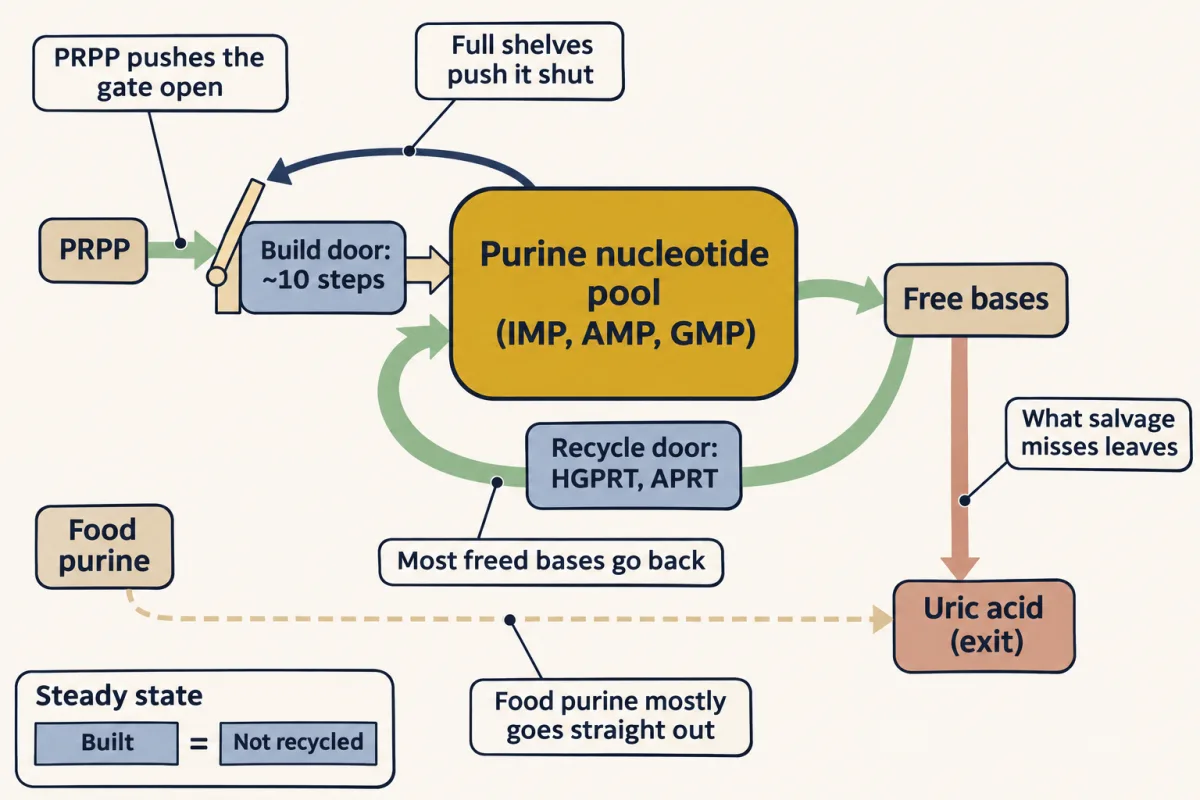
Two exceptions: a growing tissue builds extra to grow, and a sudden loss of many cells sends their nucleic acids and nucleotides into the exit (the last prediction). Food purine is mostly broken down in the gut wall and liver rather than reused, so it adds to the exit, not the pool. Pyrimidines have their own build and recycle routes (recycling runs through different enzymes) and a harmless exit, covered below.
The molecule both doors use: PRPP
Both doors start from PRPP, 5-phosphoribosyl-1-pyrophosphate: ribose-5-phosphate (from the pentose phosphate pathway) carrying a pyrophosphate, two linked phosphates, on carbon 1. PRPP synthetase takes that pyrophosphate from ATP, leaving AMP. PRPP is “activated”: when something attaches to carbon 1 the pyrophosphate leaves, and its rapid breakdown makes the attachment effectively one-way. Costs here are counted in ATP equivalents: one high-energy phosphate bond spent, as when ATP becomes ADP (a GTP becoming GDP counts the same). PRPP uses one ATP molecule, but turning it into AMP spends two bonds, and the AMP must be recharged twice to become ATP again. So one PRPP costs 2 ATP equivalents.
Door 1, build: de novo synthesis
The cell assembles the purine ring atom by atom directly on carbon 1 of PRPP, and about ten enzymatic steps later IMP appears. The ring’s nine atoms come from glycine (three), glutamine (two), aspartate (one), CO₂ (one) and two one-carbon (formyl) groups carried by tetrahydrofolate (THF), the working form of the folate vitamin. Remember those two folate carbons.
| What building spends | ATP equivalents |
|---|---|
| Making the PRPP | 2 |
| Four steps that each turn ATP into ADP (adding glycine, adding the second glutamine nitrogen, closing the first ring, adding aspartate) | 4 |
| IMP, total | 6 |
| IMP → AMP (a GTP → GDP, +1) | 7 for AMP |
| IMP → GMP (an ATP → AMP, +2) | 8 for GMP |
Door 2, recycle: salvage
Salvage attaches an existing base to PRPP in a single step, by phosphoribosyltransferases, which move the phosphoribose from PRPP onto the base. HGPRT (hypoxanthine-guanine phosphoribosyltransferase) turns hypoxanthine into IMP and guanine into GMP; APRT (adenine phosphoribosyltransferase) turns adenine into AMP.
The only cost is the PRPP, 2 ATP equivalents, with no amino acids or folate: three to four times cheaper than building. In many tissues salvage is a major source, not a backup; how much a cell relies on it also depends on how fast it is growing.
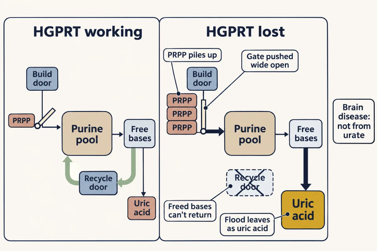
Knock out HGPRT and the recycle door shuts. Freed hypoxanthine and guanine can no longer go back into the pool, so they are oxidized to uric acid. The PRPP that salvage would have spent piles up and pushes the committed step open, so de novo building speeds up, and that extra purine leaves as uric acid too: the cell still builds what it fails to recycle. The result is severe hyperuricemia (urate crystals, kidney stones, gout). The neurological disease is a separate harm, not caused by urate.
Many tissues rely heavily on salvage — it is cheaper to glue a used base back onto PRPP than to build the nine-atom double ring (five carbons, four nitrogens) from glutamine, glycine, aspartate, CO₂, and folate-carried formyl groups. De novo is the route a rapidly dividing cell pays for when there simply aren't enough scrap bases to go around. The lesson sits in the contrast: salvage is the thrifty default, and Lesch-Nyhan shows what it costs to lose it — you pay twice, once in ATP and once in uric acid.
The gate: where the cell decides to build
PRPP itself can still go to salvage, to pyrimidines or to NAD⁺. The first step that sends it to purines only is glutamine-PRPP amidotransferase, which swaps PRPP’s pyrophosphate for an amino group from glutamine. That makes it the committed step, and so the place for control.
- Is there PRPP to spend? PRPP is the gate’s substrate and also switches it on. More PRPP, more building.
- Are the shelves full? IMP, AMP and GMP bind the enzyme away from its active site and switch it off (feedback inhibition). AMP and GMP together shut it down more than either alone.
Predict. A cell already has plenty of AMP and GMP, and it is handed free hypoxanthine. What happens to its rate of building?
Check your prediction
It falls, for two reasons. Salvaging the hypoxanthine uses up PRPP, so the gate has less to spend, and it makes IMP, which adds to the feedback. Recycling does more than save ATP: it turns building down.
The way out: uric acid
A purine that is not recycled is taken apart in three moves.
- Strip the phosphate and the amino group: AMP and adenosine funnel through to inosine (losing adenine’s amino group is what turns the adenosine into inosine, whose base is hypoxanthine).
- Remove the ribose: inosine becomes the free base hypoxanthine, which salvage can still catch.
- Oxidize twice: xanthine oxidase turns hypoxanthine into xanthine, and xanthine into uric acid. Guanine joins at xanthine, by losing its amino group.
Most mammals go further: their uricase oxidizes uric acid, the first of three steps to highly soluble allantoin. Humans and the other apes lost a working uricase during evolution, so for us sparingly soluble uric acid is the end product.
That missing enzyme is why humans are prone to gout, though who gets it depends mostly on how well the kidneys and gut excrete urate. Once serum urate (uric acid’s ionized form, the one in blood) climbs past roughly 6.8 mg/dL, it is supersaturated and can slowly crystallize as monosodium urate in and around joints. A flare is the body’s inflammatory attack on those needle-sharp crystals.
One fix is to make less urate. Allopurinol, a hypoxanthine look-alike, inhibits xanthine oxidase, so urate production falls and, over months, the crystals can dissolve. It came out of Gertrude Elion and George Hitchings’s lab, but not as a gout drug: they first tested it as a xanthine oxidase blocker to slow the breakdown of their leukemia drug 6-mercaptopurine, and its urate-lowering effect led to its use in gout. Other drugs instead help the kidneys excrete more urate.
Uric acid is where purine catabolism dead-ends in us — we lost uricase, the enzyme other mammals use to begin breaking it down further, so the poorly soluble acid is the last stop. Gout is simply that acid crossing its solubility line and crystallizing in a joint. Allopurinol, one of Elion and Hitchings's rationally designed purine analogs, is the elegant fix: it is itself oxidized by xanthine oxidase into oxypurinol, which then jams the enzyme shut — fewer urate molecules made, the serum level falls back under the line, and the crystals dissolve.
When recycling breaks: Lesch-Nyhan syndrome
Back to the opening puzzle. In Lesch-Nyhan syndrome the HGPRT gene, on the X chromosome, does not work, so nearly all patients are boys.
Predict. HGPRT is gone. Follow the freed hypoxanthine and guanine. Then ask what happens to phosphoribosyl pyrophosphate (PRPP), to the gate and to uric acid.
Check your prediction
The freed bases cannot go back into the pool, so they are oxidized to uric acid. The PRPP that salvage would have spent piles up and pushes the gate open, so building speeds up. Almost all of that extra purine leaves as uric acid too: the cell still builds what it fails to recycle, and now it fails to recycle far more. The result is severe hyperuricemia (too much urate in the blood): urate crystals in the diaper, then kidney stones and, later, gout.
Predict. Allopurinol brings these boys’ urate down. Will it also prevent the syndrome’s profound neurological disease, with severe movement problems and compulsive self-injury?
Check your prediction
No. Allopurinol protects the kidneys and joints, but the neurological disease is not caused by urate, and its mechanism is still not fully understood. Losing HGPRT does two separate kinds of harm, and allopurinol treats only the urate flood.
Pyrimidines: ring first
Pyrimidines (cytosine, uracil, thymine) mirror the purine build door: they build the ring first, from carbamoyl phosphate and aspartate, and take the ribose-phosphate from phosphoribosyl pyrophosphate (PRPP) only afterward. The carbamoyl phosphate comes from CPS-II, not the urea cycle’s carbamoyl phosphate synthetase I (CPS-I). CPS-I is mitochondrial, takes free ammonia and needs N-acetylglutamate; CPS-II is cytosolic, takes glutamine’s nitrogen and is shut off by UTP, a finished pyrimidine. Different places and signals let nitrogen disposal and nucleotide building run independently.
The exit differs too: pyrimidines break down to soluble products (β-alanine, β-aminoisobutyrate, CO₂ and ammonia), so they have no gout.
DNA’s building blocks
DNA needs deoxyribonucleotides, a branch off the pool outside the loop’s balance. In a dividing cell most new dNTPs (deoxynucleoside triphosphates) come through one gateway, ribonucleotide reductase, which reduces ribonucleotides; cells can also salvage ready-made deoxynucleosides. Its activity is therefore tied tightly to cell division, and hydroxyurea inhibits it (used in sickle-cell disease and some leukemias).
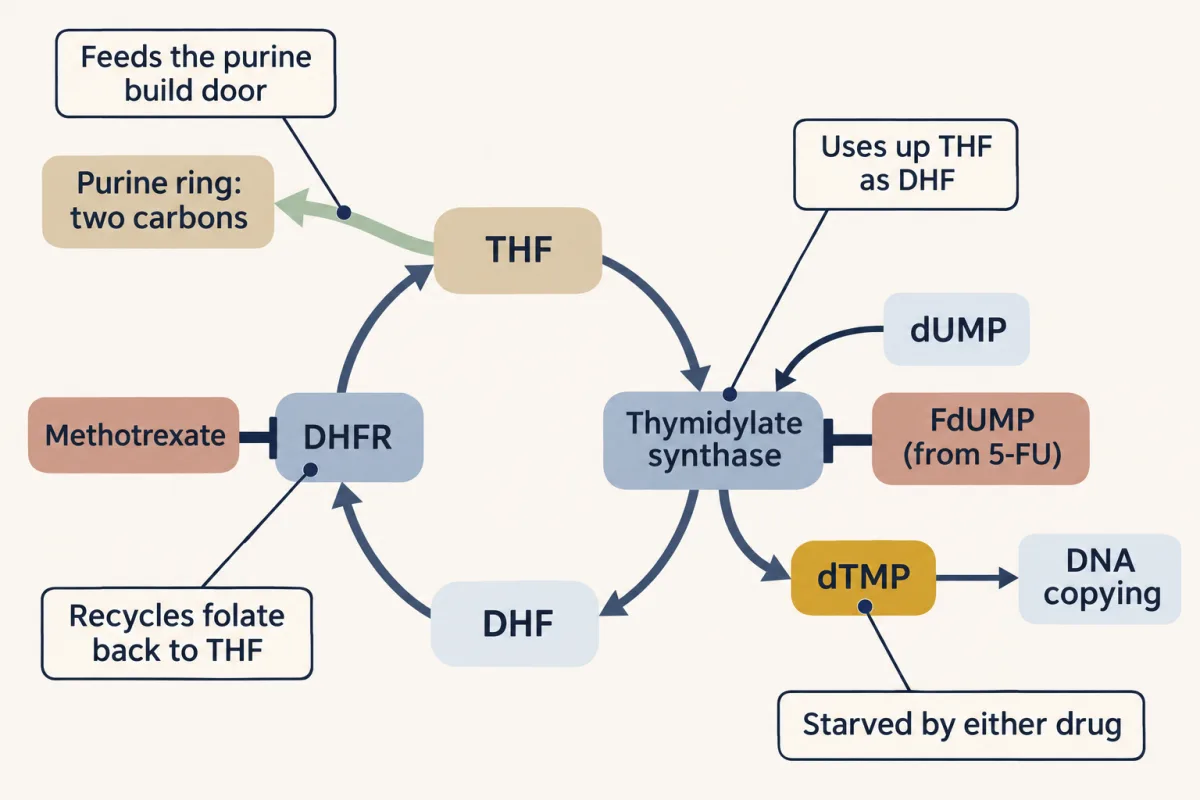
Thymine needs one more step. Thymidylate synthase adds a carbon, carried by a folate, to dUMP (uracil’s deoxynucleotide) to make thymidylate (dTMP), leaving the folate as dihydrofolate (DHF). Dihydrofolate reductase (DHFR) must turn DHF back into tetrahydrofolate (THF) before it can carry another carbon, to thymidylate or to the purine ring.
Counterfeits at each door
The drugs here run on Emil Fischer’s old principle: a molecule’s shape decides what an enzyme will accept. A counterfeit that fits the lock well enough to turn it, or jam it, is a medicine: an antimetabolite. The dividing cell is the most exposed, because it needs nucleotides fastest, though fast-dividing normal tissues are hit too.
- Methotrexate mimics folate and jams dihydrofolate reductase (DHFR), so tetrahydrofolate (THF) is not regenerated, starving both the purine ring’s two folate carbons and the carbon that makes dTMP.
- 5-fluorouracil mimics uracil. Inside the cell it becomes FdUMP, which traps thymidylate synthase itself, so it too starves the cell of the dTMP it needs to copy its DNA.
- 6-mercaptopurine (6-MP), a fake purine, enters through the recycle door: HGPRT turns it into counterfeit nucleotides. These shut down purine synthesis and, once built into DNA, damage the leukemia cells that took it up.
Predict. A leukemia cell loses its HGPRT to a mutation. What happens when it meets 6-MP?
Check your prediction
It is resistant. Without HGPRT, 6-MP is never made into nucleotides, and the cell can still build its purines de novo. That is why the related drug 6-thioguanine selects HGPRT-mutant cells in culture.
Predict. A treatment kills a large, fast-growing tumor within days. What happens to urate, and what would you give before treatment starts?
Check your prediction
Urate surges. This is the exception set aside earlier: dying cells release their DNA, RNA and nucleotides at once, their purines go down the exit to uric acid (tumor lysis), and the urate can crystallize in the kidneys. Allopurinol given beforehand blocks the exit’s last two steps, so less urate is made. Urate already in the blood needs rasburicase, a uricase made as a drug, which breaks it down.
The thread runs from Fischer naming the purine ring to Hitchings and Elion turning it into leukemia therapy, transplant drugs, the gout drug above and the antiviral acyclovir. Most aim at the doors, the exit or the DNA branch of one loop; acyclovir, a guanosine look-alike, instead jams the herpes virus’s own DNA polymerase.
Going deeper
The fine print of the tally
Phosphoribosyl pyrophosphate (PRPP) to IMP is ten steps in vertebrates (eleven counting PRPP synthetase). The CO₂ step uses no ATP in vertebrates; bacteria spend one there. IMP dehydrogenase, on the way to GMP, uses an NAD⁺ and is the target of the transplant drug mycophenolate. That AMP needs GTP and GMP needs ATP helps balance the two pools.
Variations on the loop
- An overactive PRPP synthetase pushes the gate open even with working HGPRT: urate overproduction and gout by the same logic.
- Partial HGPRT deficiency still causes urate overproduction, with gout and kidney stones, but milder or no neurological disease.
- In Lesch-Nyhan the established driver is the rise in PRPP. Weaker feedback from lower IMP and GMP may add to it, but that is secondary and debated.
- With working HGPRT, allopurinol tends to lower total purine output, not just urate, because the hypoxanthine it leaves behind is salvaged. In Lesch-Nyhan it only swaps urate for hypoxanthine and xanthine, with a risk of xanthine stones.
Pyrimidine and dNTP control
Besides UTP feedback, CPS-II is switched on by PRPP and ATP. In mammals it sits in one large protein, CAD, with the next two steps; a later enzyme, DHODH, makes orotate. In orotic aciduria, an inherited block after orotate, orotate spills into the urine. Mammalian ribonucleotide reductase works on ribonucleoside diphosphates; one allosteric site sets its overall activity and another sets which substrate it reduces, keeping the four dNTPs balanced (mis-tuning is mutagenic).
More on the drugs
- Inside cells methotrexate gains a chain of glutamates, and these forms also inhibit the purine formyl-adding enzymes directly, most strongly AICAR transformylase.
- The leucovorin paradox. Leucovorin, a ready-made reduced folate, bypasses dihydrofolate reductase (DHFR) and rescues normal cells from methotrexate. With 5-FU it does the opposite: it supplies the folate partner that locks FdUMP onto thymidylate synthase.
- TPMT (thiopurine methyltransferase) inactivates 6-MP, so people with low TPMT build up toxic amounts of its nucleotides. Xanthine oxidase also breaks 6-MP down, so giving allopurinol with 6-MP means cutting the 6-MP dose.
Your notes
Select any text in the lesson to highlight it or add a note to it. Your highlights and notes are saved in this browser and collected in your notebook.
This browser is not letting the site save anything (a private window or strict privacy settings), so highlights and notes will be lost when you leave the page.
How we measure it
Salvage (phosphoribosyltransfer)
Recycling a free base by attaching it to PRPP in a single step — HGPRT for hypoxanthine and guanine, APRT for adenine. The cheap route, and a major purine source in many tissues.
Antimetabolite drug design
Building a molecule that mimics a real metabolite closely enough to fool an enzyme, then jams it. Allopurinol (xanthine oxidase), methotrexate (DHFR), 5-FU (thymidylate synthase), 6-MP (activated by HGPRT; its nucleotides block purine synthesis and are built into DNA).
Enzyme inhibition
Blocking a single committed step to choke an entire pathway. For anticancer antimetabolites, the faster a cell divides, the harder the block bites, which is part of the therapeutic window; fast-dividing normal tissues feel it too.
Practice questions on this lesson (3) → MCAT-style, original, each with an explanation
Where this lesson fits
Before thisReview first
- The Pentose Phosphate Pathway: NADPH and Five-Carbon SugarsThis course · Lesson 24
You need it for ribose-5-phosphate, which is activated to PRPP, the molecule both purine doors start from.
- Metabolism: Basic Concepts and DesignThis course · Lesson 15
Counting costs in ATP equivalents and coupling uphill steps to ATP is used throughout the build-versus-recycle tally.
- Protein Turnover and Amino Acid Catabolism: Tag, Recognize, Destroy, RecycleThis course · Lesson 28
Glutamine and aspartate nitrogen feeding the ring, and the CPS-I versus CPS-II contrast, build on the urea cycle covered there.
After thisStudy next
- Integration of Metabolism: How the Body Switches FuelsThis course · Lesson 30
Shows how hormones switch pathways with the fed and fasting state, extending this lesson's gate and feedback control.
Elsewhere on the siteFilms, simulations and method films in other courses
- Spectrophotometry and enzyme kineticsMethod film
Xanthine oxidase inhibition by allopurinol is shown with rate measurements from absorbance changes.
- Radioactive and stable isotope tracersMethod film
Labelled precursors show whether a cell builds purines new or recycles free bases.
Suggested by an AI model from the lessons themselves, then checked. How we use AI
Finish line
Test yourself
8 questions
Practice and MCAT-style, with explained answers.
Next · Lesson 30
Integration of Metabolism: How the Body Switches Fuels
Fed, fasting, starving: led by two hormones, the body turns every pathway in this course up or down in a timed sequence.
Continue →← Lesson 28 · Protein Turnover and Amino Acid Catabolism: Tag, Recognize, Destroy, Recycle