Developmental Neuroscience · Skim version
Semaphorins and Repulsion
Read the full lesson → Skim the whole course →
The guiding idea
Pain fibres and muscle afferents enter the spinal cord side by side. One kind stops in the dorsal horn; the other runs on to the motor neurons. What sorts them?
The cue marks the place and the growth cone decides what it means: a semaphorin repels only axons whose receptors and internal state let them answer.
From the film: The Cue Marks the Place
- Sema3A, made in the ventral cord, repels NGF-dependent pain fibres but at the stage tested barely affects NT-3-dependent muscle afferents.
- The collapse assay led to collapsin, now Sema3A, a protein that folds up growth cones, though retinal growth cones did not respond to it.
- The cue marks the place and the growth cone decides what it means, which explains selectivity but not the whole map.
From the film: One Tissue, Two Axons, One Receptor
- In explants, the ventral cord repels axons grown in NGF but has little effect on axons grown in NT-3.
- Neuropilin catches Sema3A and plexin signals, and in these experiments no neuropilin-plexin pair means no response.
- A repelled growth cone typically turns away from a gradient on one side, and a high dose all round can make it collapse where the preparation supports collapse.
From the film: Same Cue, Opposite Answers
- Raising cyclic GMP turned Xenopus growth cones from repelled by a Sema3A gradient to attracted to it.
- In developing cortex, one neuron's axon is repelled by Sema3A while its apical dendrite is attracted, and soluble guanylate cyclase sits mainly in the dendrite.
- Pain fibres and muscle afferents differ in how strongly they answer the cue, while axon and dendrite differ in which way.
-
The puzzle in the spinal cord
-
A growth cone that folds up
-
Collapsin, the first vertebrate semaphorin
-
One tissue, two axons
-
Neuropilin catches, plexin signals
-
Same cue, opposite answers
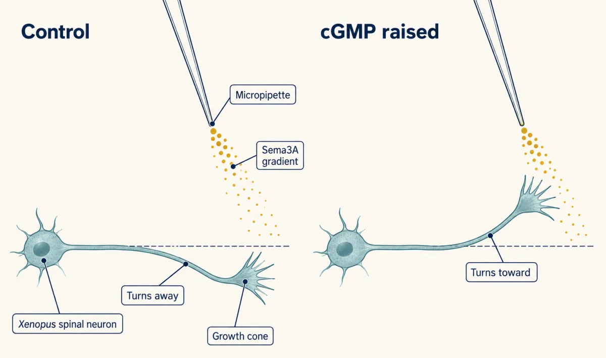
A small Sema3A gradient from a micropipette turned single Xenopus growth cones away. After the cyclic GMP pathway was raised, the same growth cones turned toward it. AI-generated illustration 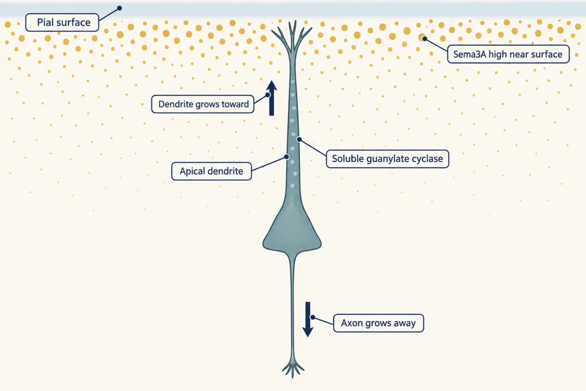
In the developing cortex Sema3A is high near the pial surface. The same pyramidal neuron's axon grows away from it and its apical dendrite toward it, and soluble guanylate cyclase sits mainly in the dendrite. AI-generated illustration -
Going deeper
Where this lesson fits
Before thisReview first
- The Growth Cone: The Tip That Steers the AxonThis course · Lesson 3
You need how the growth cone steers by building and losing actin to follow why repulsion makes it fold up.
- Pioneers, Guideposts and the Logic of Guidance CuesThis course · Lesson 22
The four kinds of cue, including contact and long-range repulsion, are used here without being re-taught.
- Netrin and DCC: One Cue, Two AnswersThis course · Lesson 23
It introduces the idea that a cue's sign depends on the growth cone's internal state, which is applied here with cGMP.
After thisStudy next
- Topographic Maps: Sperry's Eye and Eph GradientsThis course · Lesson 26
It uses repulsive cues acting on growth cones with different receptor levels to build an ordered map.
- Pruning Without Dying: Axons That Cut Themselves BackThis course · Lesson 34
Semaphorin signalling here goes on to prune axon branches after they have arrived.
- Synapses in the Brain: Adhesion, Scaffolds and AstrocytesThis course · Lesson 36
It takes the same semaphorin family on to limiting dendritic spines and shaping synapses.
Suggested by an AI model from the lessons themselves, then checked. How we use AI