Developmental Neuroscience · Skim version
Reelin and the Reeler Mouse
Read the full lesson → Skim the whole course →
The guiding idea
Why does a mouse that cannot walk straight have its cortex built in the wrong order?
Neurons are born in order, and Reelin signalling is needed to stack that order the right way up, so each new wave tends to finish its climb at the top.
From the film: Same Neurons, Wrong Order
- Reeler makes the same classes of neurons on the same schedule as normal mice, so it is a defect of placement, not production.
- In reeler the preplate fails to split, and early-born neurons tend to sit near the surface and late-born neurons deep, a roughly inverted trend with scatter.
- Reelin signalling is needed to stack birth order the right way up: with it each new wave tends to finish its climb at the top, and without it newcomers tend to stop beneath the neurons already there.
From the film: Matching Mutants, One Relay
- Reelin is a very large secreted protein made by Cajal-Retzius cells at the top of the cortex, where each new wave should stop.
- Matching reeler-like phenotypes in Dab1 mutants and in mice lacking both ApoER2 and VLDLR suggest one pathway, though they do not give its order.
- Reelin binds the receptors, Fyn and Src phosphorylate Dab1, and phosphorylated Dab1 is destroyed through Cullin 5, so normal positioning needs both the signal and its shut-off.
From the film: Needed, But What Does It Say?
- Removal shows that a signal is needed, not what it says; changing where the signal is can help tell permissive from instructive.
- Moving Reelin gave results that fit no single command: a source below the cortex rescued some defects, a small spot organized neurons around it, and an even bath organized reeler slices in a short-term assay.
- In mouse cortex, Reelin signalling through Dab1 and Rap1 has been linked to turning multipolar neurons and to the final somal translocation, which fits a switch on adhesion at particular stages.
- Helping later-born neurons settle above earlier ones does not by itself show that neurons steer toward Reelin.
-
Same neurons, wrong order
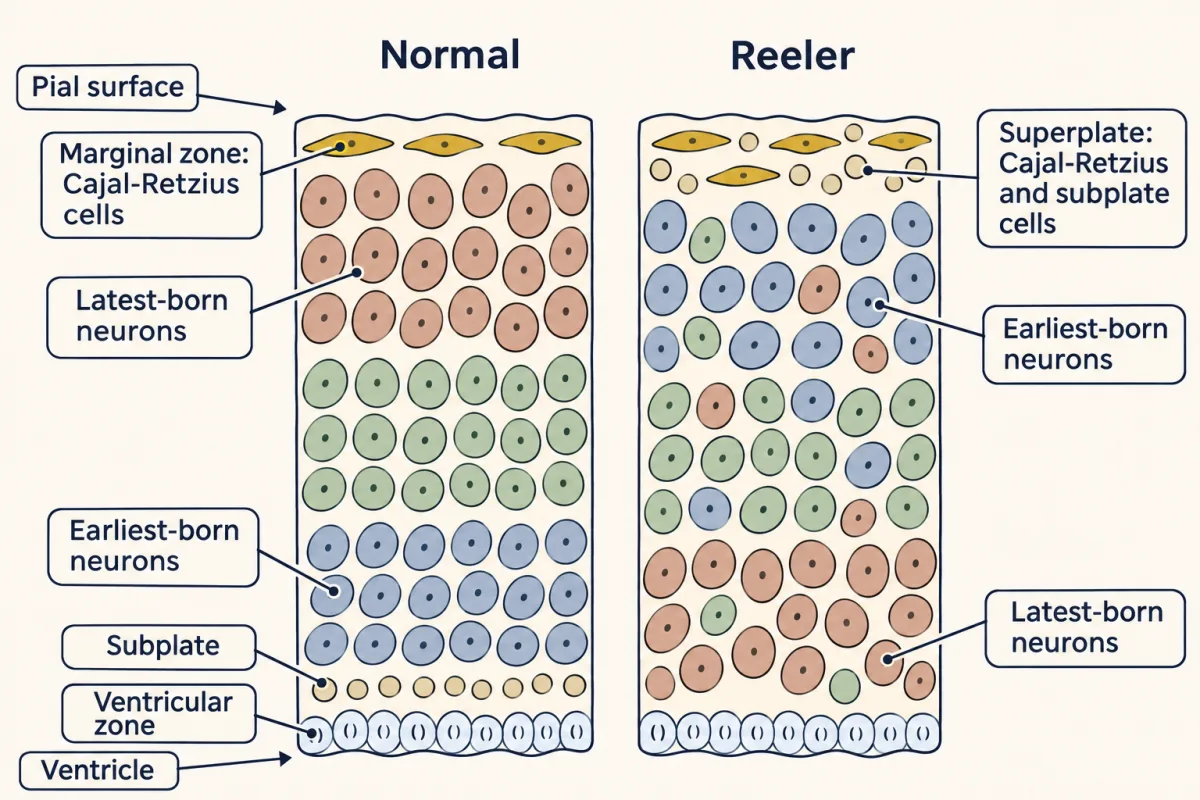
In normal cortex each new wave of neurons settles above the waves before it, so the earliest-born end up deepest. In reeler the same neurons, born on the same schedule, end up in roughly the reverse order with a lot of scatter, under a superplate where the preplate failed to split. AI-generated illustration -
Where the signal comes from, and who reads it
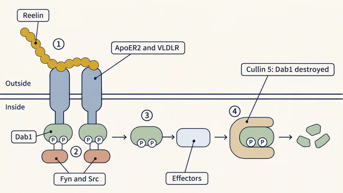
Reelin binds ApoER2 and VLDLR, Fyn and Src phosphorylate the adaptor Dab1 on tyrosine, phosphorylated Dab1 recruits effectors, and the same phosphorylated Dab1 is then destroyed through Cullin 5. Normal positioning needs both the signal and its shut-off. AI-generated illustration -
Needed, but what does it say?
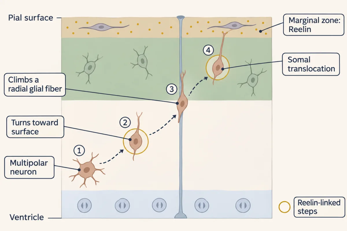
In mouse cortex, Reelin signalling through Dab1 has been linked to two stages of one neuron's climb: turning the multipolar neuron toward the surface, well below the marginal zone, and the final somal translocation. Reelin is not read only at the top. AI-generated illustration -
Putting it together
-
Going deeper
Where this lesson fits
Before thisReview first
- Birth Order and Fate: Temporal Identity and the Inside-Out CortexThis course · Lesson 16
You need the inside-out order of cortical layers from there to see why reeler's roughly inverted order is wrong.
- Radial Migration: Climbing the Glial LadderThis course · Lesson 17
You need the neuron's climb along a radial glial fiber from there to understand where Reelin acts.
- Adaptors, Docking Proteins and ScaffoldsCell Signaling · Graduate school and beyond
Dab1 is an adaptor, and this lesson uses the idea of docking proteins without re-teaching it.
After thisStudy next
- When Building Goes Wrong: Microcephaly, Zika and LissencephalyThis course · Lesson 48
Human RELN mutations give a smooth cortex, taking the Reelin pathway into disease.
- The Neurotrophic Hypothesis TestedThis course · Lesson 29
It reuses the permissive-versus-instructive test on a survival signal, moving the signal rather than removing it.
- Tangential Migration: Interneurons and the Long Way RoundThis course · Lesson 19
Interneurons arrive by another route and settle among the layers that Reelin signalling helps stack.
Elsewhere on the siteFilms, simulations and method films in other courses
- Forward genetic screensMethod film
Scrambler and yotari were found as mutants with a reeler-like walk, the way a screen finds pathway genes.
- Complementation (cis-trans) testMethod film
Matching mutant phenotypes put genes in one pathway, and combining mutations tests whether they hit the same gene.
- Tissue grafting and transplantationMethod film
Moving a Reelin source to a new place tests permissive against instructive signals, as in grafting.
- Cell-free extracts and reconstitutionMethod film
Adding Reelin as an even bath to reeler slices rebuilds the signal in a simple dish assay.
Suggested by an AI model from the lessons themselves, then checked. How we use AI