Developmental Neuroscience · Skim version
Local Organizers: The Midbrain-Hindbrain Boundary
Read the full lesson → Skim the whole course →
The guiding idea
How does one protein on a bead make a second midbrain grow, built back to front?
A line becomes a source: where Otx2 meets Gbx2, boundary cells send out FGF8 and Wnt1, and on the simplest reading the signal a neighbour gets tells it which part to build.
From the film: Where the Line Falls
- A local organizer is a small signaling center that tells nearby cells what to build.
- A line becomes a source: where Otx2 meets Gbx2, boundary cells make signals such as FGF8 and Wnt1.
- On the simplest reading, the signal a cell gets tells it which part to build, and the repressor it carries sets which region it can become.
From the film: One Bead Builds a Midbrain
- An FGF8 bead in the chick caudal forebrain induced an extra midbrain built as a mirror image.
- This shows the line can act as a source, since adding it somewhere new instructs the neighbouring cells.
- The result may also depend on an organizer the bead induced, and on the dose and duration of FGF8.
From the film: Started, Then Lost
- In Wnt1-null mice Engrailed comes on and then fades, so Wnt1 is needed to keep the program on, not to start it.
- Fgf8 loss gives the same started-then-lost signature, in zebrafish and in mice.
- The signals are needed to keep the program running, and in the standard model the transcription factors help keep the signals on.
-
Beyond the Hox border
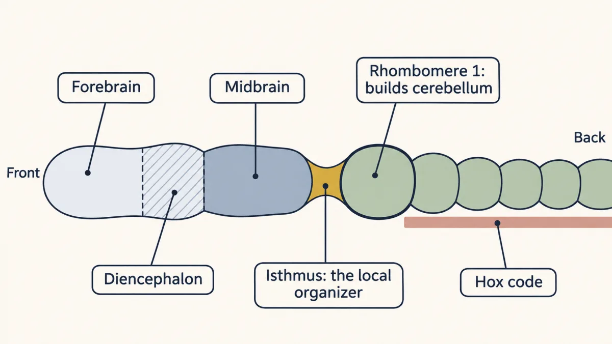
The Hox code stops near the front of the hindbrain, so the midbrain and forebrain need another address. A local organizer at the isthmus, where midbrain meets hindbrain, supplies it: a small signaling center that tells the tissue around it what to build. AI-generated illustration -
Where the line falls
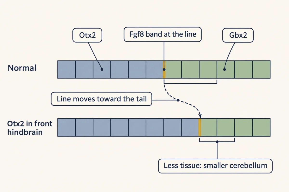
A move-it test: when Otx2 is made in the front of the hindbrain, the line and its Fgf8 band shift toward the tail and less tissue is left behind them, so the cerebellum is smaller. The line follows the edge of each repressor. AI-generated illustration -
The line becomes a source
-
Add it: a graft, then a bead
-
Take it away: started, then lost
-
Why the source stays on
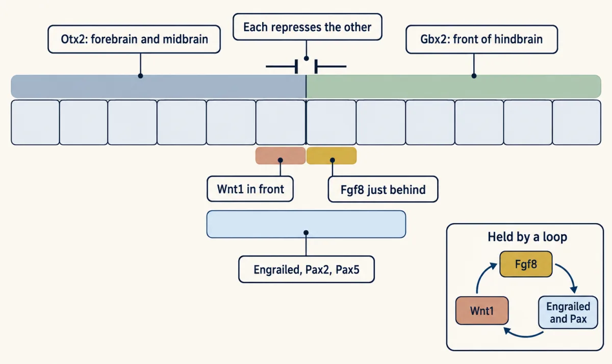
In the resolved pattern, Otx2 and Gbx2 repress each other and help position a sharp boundary. Wnt1 is expressed just in front, Fgf8 just behind, and Engrailed and Pax genes across it. In the standard model, these genes help maintain one another; some single losses are milder because a related gene can compensate. The inset loop is schematic, not a defined wiring diagram. AI-generated illustration -
Other organizers along the tube
-
Putting it together
-
Going deeper
Where this lesson fits
Before thisReview first
- The Organizer: Spemann and MangoldThis course · Lesson 5
Spemann's organizer is the first organizer, and this lesson sets local organizers apart from it.
- Head to Tail: Hox Genes, Retinoic Acid and RhombomeresThis course · Lesson 8
Hox genes address the hindbrain and stop near its front, which leaves the midbrain needing a different address.
- Tools for Following Cells and GenesThis course · Lesson 4
The grafts, bead implants and conditional deletions here rely on the tools for following cells and genes described there.
After thisStudy next
- Ventral to Dorsal: Sonic Hedgehog and BMP in the Neural TubeThis course · Lesson 10
Another secreted source patterns the tube by how much signal a cell receives, now across ventral to dorsal.
Suggested by an AI model from the lessons themselves, then checked. How we use AI