Cell Signaling · Skim version
Lipids as Anchors and Addresses
Read the full lesson → Skim the whole course →
The guiding idea
How does a protein with no membrane-spanning segment end up stuck to a membrane, and how does a lipid label tell it where?
Lipids control signaling protein location by supplying grip, address, or both.
From the film: Why One Farnesyl Tail Is Usually Not Enough
- A second signal turns weak membrane contact into a regulated hold.
- Lipids control signaling protein location by supplying grip, address, or both.
- A permanent lipid-protein bond does not make membrane contact permanent.
- Removing palmitate or neutralizing a positive patch releases the protein without breaking its permanent anchor.
From the film: How a Membrane Writes an Address
- Writers, readers, and erasers turn membrane lipids into changing addresses.
- PIP3 production, spread and removal together set where the address sits.
- PI3-kinase writes PIP3, matching PH domains read it, and PTEN erases it.
- A PH-domain reporter measures its own recruitment, so controls establish what that glow means.
-
Covalent anchors: what is attached, and where?
-
Two contacts make residence strong and regulatable
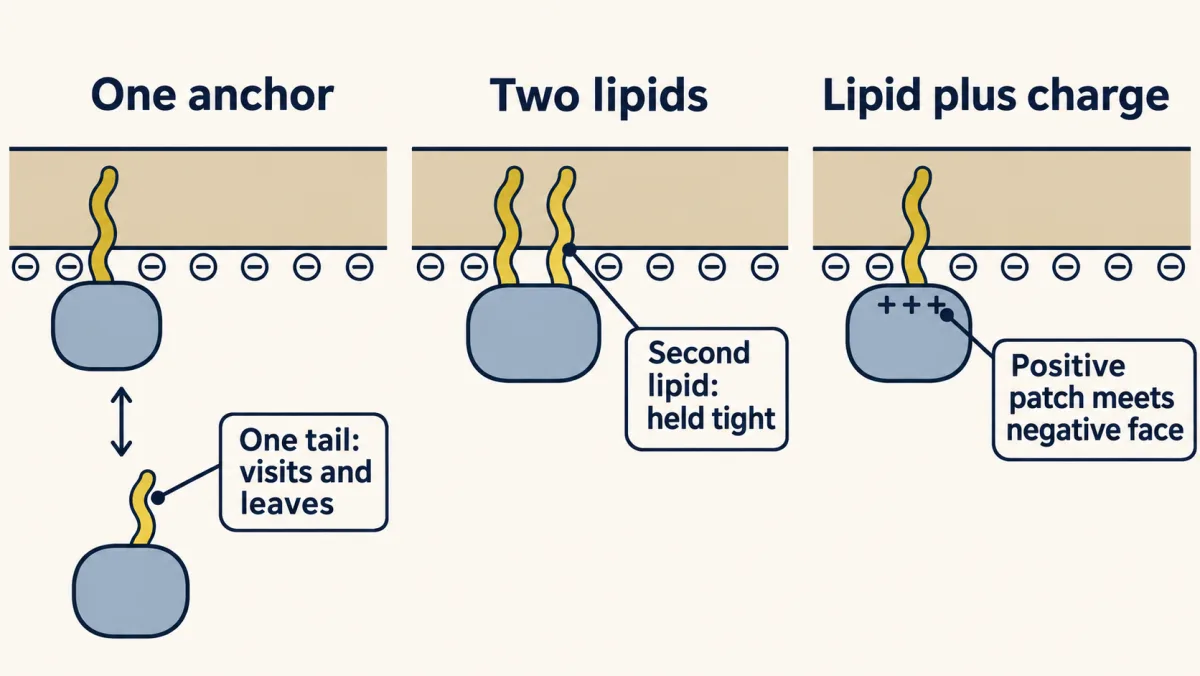
One lipid lets a protein sample membranes; a second lipid or a compatible charge patch makes residence much stronger and creates a point of regulation. AI-generated illustration -
Palmitate turns a hold into a cycle
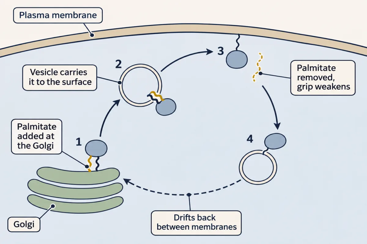
Reversible palmitoylation creates a kinetic trap: Golgi capture and vesicle transport enrich H-Ras at the plasma membrane, while depalmitoylation resets sampling; the exact cellular removal sites are not all settled. AI-generated illustration -
Can blocking an anchor block cancer signaling?
-
Phosphoinositides make dynamic addresses
-
Readers and erasers
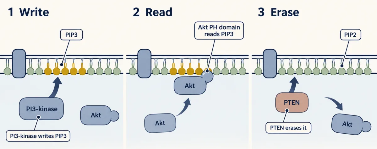
PI3-kinase writes a PIP3 address, a matching PH domain recruits Akt, and PTEN erases the 3-phosphate; lateral diffusion and alternate phosphatases make the real pattern dynamic. AI-generated illustration -
Turn recruitment into a measurement
-
Going deeper
Where this lesson fits
Before thisReview first
- Membranes: The Impermeable Sheet That Builds ItselfBiochemistry · College: going further
It explains why a long hydrocarbon chain sits happily in a bilayer, which is the basis of every lipid anchor here.
- GTPase Switches: The TimerThis course · Lesson 7
Ras is the GTP-binding switch whose membrane position this lesson explains, so you need how it cycles first.
- Protein Modules: SH2, SH3, PTB, PDZ and PHThis course · Lesson 9
PH domains are introduced there as readers of marks, and this lesson uses them to read PIP3.
After thisStudy next
- PI 3-Kinase, Akt and InsulinThis course · Lesson 20
It continues from PIP3 and the Akt PH domain to what Akt does once it reaches the membrane.
- Ras to MAPK: The Kinase CascadeThis course · Lesson 19
It uses Ras sitting on the inner membrane face to recruit Raf and start the kinase cascade.
- PLC, IP3, DAG and Protein Kinase CThis course · Lesson 14
It follows the PIP2 lipid cut by PLC, whose product DAG recruits kinase C to the membrane.
Elsewhere on the siteFilms, simulations and method films in other courses
- Which Side Is Outside?Film · Cell Biology
It shows the two faces of a membrane, where lipid-anchored proteins sit on the cytosolic side.
Suggested by an AI model from the lessons themselves, then checked. How we use AI