Developmental Neuroscience · Skim version
Head to Tail: Hox Genes, Retinoic Acid and Rhombomeres
Read the full lesson → Skim the whole course →
The guiding idea
Rhombomeres r2 and r4 start out looking alike. What tells one to build jaw motor neurons and the other facial ones?
Before the neural tube makes neurons, its hindbrain and spinal cord are given an address written largely in Hox transcription factors; the front of the brain, including r1, uses other genes.
From the film: Two Segments, Two Jobs
- Rhombomeres look alike but each carries its own code of transcription factors, mostly from Hox genes.
- Neighbouring segments can share many Hox genes, and segment genes such as Krox20 help tell them apart.
- More early retinoic acid gives a more posterior code, and the code helps set which neurons a segment makes.
From the film: Remove One Letter
- Without Hoxb1, rhombomere 4 stays in the mouse but loses parts of its identity.
- Its motor neurons then behave like trigeminal neurons, and the facial motor nucleus fails to form normally.
- Without Krox20, by contrast, r3 and r5 do not persist as distinct segments, so the code acts as a cause, not just a marker.
From the film: A Tap at the Back, a Drain at the Front
- Retinoic acid, made from vitamin A, is made toward the back of the hindbrain and destroyed toward the front by Cyp26 enzymes.
- Extra early retinoic acid shifts Hox domains forward, and less of it leaves the posterior hindbrain poorly patterned.
- The signal sets the direction of the code, not the exact edges, which other inputs help draw.
- Retinoids are teratogens in mouse and chick, and in humans this mechanism is inferred from animal work.
-
Two segments that look alike
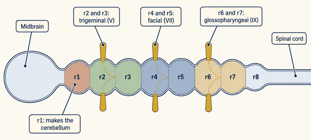
Schematic dorsal view of the chick embryonic hindbrain between midbrain and spinal cord. Branchiomotor neurons contributing to nerves V, VII and IX arise in r2-r3, r4-r5 and r6-r7, respectively; their axons leave at r2, r4 and r6. Gold stubs mark these exit sites schematically; other cranial nerves and detailed axon trajectories are omitted. Rhombomere 1 contributes to the cerebellum. AI-generated illustration 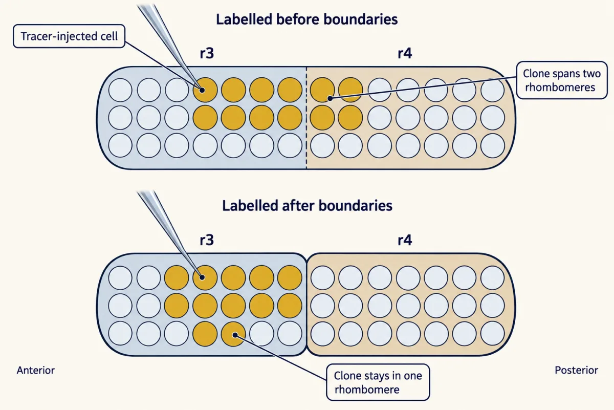
In chick hindbrain, a single neuroepithelial progenitor was labelled and its descendants examined later. Clones labelled before boundaries appeared could span neighbouring rhombomeres; clones labelled after boundaries appeared remained within one rhombomere. This demonstrates progenitor lineage restriction, not a barrier to all later neuronal migration. Gold cells illustrate clone distribution, not measured clone size. AI-generated illustration 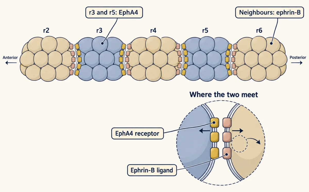
In fish and frog embryos, r3 and r5 express EphA4 and neighbouring rhombomeres express ephrin-B ligands. Contact between receptor-bearing and ligand-bearing cells promotes sorting and helps restrict mixing of hindbrain progenitors across boundaries; this does not prevent all later neuronal migration. AI-generated illustration -
The code each segment carries
-
Remove one letter: Hoxb1
-
Where the edges come from: a tap at the back, a drain at the front
-
Order on the chromosome, order along the body
-
Putting it together
-
Going deeper
Where this lesson fits
Before thisReview first
- Neural Induction: The Default ModelThis course · Lesson 6
You need the neural plate and its default posterior-to-anterior patterning from there to follow how the hindbrain gets an address.
- Neurulation and Neural Tube DefectsThis course · Lesson 7
This lesson assumes the closed neural tube and its early regions, which that lesson builds.
- The Organizer: Spemann and MangoldThis course · Lesson 5
Graded signals from a source that pattern the neural plate are introduced there, and retinoic acid extends that logic.
After thisStudy next
- Local Organizers: The Midbrain-Hindbrain BoundaryThis course · Lesson 9
It shows how a boundary signal holds Hoxa2 out of r1, an edge only mentioned here.
- Transcription Factor Codes: Building a Motor NeuronThis course · Lesson 11
It takes the idea that a transcription factor code sets neuron identity and works it out for spinal motor neurons.
Elsewhere on the siteFilms, simulations and method films in other courses
- Why Far Signals Ride the BloodFilm · Cell Signaling
A retinoic acid gradient is a morphogen-style signal whose range depends on how far it travels.
Suggested by an AI model from the lessons themselves, then checked. How we use AI