Sensory Systems · Skim version
Hidden Hearing Loss, Gene Therapy and Regeneration
Read the full lesson → Skim the whole course →
The guiding idea
A mouse can pass its hearing test after loud noise and still have lost about half the ribbon synapses at one place in the cochlea. What does a normal threshold actually tell you?
A hearing test can come back normal while a cochlea has lost many of its synapses.
From the film: A Threshold Is Not a Count
- In these mice, a cochlea lost many synapses and still passed a hearing test.
- A threshold depends mostly on the most sensitive fibres and does not count them.
- A normal threshold is good news about sensitivity, but it is not a census of synapses.
From the film: The Link That Must Fire, and How to Put It Back
- A knockout shows that a protein is needed, and it does not by itself show that the protein is the sensor.
- In severe otoferlin deafness the inner hair cells are present, but their synapses release almost nothing.
- Replacing a missing link can work because the cells that need it are still alive.
From the film: Reading the Trial, and When the Cell Is Gone
- Nine of twelve is a share of children meeting an endpoint, not a share of hearing.
- When the hair cell itself is gone, a new hair cell has to be made, and the adult mammalian cochlea does not do this.
- In newborn mice, reprogramming works less well a week later, so age is a barrier.
-
A hearing test passed by a damaged cochlea
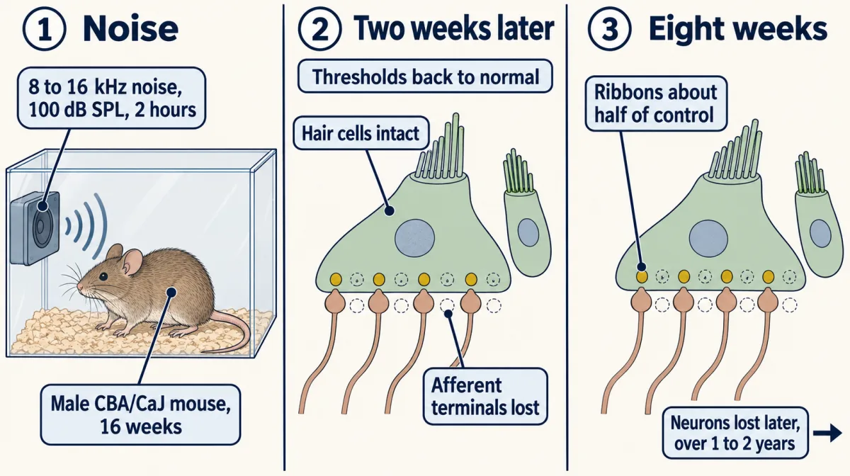
After the noise, thresholds recovered by two weeks and hair cells stayed intact despite acute afferent-terminal loss. At eight weeks, ribbons in the 32 kHz region were about half of control, and neurons were lost much later, over about one to two years. Ribbon symbols illustrate the relative count, not the measured number in an individual cell. AI-generated illustration -
Why a threshold can miss half the synapses
-
Finding the lesion in people
-
The link that has to fire: what makes the synapse release
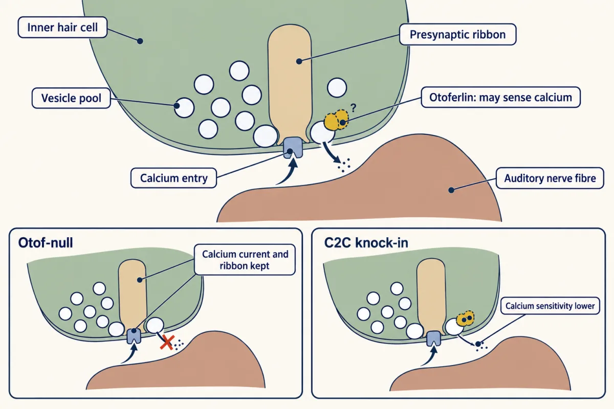
In the Otof-null mice studied by Roux and colleagues, calcium current and ribbon morphogenesis were preserved at the age recorded, but exocytosis was almost abolished. The inset label applies only to that age: by postnatal days 26 to 29, Otof-null inner hair cells studied by Al-Moyed and colleagues had about 40 percent fewer ribbons than wild-type cells. In the C2C knock-in mice studied by Michalski and colleagues, otoferlin was present but the calcium sensitivity of fast and sustained exocytosis was reduced. AI-generated illustration -
Putting the link back: a gene too big for its vector
-
Reading an early-phase trial without inflating it
-
When the cell itself is gone: regeneration
-
Going deeper
Where this lesson fits
Before thisReview first
- The Hair Cell's Transduction ChannelThis course · Lesson 22
You need the hair cell's transduction step from there, since the chain here starts with that cell and ends at the synapse.
- The Cochlear Amplifier and Otoacoustic EmissionsThis course · Lesson 23
Outer hair cell amplification explains why otoacoustic emissions stay present in otoferlin deafness while the brainstem response is absent.
After thisStudy next
- Restoring Senses: Implants, Prostheses and Optogenetic VisionThis course · Lesson 30
Cochlear implants bypass lost hair cells and talk to the nerve, the alternative when gene replacement cannot work.
Elsewhere on the siteFilms, simulations and method films in other courses
- Counting failuresSimulation · Neuroscience
It shows how calcium entry triggers vesicle release at a synapse, the step that otoferlin loss removes.
- One Cell, One SwitchFilm · Neuroscience
It shows how hair cells turn sound into signals sent to the nerve, the chain that fails here.
Suggested by an AI model from the lessons themselves, then checked. How we use AI