Sensory Systems · Skim version
Computing Motion in the Retina
Read the full lesson → Skim the whole course →
The guiding idea
A cell in the eye fires to a spot moving left and stays quiet for the same spot moving right. Which neuron makes the difference?
In mouse and rabbit retina, each starburst dendrite prefers outward motion, and a direction-selective ganglion cell is wired mainly to the dendrites pointing in its null direction, so null-direction motion mostly drives inhibition.
From the film: A Veto From the Null Side
- A direction-selective ganglion cell fires to its preferred direction and little to its null direction, and Barlow and Levick found a deficit for null-order flashes, so the main operation they inferred is a veto.
- Each starburst dendrite is its own small direction detector, most active when motion runs outward from cell body to tip.
- The dendrites that give a ganglion cell its strongest inhibition prefer roughly the direction that cell ignores.
From the film: Reading the Wiring
- In rabbit starburst cells, dendritic calcium was larger for outward motion, while the somatic voltage did not show that preference, so the branch is the unit.
- In mouse retina, most starburst input to a direction-selective ganglion cell came from branches pointing roughly in its null direction.
- An electron microscopy contact shows wiring, not synaptic strength.
From the film: Built by Strengthening, and Where the Idea Stops
- In paired recordings in mouse retina, null-side inhibition grew while preferred-side inhibition stayed about constant, so development strengthened one side and did not delete the other.
- The authors concluded that null-side pairs had more inhibitory synapses, not stronger single synapses.
- When only starburst GABA release was cut in mice, inhibition lost its direction tuning but selectivity survived in a subset of ON-OFF cells, so tuned inhibition is important but not indispensable.
-
What the mechanism must do: a veto on the null direction
-
The circuit in one picture
-
Each branch is its own detector (2002)
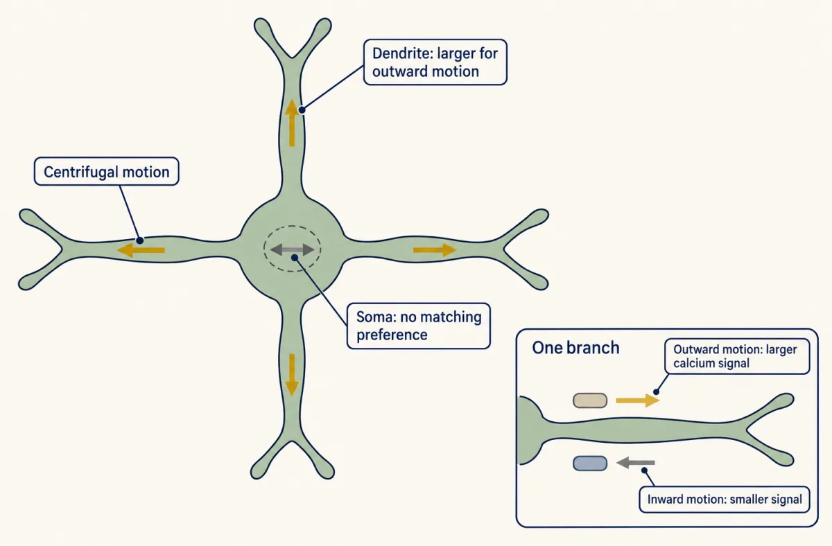
In ON starburst amacrine cells of isolated rabbit retina, Euler, Detwiler and Denk found dendritic calcium signals larger for motion outward from the soma than inward, while somatic voltage did not show the same preference under the conditions tested. Arrows show stimulus motion. Calcium signals are not transmitter release, so the result shows a local directional signal and leaves its mechanism open. AI-generated illustration -
Reading the wiring (2002, 2011)
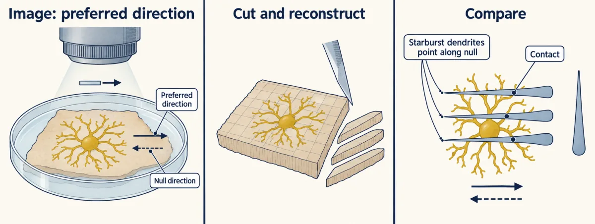
Briggman, Helmstaedter and Denk read each ganglion cell's preferred direction by calcium imaging in mouse retina, then reconstructed the same tissue by serial block-face electron microscopy. Most starburst input onto a direction-selective ganglion cell came from dendrites pointing roughly in its null direction. The reconstruction is an illustrative subset; contacts do not measure synaptic strength. AI-generated illustration -
Built by strengthening, not pruning (2011)
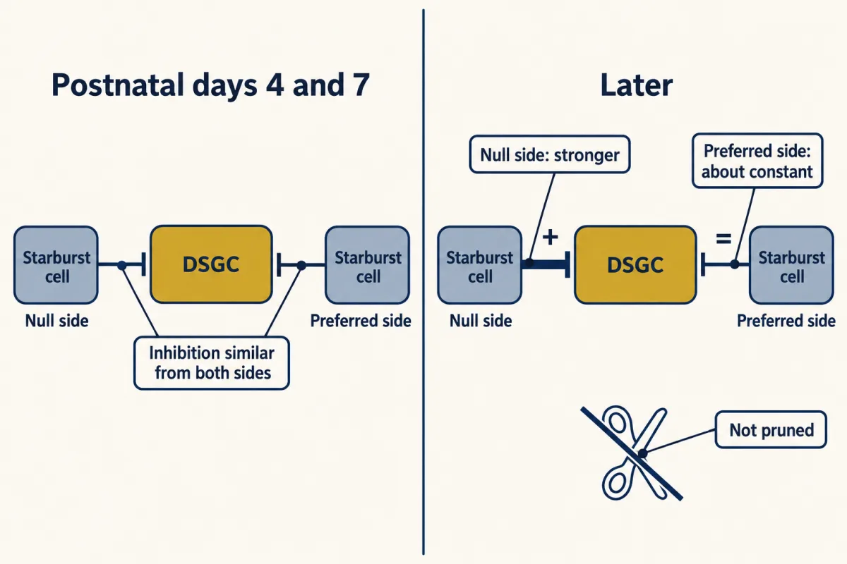
In paired recordings in mouse retina, Wei, Hamby, Zhou and Feller found that inhibition onto the direction-selective ganglion cell was similar from the two sides at postnatal days 4 and 7. Later the null-side inhibition increased while the preferred-side inhibition stayed about constant and was still detectable in adult retina, so development strengthened one side rather than pruning the other. AI-generated illustration -
Where the idea stops
-
Going deeper
Where this lesson fits
After thisStudy next
- Melanopsin Cells: Light for the Clock and the PupilThis course · Lesson 20
It uses the same balance-point rule for conductances, applied to another ganglion cell type that reads light differently.
- Combining Senses and Predicting ThemThis course · Lesson 29
It takes motion signals further, asking how the brain weights and predicts movement signals that disagree or arise from the self.
Elsewhere on the siteFilms, simulations and method films in other courses
- One More RopeFilm · Neuroscience
Null-direction inhibition vetoing excitation follows the same rules as inhibition shunting a cell's firing.
- Shunting inhibitionSimulation · Neuroscience
It lets you see how an inhibitory input can block firing without changing voltage, the same arithmetic as the null-side veto.
- One Cell CountsFilm · Neuroscience
It shows how retinal circuits process light before the optic nerve, the setting for the starburst veto.
Suggested by an AI model from the lessons themselves, then checked. How we use AI