Biochemistry · Metabolism: Foundations
Metabolism: Basic Concepts and Design
Rewritten on : new text, figures and flashcards. Your notes and highlights stay, but the text around them may have moved.
Metabolism is an energy economy; learn its grammar before any pathway teaches its vocabulary.
Pause when the reading reaches a film, and offer to play it. Saved on this device.
Watch the film now, or keep listening
Builds on:Basic Concepts of Enzyme ActionWater, Weak Bonds, and the Generation of OrderWhy these?
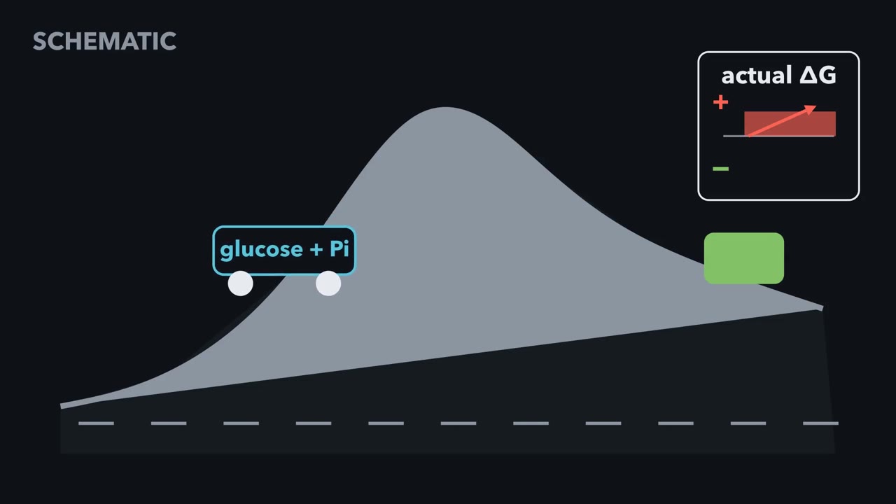 ▶ Watch How cells pull reactions uphill · 3:18
Read the transcript of “How cells pull reactions uphill”
Film transcript: How cells pull reactions uphill
How can a cell make a reaction run uphill without breaking thermodynamics? Watch what happens when glucose receives a phosphate. At cellular concentrations, glucose plus free phosphate favors the starting side. Its actual delta ΔG is positive, so the cart rolls backward.
Here is the rule that governs every case. A reaction runs forward only when its actual delta ΔG is negative. Enzymes lower activation barriers and speed traffic. They cannot change the sign of delta ΔG. The cell must change the chemical journey or the concentrations along it. In simplest form, the cell ties an uphill reaction to a downhill one. Hexokinase directly transfers ATP's end phosphoryl to glucose, not through free phosphate. ATP becomes ADP as glucose becomes glucose-6-phosphate. This is one chemical reaction, so the free-energy changes add. Its actual delta ΔG is negative, and the joined carts move forward. Now predict what happens if a mutant enzyme separates the jobs.
One site hydrolyzes ATP. Another site tries to attach free phosphate to glucose, with no shared intermediate or conformational link. The standard free-energy changes still add on paper. Will glucose-6-phosphate form? No. ATP hydrolysis runs downhill, but its free energy leaves as heat. The glucose cart still rolls backward. Arithmetic alone is not coupling.
Chemistry must be joined by direct transfer, a shared intermediate, or linked shape changes. Cells also change concentrations. Here, A → B + C has standard delta ΔG°' = +21 kJ/mol. B → D has ΔG°' = −34 Draining B pulls the first step's actual delta ΔG below zero. Although the pathway total is minus −13 that is not enough. Every step carrying forward flux must have negative actual delta ΔG. B to D has negative actual delta G. Take a separate standard example. Coupling to ATP shifts K from about 0.001 to about 267. That moves equilibrium toward products by more than 200,000-fold. That is the answer to our puzzle. Hexokinase's direct phosphoryl transfer changes the journey, so glucose phosphorylation can run forward.
A following step may instead keep a product scarce. The same rule governs ATP and other activated carriers. Uphill chemistry runs only when coupling makes its actual delta ΔG negative.
From the film
- A reaction runs forward only when its actual ΔG is negative.
- ATP pays only when its downhill chemistry is joined to the uphill reaction.
- A following step can pull a reaction forward by keeping its product scarce.
- Every step carrying forward flux must have a negative actual ΔG.
Once glucose is inside a cell, the first step attaches a phosphate to it, and written with free phosphate, that attachment runs backward.
Hexokinase turns glucose into glucose-6-phosphate, which traps the sugar in the cell and starts glycolysis. Yet at a cell’s phosphate level, glucose and free phosphate left to reach equilibrium would put a phosphate on only about one glucose in 50,000 (worked out below). How the cell makes this uphill step anyway, without breaking thermodynamics, is the design idea behind all of metabolism.
The frame is an energy economy. A cell earns energy by oxidizing fuel (catabolism) and spends it on three jobs: mechanical work (a muscle contracting, a cell crawling), active transport (the Na⁺/K⁺ pump fighting a gradient) and biosynthesis (building proteins, lipids and nucleic acids), the building being anabolism. Plants and other phototrophs capture sunlight; we are chemotrophs and oxidize carbon fuels. Energy is conserved; it is transformed from electrons in food’s carbons into a currency the cell can spend.
The one idea to carry through this lesson. A reaction runs forward only when its own ΔG, at the concentrations in the cell, is negative. The film above shows the two ways a cell makes an uphill step’s actual ΔG negative; the sections below supply the mechanisms and numbers.
The rule says whether a reaction can run, not how fast; enzymes decide that, and regulation (the last section) decides which reactions that can run actually do.
The rule: ΔG, not ΔG°’
Written the simplest way, attaching free phosphate to glucose runs uphill:
glucose + Pi → glucose-6-phosphate + H₂O ΔG°’ = +13.8 kJ/mol
ΔG°’ is the free-energy change under biochemists’ standard conditions: everything at 1 M, pH 7, 25 °C. Positive means that, from those conditions, the reaction runs backward. A cell is never at standard conditions; what decides direction there is ΔG, at the concentrations actually present.
In words The real free-energy change equals the standard one, plus a correction for how far the actual concentrations are from 1 molar.
- ΔG“delta G”
- the free-energy change at the concentrations present right now. Negative means the reaction can run forward.kJ/mol
- ΔG°'“delta G naught prime”
- the free-energy change at 1 M of everything, pH 7kJ/mol
- R“R”
- the gas constant, 0.008314kJ/(mol·K)
- T“T”
- temperature in kelvin; at 25 °C, RT is about 2.48 kJ/mol, and at 37 °C about 2.58K
- Q“Q”
- products over reactants, using the concentrations present right now, in molar. Water is left out by convention.depends on the reaction
Sanity check At equilibrium nothing changes, so ΔG = 0 and Q equals the equilibrium constant K'eq. That gives ΔG°' = −RT ln K'eq. A small Q (little product) makes ln Q negative and pulls ΔG down.
Try one number
For glucose + Pi → glucose-6-phosphate: K’eq = e−13.8 / 2.48 = e−5.57 ≈ 0.0038.
Here Q = [G6P] / ([glucose] × [Pi]). A cell holds about 5 mM phosphate, which is 0.005 M.
At equilibrium Q = K’eq, so [G6P] / [glucose] = K’eq × [Pi] = 0.0038 × 0.005 ≈ 0.000019. About one glucose in 50,000 would carry a phosphate.
Need a refresher? e and ln · units and prefixes
Predict. Could the cell make glucose-6-phosphate from free phosphate just by piling up more phosphate?
Check your prediction
Not usefully. To get as much glucose-6-phosphate as glucose at equilibrium, [Pi] would have to reach 1 / 0.0038, about 260 M. That is not a concentration any cell, or any beaker, can hold.
Coupling, the first way: one enzyme, one reaction
As the film shows, hexokinase bypasses free phosphate and transfers ATP’s end γ-phosphoryl straight to glucose in one active site:
glucose + ATP → glucose-6-phosphate + ADP
Free energy depends only on where a reaction starts and ends, so add two bookkeeping reactions that together have the same start and end:
| Step | ΔG°’ (kJ/mol) |
|---|---|
| glucose + Pi → glucose-6-phosphate + H₂O | +13.8 |
| ATP + H₂O → ADP + Pi | −30.5 |
| glucose + ATP → glucose-6-phosphate + ADP | −16.7 |
Pi and water appear on both sides and cancel. Because hexokinase performs the direct group transfer as one chemical reaction, the cancellation is legitimate and free phosphate is never made. Other enzymes pass the group through a short-lived covalent intermediate, which textbooks call a shared (common) intermediate: glutamine synthetase first puts ATP’s phosphoryl group on glutamate, then ammonia displaces it.
The payoff: the coupled K’eq is e16.7 / 2.48, about 840. Adding −30.5 kJ/mol multiplied the equilibrium constant by e30.5 / 2.48, about 2 × 10⁵. And because Q is now [G6P][ADP] / ([glucose][ATP]), the cell’s high ATP-to-ADP ratio helps too. With ATP ten times ADP, equilibrium sits at 840 × 10, about 8,400 glucose-6-phosphates per glucose, up from one per 50,000.
The film above turns this coupling into two joined wagons, with hexokinase as the rope.
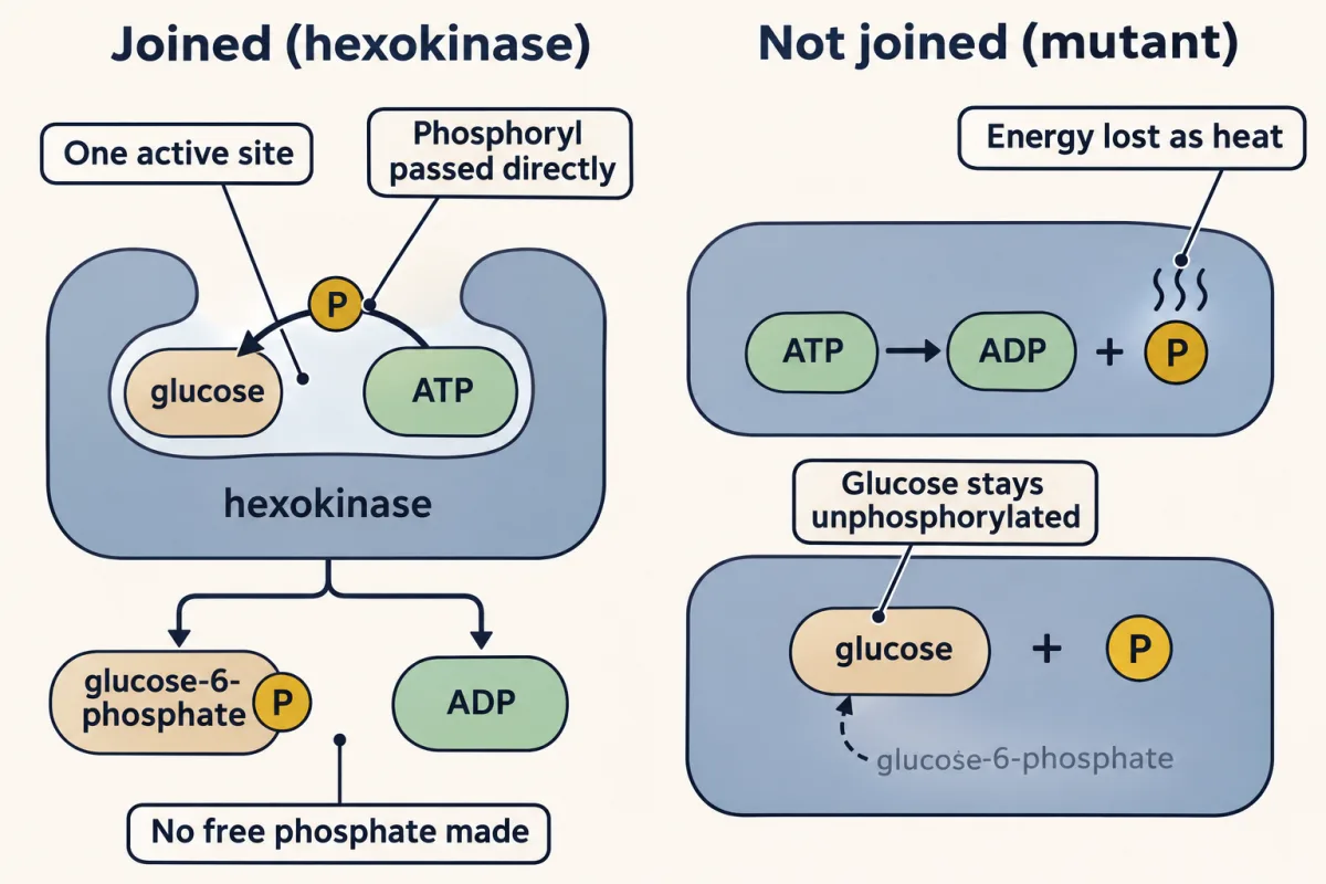
Predict. A mutant enzyme hydrolyzes ATP at one site, and at a second site, with no link to the first, makes glucose-6-phosphate from glucose and free phosphate. The ΔG°’ values still add to −16.7 kJ/mol. Does glucose-6-phosphate form?
Check your prediction
No. ATP is hydrolyzed and its free energy leaves as heat. The glucose reaction still faces +13.8 kJ/mol with the same Q, so it still runs backward. Free energies add only when the chemistry is joined.
Coupling, the second way: keep the product scarce
The film above shows B being drained by the next step. For A → B + C, ΔG°’ = +21 kJ/mol; for B → D, it is −34, so the pathway total is −13 kJ/mol. B is the shared intermediate, a real molecule passed between enzymes. The first step itself runs forward only while the second keeps [B] low. With A and C at 1 mM, Q is [B], and +21 + 2.48 ln[B] stays below zero only while B is below about 0.2 mM. Thus every step in a pathway carrying flux must have negative ΔG, even if only slightly.
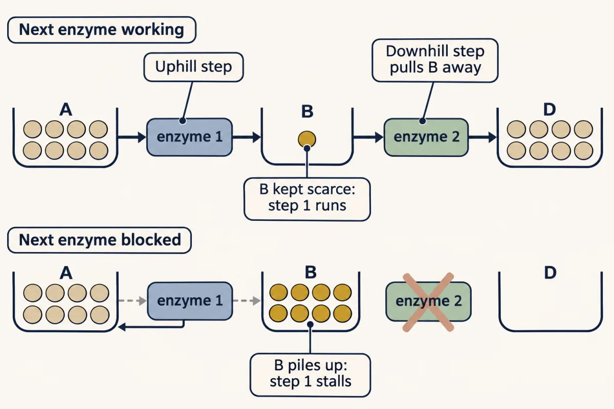
For chemistry that passes a group between molecules, those two are all coupling is. Pumps, motors and ATP synthase couple through changes of protein shape and through gradients, though the ΔG test still applies to the whole coupled process.
In the bench’s first module, couple an uphill reaction to ATP hydrolysis and watch K’eq jump. The bench uses standard-state values at 37 °C, so its multiplier reads about 1.4 × 10⁵ rather than 2.2 × 10⁵ at 25 °C.
Couple it: ATP pays for the uphill climb
- ΔG°' of target
- +13.8 kJ/mol
- ΔG°' summed
- +13.8 kJ/mol
- K'eq (310 K)
- 4.7 × 10⁻³
The sum is real only because one enzyme joins the two: ATP's phosphoryl group goes straight to the substrate (hexokinase) or through a short-lived covalent intermediate (glutamine synthetase).
Rank the donors: under standard conditions, phosphoryl groups go downhill
The phosphoryl group (orange token) starts on ATP. Drag it to another rung, or focus it and press ↓ / ↑. Which moves does the cell allow?
Transfer potential = −ΔG°' of hydrolysis. ATP sits in the middle on purpose: high-energy donors (PEP, 1,3-BPG, creatine-P) can charge it; it in turn can phosphorylate glucose. Glucose-6-P can't pay ATP back.
Read the battery: energy charge gates spending
Drag ATP down and watch AMP. Adenylate kinase (2 ADP ⇌ ATP + AMP) stays near equilibrium, so as ATP is spent the leftover ADP is partly converted to AMP: AMP rises far faster, in relative terms, than ATP falls. That makes AMP the cell's sensitive alarm (it is what AMPK reads). Concentrations are relative, total adenylate = 100.
Try the lesson's case: drag ATP from 90 to 81.
Run the first bench and the whole chapter collapses into one sentence: alone, glucose + Pi → glucose-6-P (+13.8 kJ/mol) has a K'eq near 0.0047 at 37 °C, about one product for every 200 glucose; join it to ATP hydrolysis in hexokinase and the summed ΔG°' goes to −16.7 — K'eq jumps to about 650, and the reaction runs. (At 25 °C, as in the text above, the same numbers are 0.0038 and 840.) The arithmetic holds only because one enzyme joins the two reactions; hydrolyze ATP somewhere else and its free energy is just heat. The third bench tells you when the cell is willing to pay. It does not spend its last dollar; it holds energy charge near 0.85–0.90, throttling fuel-burning and fuel-building against the same gauge, the way you'd manage a checking account you can never let hit zero.
Why ATP can pay
ATP is adenine plus ribose plus three phosphates, named α, β and γ from the ribose outward; most kinases transfer the γ-phosphoryl. Hydrolysis of that terminal phosphate has ΔG°’ ≈ −30.5 kJ/mol. Three properties make ATP good at paying.
The products are more stable; there is no high-energy bond. ADP and Pi carry less crowded negative charge (less electrostatic repulsion), the released phosphate gains resonance stabilization, two molecules have more entropy than one, and the products are better hydrated than ATP was. Textbooks add a fifth factor: ionization of the products at pH 7 releases H⁺. When you hear “spending a bond,” read it as “transferring the γ-phosphoryl group to products that are more stable.”
The cell holds ATP far from equilibrium. Q for hydrolysis is tiny, so the actual ΔG is well below the standard −30.5. In a red blood cell at 37 °C, with about 2.25 mM ATP, 0.25 mM ADP and 1.65 mM Pi:
ΔG = −30.5 + 2.58 × ln(0.00025 × 0.00165 / 0.00225) ≈ −30.5 − 22.2 ≈ −52.7 kJ/mol
Across cell types it runs from about −45 to −65 kJ/mol, so about −50 is a fair working number.
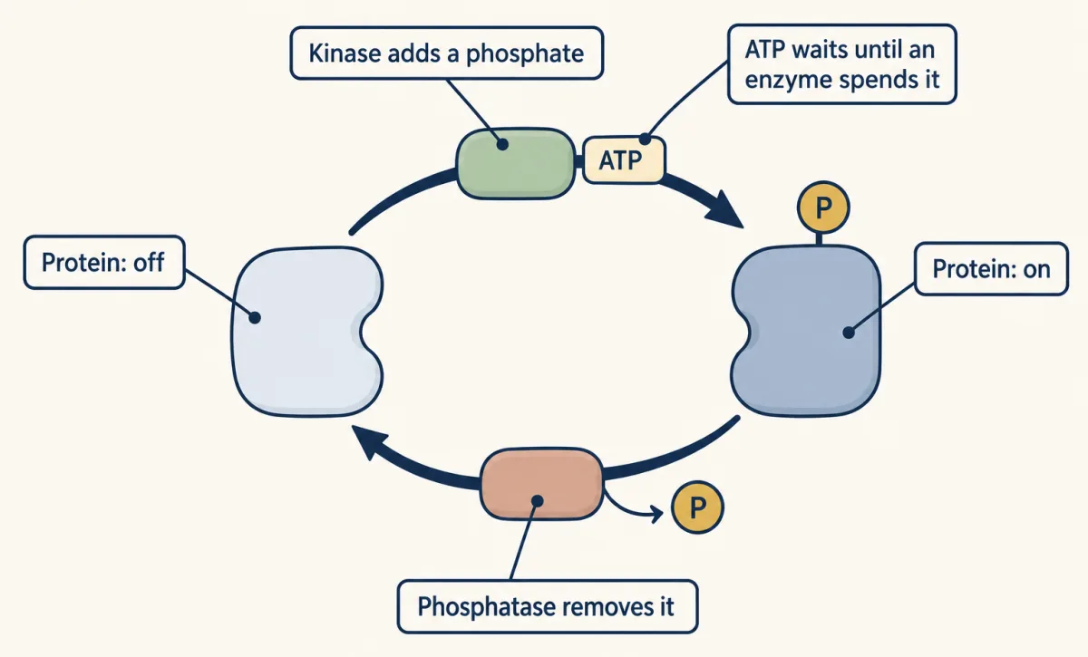
ATP is kinetically stable. ATP, and the phosphate esters it puts on proteins, are thermodynamically unstable but kinetically stable: they will not hydrolyze at any useful rate without an enzyme. That makes phosphorylation an ideal on/off switch: a kinase adds a phosphate to flip a protein’s state (on for some proteins, off for others), a phosphatase removes it to flip it back, and nothing happens in between.
The phosphoryl-transfer ladder
The phosphoryl-transfer ladder ranks phosphorylated compounds by how much free energy is released when each transfers its phosphoryl group to water (its ΔG°’ of hydrolysis). It ranks energy released, not speed.
Predict. Which of phosphoenolpyruvate (PEP), creatine phosphate and glucose-6-phosphate can make ATP from ADP, under standard conditions?
Check your prediction
PEP and creatine phosphate rank above ATP and can, as can 1,3-bisphosphoglycerate (1,3-BPG); glucose-6-phosphate ranks below and cannot. The ΔG°’ of handing each phosphoryl group to ADP (its hydrolysis value plus 30.5, the cost of making ATP):
| Donor + ADP → product + ATP | ΔG°’ (kJ/mol) |
|---|---|
| PEP | −31.4 |
| 1,3-BPG | −18.9 |
| creatine phosphate | −12.6 |
| glucose-6-phosphate | +16.7 |
Under standard conditions, transfers run downhill, from higher on the ladder to lower. ATP sits in the middle, and that is what makes it good currency: compounds above it recharge ADP, and ATP phosphorylates compounds below, like glucose. Drag the token in the bench’s ladder module to test the rule. In muscle, creatine phosphate recharges ADP from the first contraction, holding ATP nearly steady for several seconds of hard effort until creatine phosphate itself runs low.
But the ladder is a standard-state ranking, and the rule is about actual ΔG.
Predict. Creatine phosphate sits above ATP. Why, then, can muscle use ATP to rebuild creatine phosphate after a sprint?
Check your prediction
Creatine kinase sits so close to equilibrium that ΔG is within about 1 kJ/mol of zero. When ATP is plentiful and free ADP scarce (as in recovery, when respiration refills ATP), Q for creatine phosphate + ADP → creatine + ATP rises above K’, ΔG turns slightly positive and net flux runs toward creatine phosphate. In exercise ADP rises, Q falls, and the reaction flips to making ATP (numbers in Going deeper).
Earning: how fuels recharge the carriers
The cell recharges ATP by oxidizing fuel, in three stages. Digestion breaks large molecules into small ones: proteins to amino acids, starch to sugars, fats to fatty acids. Those small molecules funnel into a handful of key intermediates, above all acetyl-CoA. Then acetyl-CoA is oxidized completely, and that oxidation drives ATP synthesis.
Whether you eat a steak, a potato or olive oil, most of the carbon reaches your mitochondria as acetyl-CoA; some amino acids join at pyruvate or the citric acid cycle itself. Different highways, one city center. So one central pathway, the citric acid cycle (its own lesson later), serves every fuel you eat. The cycle is amphibolic: it oxidizes fuel and also supplies starting materials for biosynthesis.
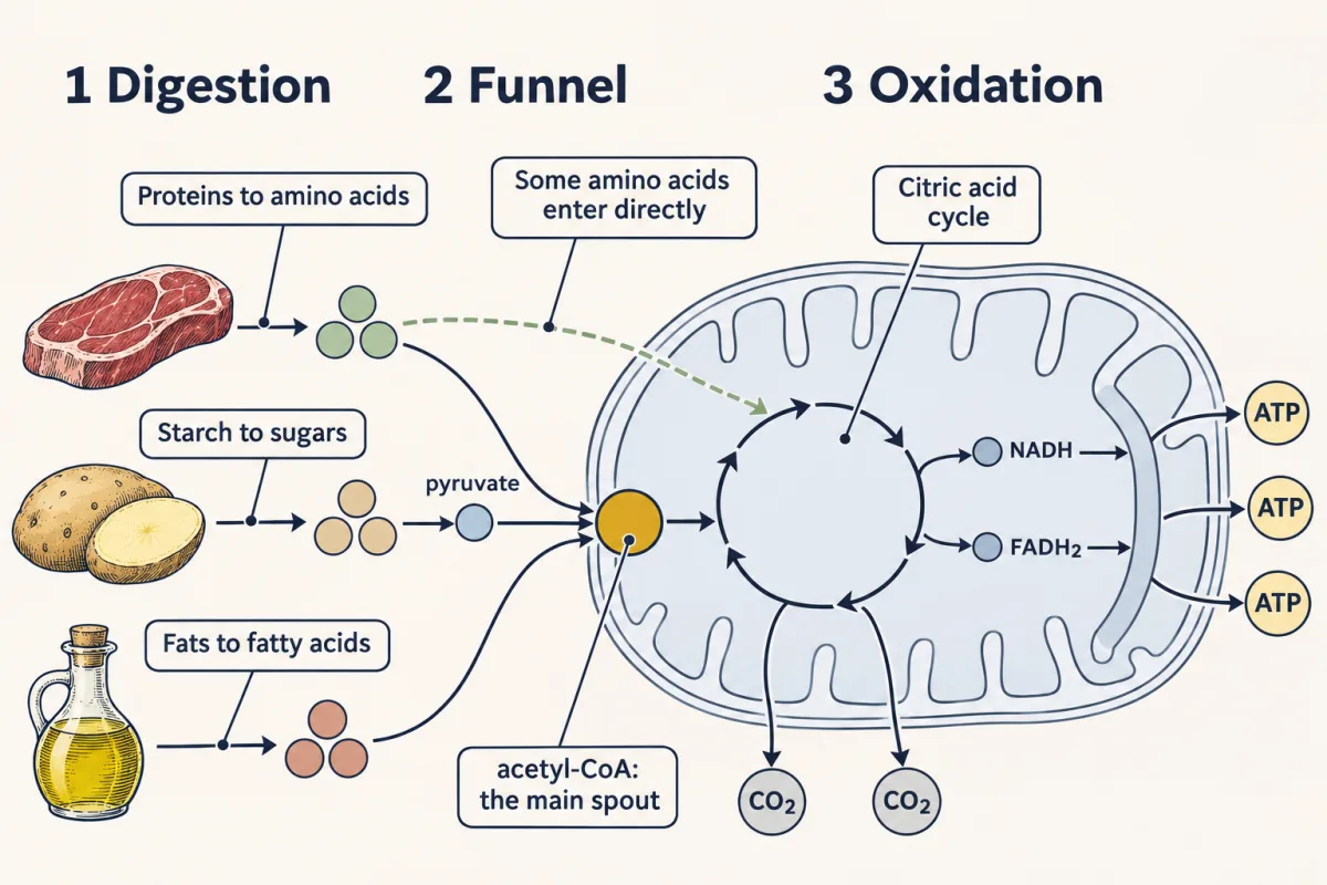
Burning a fuel means stripping electrons from its carbons and handing them, in the end, to oxygen. A carbon loaded with hydrogens (the long chain of a fatty acid) has many electrons to give; a carbon already bonded to oxygen (every carbon in glucose) has fewer; CO₂ has none left. That is the main reason fat yields about 9 kcal per gram and carbohydrate about 4, and one reason fat is the body’s main long-term fuel store. The other is that fat is stored nearly water-free, while glycogen carries about twice its weight in water, which matters more for storage density than 9 versus 4. Click through the fuels below and watch energy density climb the farther a fuel sits from CO₂.
Burning a fuel means stripping electrons off its carbons and handing them, ultimately, to oxygen — and every electron handed down that chain pays out ATP. A carbon already at +4 (CO₂) has nothing left to give, so it is metabolic ash. A carbon near −2 (the long reduced chain of a fatty acid) is loaded with electrons, which is why a gram of fat yields more than twice the energy of a gram of carbohydrate or protein. Distance from CO₂ is the main reason fat yields about 9 kcal/g and carbohydrate about 4.
Fuel’s electrons, like ATP’s phosphoryl groups, ride on a small set of activated carriers, the cell’s delivery trucks. Each is ATP’s idea again: a molecule that holds one kind of group (phosphoryl, electrons, acyl) at high transfer potential and hands it over when an enzyme asks. They are kinetically stable, few in number, and mostly built from vitamins, which is why a vitamin deficiency hits metabolism hard.
| Carrier | What it carries | Where it is spent | Built from |
|---|---|---|---|
| ATP | phosphoryl groups | kinases, pumps, motors, biosynthesis | (not a vitamin) |
| NADH | electrons from carbon oxidations | the electron transport chain, roughly 2.5 ATP each | niacin (B3) |
| FADH₂ | electrons | the electron transport chain, entering lower (at ubiquinone), about 1.5 ATP | riboflavin (B2) |
| NADPH | electrons for building | reductive biosynthesis and antioxidant defense | niacin (B3) |
| Coenzyme A (as acyl-CoA) | acyl groups, through a thioester | acyl transfers, such as acetyl into the citric acid cycle | pantothenate (B5) |
NADPH shares NADH’s chemistry. CoA’s thioester has high transfer potential because a thioester is less resonance-stabilized than an oxygen ester, so its hydrolysis or transfer releases more free energy. A broken carrier is a disease: inherited loss of pantothenate kinase 2 (PANK2), the first enzyme in making CoA, causes PKAN, with brain iron accumulation and neurodegeneration.
Regulation: what can run versus what does
Regulation picks which possible reactions run, in three ways on three timescales. The cell changes the amount of an enzyme by transcription (hours to days). It changes the activity of existing enzymes by allostery and reversible covalent modification (seconds to minutes), which is how hormones like insulin coordinate the whole body fast. And it changes substrate accessibility by compartmentation, keeping fatty-acid synthesis in the cytosol and fatty-acid oxidation in the mitochondrion so the two do not run at once.
Most steps of a pathway sit near equilibrium and run either way on the same enzyme: seven of glycolysis’s ten steps run backward in gluconeogenesis, which makes glucose. The few steps far from equilibrium cannot simply be reversed, so each direction gets its own, separately regulated enzyme there, and those steps are where most regulation acts.
A key gauge is energy charge, the cell’s battery meter, normally held near 0.85–0.90:
energy charge = ([ATP] + 0.5[ADP]) / ([ATP] + [ADP] + [AMP])
Adenylate kinase (2 ADP ⇌ ATP + AMP) holds the three nucleotides near equilibrium, so [AMP] = K × [ADP]² / [ATP]. Because ADP is squared, a small rise in ADP gives a bigger rise in AMP.
Predict. A very well-charged cell (energy charge about 0.95, a little above the usual range) has ATP 9, ADP 0.9 and AMP 0.1 (relative units, total 10). ATP falls by 10%, to 8.1, and adenylate kinase re-equilibrates. Roughly what happens to AMP?
Check your prediction
It rises about 3.4-fold, from 0.10 to about 0.34, while energy charge falls only from about 0.95 to 0.89. (K = 0.1 × 9 / 0.9² ≈ 1.1; with the total held at 10, ADP and AMP share 1.9, which works out to ADP ≈ 1.56 and AMP ≈ 0.34.) So AMP is a sensitive alarm: it activates AMPK, which switches on catabolism and shuts down anabolism. The gain is smaller in a pool at the low end of the normal range: at 7.5, 2 and 0.5 (energy charge 0.85), the same 10% fall in ATP raises AMP only about 1.6-fold.
In the bench’s third module, watch the battery needle redline as ATP falls.
When regulation fails, you get disease: pathophysiology is biochemistry in a clinical context.
- Diabetes is a failure of insulin-based regulation: too little insulin (type 1), or tissues that respond poorly to insulin plus beta cells that cannot keep up (type 2).
- The Warburg effect. Many cancer cells run aerobic glycolysis, taking up glucose fast and making lactate even with oxygen present. A leading idea is that this flux feeds biosynthesis rather than maximal ATP yield, though the reasons are still debated. The high glucose uptake is why many tumors light up on an FDG-PET scan (inflamed tissue can too).
- Mitochondrial disease is oxidative-phosphorylation failure, often presenting as lactic acidosis.
In Chapter 16, Glycolysis, you will watch every one of these ideas (coupling, substrate-level phosphorylation, which makes ATP by direct transfer from a donor such as phosphoenolpyruvate (PEP), activated carriers, energy-charge regulation) at work in a single ten-step pathway.
Going deeper
The main line above does not depend on these details.
Six reaction types
Beneath the thousands of named reactions, metabolism uses only about six reaction types: redox, ligation (ATP-driven joining), isomerization, group transfer, hydrolysis and lyase reactions. Three have appeared here: group transfer (hexokinase), ligation (glutamine synthetase joining ammonia to glutamate) and hydrolysis (ATP).
Where FADH₂’s electrons enter
FADH₂’s working part is the isoalloxazine ring. Its electrons reach ubiquinone via Complex II from succinate, or via electron-transferring flavoprotein (ETF) from fatty acids.
More vitamin diseases
Niacin deficiency (NADH, NADPH) is pellagra. Thiamine forms the carrier TPP (thiamine pyrophosphate), and thiamine deficiency causes beriberi and Wernicke encephalopathy. Riboflavin deficiency (FADH₂) is ariboflavinosis. Each disease is a missing or broken truck.
Reciprocal control of fat
Compartmentation works with reciprocal control: malonyl-CoA, made during fatty-acid synthesis, blocks fatty-acid entry into the mitochondrion, so synthesis and oxidation cannot run together as a futile cycle.
The muscle’s energy timeline
On its own, stored ATP would last only seconds of hard effort, and the creatine phosphate buffer only several seconds more. Glycolysis runs from the first seconds and soon carries more of the load, and aerobic respiration, also on from the start, carries the long haul. When muscle is injured, creatine kinase leaks into the blood, which is why elevated serum CK is a clinical marker of muscle damage.
Creatine kinase in numbers
At 37 °C, K’ for creatine phosphate + ADP → creatine + ATP is about 132 (measured values shift with pH and free Mg²⁺). With resting-muscle values of about 8 mM ATP, 13 mM creatine, 25 mM creatine phosphate and 20 µM free ADP, Q = (13 × 8) / (25 × 0.020) ≈ 208. Q is above K’, so ΔG = RT ln(Q/K’) ≈ +1.2 kJ/mol for the direction that makes ATP. Read the sign loosely: free ADP in muscle is usually calculated by assuming creatine kinase is at equilibrium, so the numbers show only that ΔG is near zero and small swings in ADP set the direction.
ATP’s actual ΔG in other cells
The −45 to −65 kJ/mol range above comes from concentrations like these. Beside the red cell’s −52.7 kJ/mol (all at 37 °C), liver-like concentrations (about 3.4 mM ATP, 1.3 mM ADP, 4.8 mM Pi) give about −47, and resting muscle, counting only free ADP (about 8 mM ATP, 20 µM ADP, 3 mM Pi), about −61.
The film’s numbers
Both of the film’s numerical examples (the +21/−34 pair above, then a K rising from about 1/1000 to about 267) are separate textbook reactions with rounded, standard-state values. In a cell, ATP pushes harder.
Where the “high-energy bond” came from
Fritz Lipmann introduced the “energy-rich phosphate bond” and the ~P squiggle. His “group potential” put the emphasis on the free energy of transferring the group, the idea the ladder is built on. The explanation in terms of product stability came largely from later physical-chemical work.
Your notes
Select any text in the lesson to highlight it or add a note to it. Your highlights and notes are saved in this browser and collected in your notebook.
This browser is not letting the site save anything (a private window or strict privacy settings), so highlights and notes will be lost when you leave the page.
How we measure it
Phosphoryl-transfer potential (the ΔG°' of hydrolysis)
Rank a phosphorylated compound by how much free energy its hydrolysis releases under standard conditions. Measured against a common reference, these values build the ladder that tells you which molecule can phosphorylate which: PEP and creatine phosphate sit above ATP and can charge ADP; glucose-6-phosphate sits below and cannot. The whole logic of substrate-level phosphorylation falls out of this one measured quantity.
Energy charge
A single number, ([ATP] + 0.5[ADP]) / ([ATP] + [ADP] + [AMP]), that reports how 'charged' the cell's adenylate pool is, normally near 0.85–0.90. Because adenylate kinase interconverts the three nucleotides, a 10% drop in ATP can raise AMP several-fold in a well-charged cell, so energy charge is a sensitive, dimensionless readout of metabolic state — the experimental basis for understanding AMPK signaling.
Isotopic and PET tracing of fuel use
Label a fuel carbon and follow where it lands, or scan a patient with an 18F-glucose analog and watch which tissues take up sugar fastest. Tracing is how convergence on acetyl-CoA was established and how the Warburg effect is read clinically — many tumors light up on a PET scan because they take up glucose fast even when oxygen is plentiful (inflamed tissue can light up too).
Practice questions on this lesson (3) → MCAT-style, original, each with an explanation
Where this lesson fits
Before thisReview first
- Basic Concepts of Enzyme ActionThis course · Lesson 7
You need free energy, equilibrium and why enzymes speed only possible reactions before coupling makes sense.
- Water, Weak Bonds, and the Generation of OrderThis course · Lesson 3
Hydration and charge repulsion in ATP's products come from the water and weak-bond ideas taught there.
After thisStudy next
- Glycolysis: The Investment-and-Payoff PathwayThis course · Lesson 16
Hexokinase coupling, substrate-level phosphorylation and energy-charge control are all seen working in one ten-step pathway.
- Oxidative Phosphorylation: The Electron-Transport ChainThis course · Lesson 20
NADH and FADH2 electrons recharged here are spent in the chain, which makes most of the ATP.
- Integration of Metabolism: How the Body Switches FuelsThis course · Lesson 30
The three modes of regulation and energy charge are applied to whole-body fuel switching.
Elsewhere on the siteFilms, simulations and method films in other courses
- Build an energy pyramidSimulation · Biology
The idea that energy transfers are never complete is a simple everyday version of energy accounting in cells.
- Spending ATP, Rebuilding ATPFilm · Biology
It shows cells spending ATP to do work and rebuilding it, the currency idea developed here in more depth.
- A cell’s energy budgetSimulation · Biology
It lets you balance a cell's ATP budget, which the energy charge and earning-versus-spending ideas here develop further.
Suggested by an AI model from the lessons themselves, then checked. How we use AI
Finish line
Test yourself
8 questions
Practice and MCAT-style, with explained answers.
Play it · about 3 min
Ledger Rush: Glycolysis
Keep the ATP and NADH books for one glucose. Bank the real yields, toss the myths.
Play →Next · Lesson 16
Glycolysis: The Investment-and-Payoff Pathway
Why does a cell spend ATP to make ATP? The first steps are a deliberate investment.
Continue →Also in the arcade for this lesson: Flux Foundry
← Lesson 14 · Digestion and Absorption: Turning a Meal into Molecules Your Cells Can Use