Biochemistry · Skim version
Digestion and Absorption: Turning a Meal into Molecules Your Cells Can Use
Read the full lesson → Skim the whole course →
The guiding idea
Digestion has two halves that only work together: enzymes cut a meal into small pieces, and the gut wall carries those pieces into the blood or lymph.
Digestion couples breakdown to absorption.
-
One reaction, downhill but slow
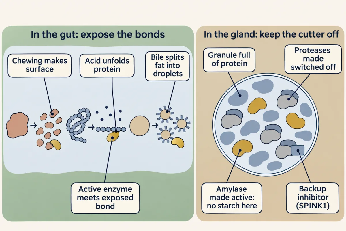
Two meetings decide where a food bond is cut. In the gut, chewing, acid and bile expose bonds to enzymes that are already on. In the pancreas, the granule is full of protein, so every protease is made switched off; amylase, with no starch in the granule, is made switched on. AI-generated illustration -
Carbohydrate: the enzyme’s shape picks the bond
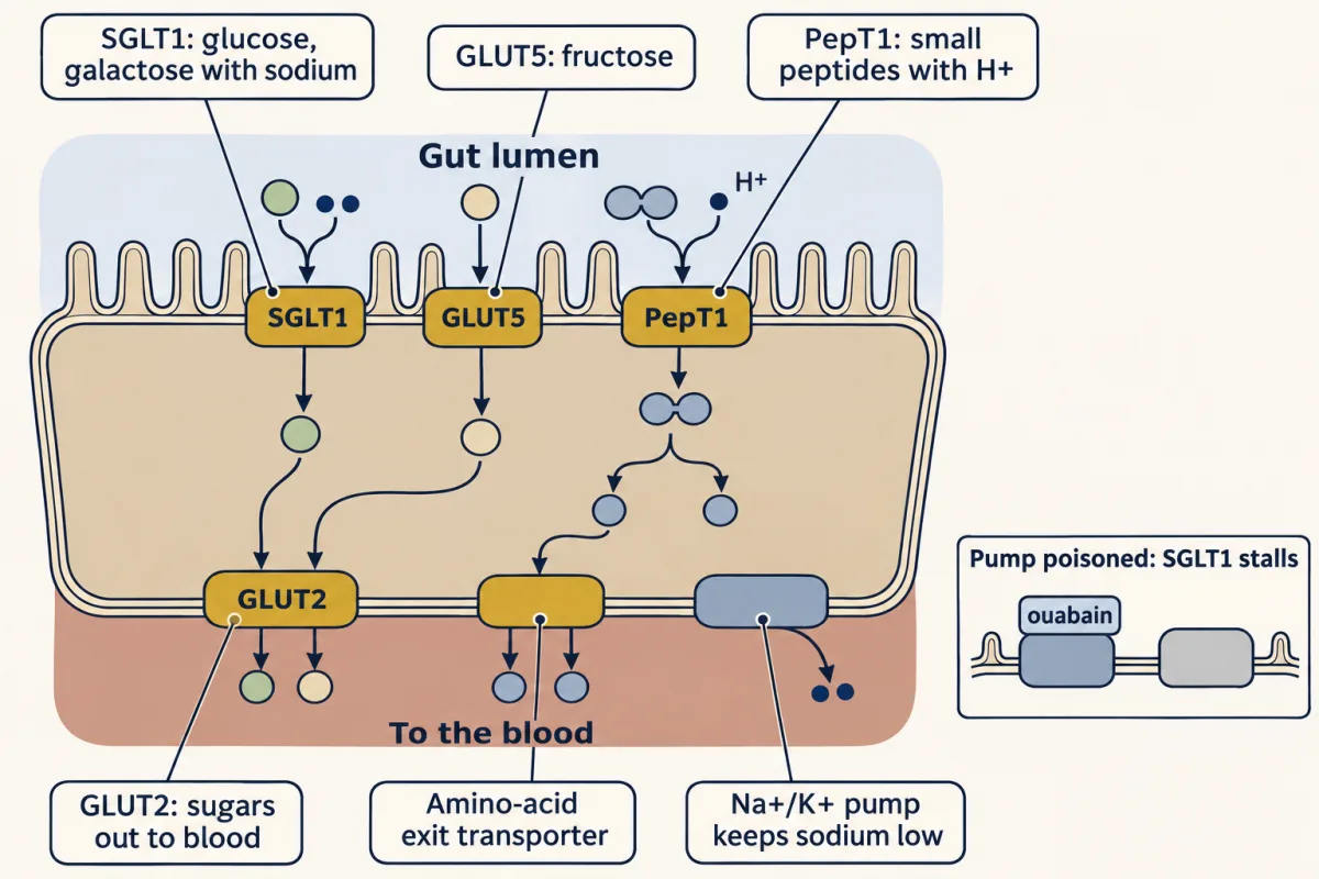
Doors in and out of the enterocyte. Glucose and galactose enter with sodium through SGLT1, fructose through GLUT5, and small peptides with a proton through PepT1; the sugars leave through GLUT2 and the amino acids through basolateral transporters. The Na+/K+ pump keeps sodium low inside, which is what powers SGLT1: poison the pump with ouabain and SGLT1 stalls. AI-generated illustration -
Protein: switch the enzyme on only in the gut
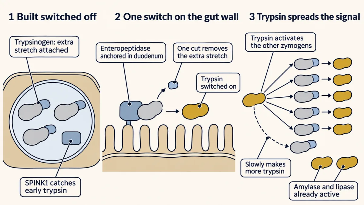
The pancreas's three-step answer. Proteases are stored switched off; the one switch, enteropeptidase, sits on the gut wall; trypsin then switches on the other zymogens and, slowly, more trypsin. Amylase and lipase skip the relay because they are secreted active. AI-generated illustration -
Fat: the exposure problem
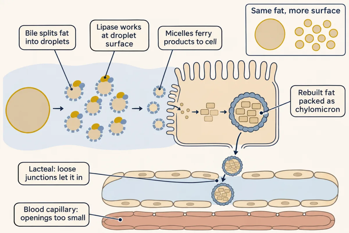
Fat's exposure problem and its solution. Bile turns one drop into many, creating the surface where lipase works; micelles carry the products to the cell, which rebuilds them into chylomicrons. Too large for blood capillaries, chylomicrons enter the lymph through the lacteal's loose junctions. AI-generated illustration -
Taking it up: where each piece goes
-
Clinical connections
-
Going deeper
Where this lesson fits
Before thisReview first
- Basic Concepts of Enzyme ActionThis course · Lesson 7
You need to know that enzymes speed up possible reactions without changing their direction to follow why hydrolysis is downhill but slow.
- Carbohydrates: One Bond Decides Fuel or FiberThis course · Lesson 10
The alpha versus beta glycosidic bonds from there explain why amylase cuts starch but not cellulose.
- Lipids: The Building Blocks That Refuse to PolymerizeThis course · Lesson 11
Triacylglycerol structure and the ester bonds from there are what lipase cuts and the enterocyte rebuilds.
After thisStudy next
- Glycolysis: The Investment-and-Payoff PathwayThis course · Lesson 16
The monosaccharides absorbed here are what enter glycolysis as the first step of fuel use.
- Integration of Metabolism: How the Body Switches FuelsThis course · Lesson 30
Absorbed sugars, amino acids and chylomicron fats feed the fed-state fuel switching described there.
- Fatty Acid Degradation: The β-Oxidation SpiralThis course · Lesson 25
Fats delivered by chylomicrons and fatty acids from digestion are the fuel burned by this pathway.
Elsewhere on the siteFilms, simulations and method films in other courses
- The gut's daily water budgetSimulation · Organismal Biology
This lesson's own gut-tube ideas come from the animal-level view of how a one-way gut specializes.
- Why Order Matters in a GutFilm · Organismal Biology
It shows why a one-way gut lets each stretch work on what the previous one hands on.
- Take It Apart, Build It AgainFilm · Biology
It shows digestion as hydrolysis splitting starch and protein into small pieces by adding water.
- The gut keeps its own timeFilm · Physiology
It shows how the gut times its secretions and emptying, which controls when digestive enzymes act.
Suggested by an AI model from the lessons themselves, then checked. How we use AI