Biochemistry · Enzymes and Bioenergetics
New to this? Review it at high school level: Enzymes at Work
Basic Concepts of Enzyme Action
Rewritten on : new text, figures and flashcards. Your notes and highlights stay, but the text around them may have moved.
Can an enzyme make an impossible reaction happen? No — it can only make a possible one fast.
Pause when the reading reaches a film, and offer to play it. Saved on this device.
Watch the film now, or keep listening
Builds on:Water, Weak Bonds, and the Generation of OrderProtein Three-Dimensional Structure: The Sequence Sets the ShapeWhy these?
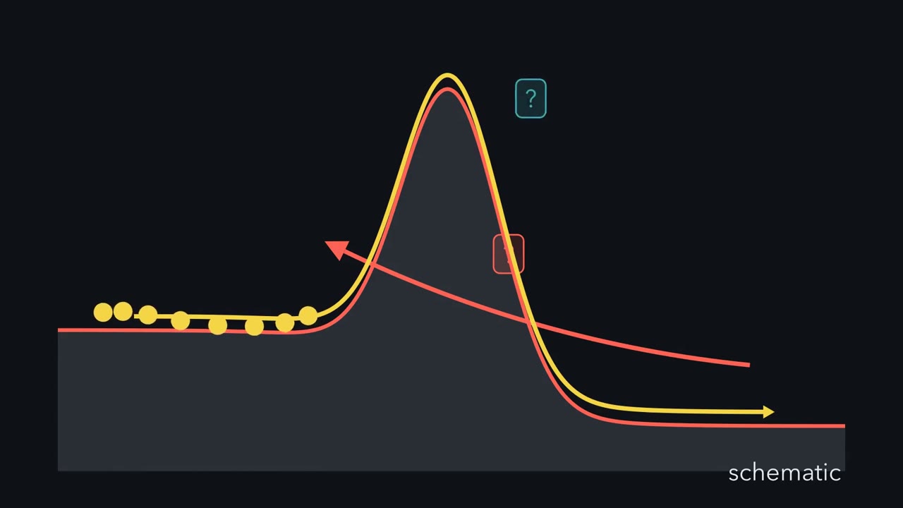 ▶ Watch Lower the Ridge, Not the Valleys · 3:14
Read the transcript of “Lower the Ridge, Not the Valleys”
Film transcript: Lower the Ridge, Not the Valleys
Can an enzyme make an impossible reaction happen? Or can it only make a possible one fast? Start with a reaction your cells need to make RNA. On its own, this step is painfully slow. Half the molecules would take 78 million years to react. With its enzyme, the step is done in milliseconds. That is a speed-up of about 10¹⁷. to the 17. So could an enzyme that strong push a reaction uphill? No. To see why, split one question into two. Which valley is lower decides which way the reaction runs. That is thermodynamics, delta ΔG. How high the ridge is decides how fast. That is kinetics. That climb is the activation energy, delta G double dagger. At the very top sits the transition state.
Its bonds are partly breaking and forming, and it lasts femtoseconds. Here is the whole idea. An enzyme lowers the ridge. It never moves the valleys. It opens a lower pass over the same ridge, still a highest point, just lower. A little lower buys a lot of speed. At 25 °C, every 5.7 kJ/mol taken off makes it ten times faster. Take off about 97, and you reach that 10¹⁷. to the 17. Now flip the trip, from product back to substrate. Say the enzyme speeds the forward trip a millionfold. Watch both arrows cross the same lowered top. What happens to the backward trip?
Also a millionfold. Counted from the two valleys, both trips cross the same lowered top. So their ratio, the equilibrium constant, stays the same. The enzyme reaches the same equilibrium, only sooner. It cannot push a reaction uphill. If the far valley is higher, the net reaction runs the other way. So how does a protein lower the top? Its active site better matches the transition state than the substrate. It holds the substrate more loosely and the transition state more snugly. That extra grip pays back energy right at the top. The climb from the bound substrate shrinks. That bound-substrate speed-up equals how many times tighter it grips the transition state.
An active site that gripped the substrate just as hard would gain nothing. Both would sink together, and the climb would stay the same. So the valleys decide which way a reaction runs. The ridge decides how fast. An enzyme touches only the ridge, by gripping the top hardest.
From the film
- An enzyme lowers the ridge between substrate and product; it never moves the valleys.
- Which valley is lower (ΔG) decides which way a reaction runs; the ridge height (ΔG‡) decides how fast.
- At 25 °C, every 5.7 kJ/mol taken off the barrier makes a reaction ten times faster.
- An enzyme speeds the forward and backward trips by the same factor, so the equilibrium constant stays the same.
- It lowers the top by gripping the transition state more tightly than the substrate.
An enzyme cannot make an impossible reaction happen. It can only make a possible one fast, by lowering the energy barrier the reaction has to cross.
The film above opens on OMP decarboxylase, the enzyme that finishes making UMP (a building block of RNA): about 10¹⁷ times faster than the uncatalyzed step, whose half-life is 78 million years, longer than the time since the dinosaurs vanished. Why can’t a protein that strong push a reaction uphill? The film answers with two valleys, the free substrate and the free product, and a ridge between them.
The one idea to carry through this lesson. An enzyme, like any catalyst, lowers only the ridge, never the two valleys. Of the valleys, the drop between them at the actual concentrations (ΔG) sets which way the reaction runs, and the drop at standard conditions (ΔG°′) sets where it stops (Keq).
Where a reaction ends up: ΔG, ΔG°′ and Q
The currency of thermodynamics is Gibbs free energy, G, the energy in a system available to do work. The sign of the change, ΔG, gives the direction. A negative ΔG means the reaction releases free energy and can go forward on its own: it is exergonic, or spontaneous. A positive ΔG means it needs an input of energy: it is endergonic. Spontaneous means downhill, not quick, as uncatalyzed OMP decarboxylation shows. And exoTHERMIC is not exoGONIC: exothermic is about heat (ΔH), and a reaction can release free energy without releasing much heat.
ΔG also depends on how much product and reactant are present right now; ΔG°′ is the standard reference value, and ΔG the value for the mixture actually in the tube or the cell.
In words The actual free-energy change equals the standard one, plus RT times the natural log of the current products-to-reactants ratio.
- ΔG“delta G”
- the actual free-energy change at the current concentrationskJ/mol
- ΔG°′“delta G naught prime”
- the standard free-energy change: every reactant and product at 1 M, 25 °C, and pH 7 (the prime)kJ/mol
- R“R”
- the gas constant, 8.314 J/(mol·K)J/(mol·K)
- T“T”
- temperature in kelvin; 298 K is 25 °CK
- Q“Q”
- the reaction quotient: products over reactants right now, in the form of Keq, each concentration in M divided by the 1 M standardnone
Sanity check When everything is at 1 M, Q = 1, ln Q = 0, and ΔG = ΔG°′. When Q reaches Keq, ΔG = 0 and the reaction is at equilibrium.
Try one number
RT at 298 K is 0.008314 × 298 = 2.48 kJ/mol.
Glucose + phosphate → glucose-6-phosphate (G6P), ΔG°′ = +13.8 kJ/mol. At illustrative cell levels (5 mM glucose, 1 mM phosphate, 0.1 mM G6P), in M: Q = [G6P] / ([glucose][phosphate]) = 0.0001 / (0.005 × 0.001) = 20, RT ln 20 = 2.48 × 3.0 = 7.4, so ΔG ≈ +21 kJ/mol, still uphill.
ATP hydrolysis, ΔG°′ = −30.5 kJ/mol. At illustrative red-cell levels (ATP 2.25, ADP 0.25, phosphate 1.65 mM): Q = (0.00025 × 0.00165) / 0.00225 = 1.8 × 10⁻⁴, RT ln Q = 2.48 × (−8.6) = −21.3, so ΔG ≈ −52 kJ/mol.
Need a refresher? e and ln · units and prefixes
So ATP hydrolysis is the textbook exergonic reaction and direct glucose-6-phosphate (G6P) formation the textbook endergonic one, in standard conditions and in the cell.
As a reaction runs, Q moves toward Keq and ΔG creeps toward zero; at equilibrium, going nowhere net, ΔG = 0 and Q = Keq. Put those into the card:
ΔG°′ = −RT ln Keq
The more negative ΔG°′, the larger Keq: ΔG°′ < 0 means Keq > 1, and ΔG°′ > 0 means Keq < 1. Work an example: ΔG°′ = −17 kJ/mol, so Keq = e−ΔG°′/RT. Convert kJ to J first: 17,000 / (8.314 × 298) = 6.86, and e6.86 ≈ 950, roughly a thousand-to-one in favor of products. Quicker: every 5.7 kJ/mol is a factor of ten (the next section shows why), so 17 kJ/mol is about three. Because the relationship is logarithmic, a modest change in free energy moves the equilibrium a lot.
Predict. You add an enzyme that joins glucose directly to phosphate to a cell-like mix. Does G6P pile up?
Check your prediction
No. In the cell ΔG is about +21 kJ/mol, and the enzyme cannot change that. Keq is about 0.004, so equilibrium is mostly glucose and phosphate. This mix (Q = 20) is already far past it, so the enzyme would only speed the net reaction backward, breaking G6P down. Cells make G6P with hexokinase, which runs a different reaction: glucose + ATP → G6P + ADP, ΔG°′ = +13.8 − 30.5 = −16.7 kJ/mol, downhill. That is coupling: a change of reaction, not a moved equilibrium.
How fast it gets there: the height of the hill
Now the ridge, which the film animates: the enzyme opens a lower pass over the same ridge, still a highest point, and molecules cross far more often.
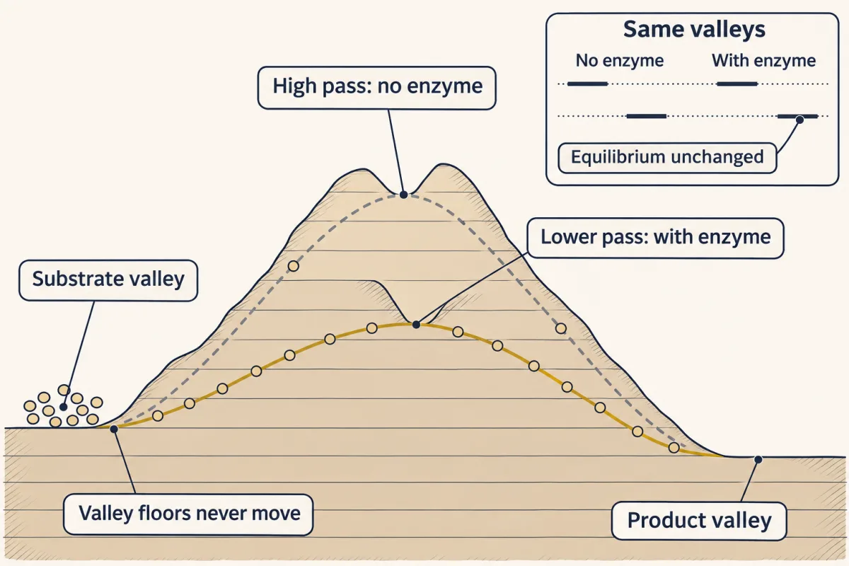
Each chemical step passes through a transition state, written X‡, the femtosecond-lived top of the pass shown in the film: neither substrate nor product, a peak rather than a molecule you can isolate (a stable stop between two peaks is an intermediate). Its height above the substrate is the activation energy, ΔG‡, and it sets the rate.
In words The rate constant shrinks exponentially as the barrier rises.
- k“k”
- the rate constant: how often a molecule crosses the barriers⁻¹
- ΔG‡“delta G double dagger”
- the activation energy: the height of the transition state above the substratekJ/mol
- R“R”
- the gas constant, 8.314 J/(mol·K)J/(mol·K)
- T“T”
- temperature in kelvin; 298 K is 25 °CK
Sanity check RT × ln 10 at 298 K is 2.48 × 2.30 = 5.7 kJ/mol. So 5.7 kJ/mol off the barrier is 10 times faster, 11.4 is 100 times faster.
Try one number
An enzyme turns over its substrate at 540 s⁻¹. Without it, the same step runs at 2.9 × 10⁻⁶ s⁻¹.
Speed-up: 540 ÷ (2.9 × 10⁻⁶) = 1.9 × 10⁸, about 8.3 factors of ten. Barrier lowered: 8.27 × 5.7 ≈ 47 kJ/mol.
OMP decarboxylase, at about 10¹⁷: 17 × 5.7 ≈ 97 kJ/mol.
Need a refresher? e and ln · units and prefixes
Predict. The enzyme lowers the top of the ridge. What happens to the barrier for the reverse reaction, product back to substrate?
Check your prediction
It falls too. Count both trips from the free substrate and the free product, the valleys the enzyme cannot move: both cross the same lowered top, so forward and reverse speed up by the same factor and their ratio, Keq, does not change. An enzyme cannot change ΔG, ΔG°′, or Keq; it reaches the same equilibrium sooner. (The 47 kJ/mol above is counted from the bound substrate; counted from the bound product, the reverse drop matches it only if the enzyme holds product and substrate equally tightly.)
In the explorer below, slide ΔG°′ to move the product up or down, then raise how many kJ/mol the enzyme takes off the barrier and watch which part of the curve changes and which never moves.
An enzyme lowers ΔG‡. It never changes ΔG, ΔG°′ or Keq.
How an enzyme lowers the top: grip the transition state
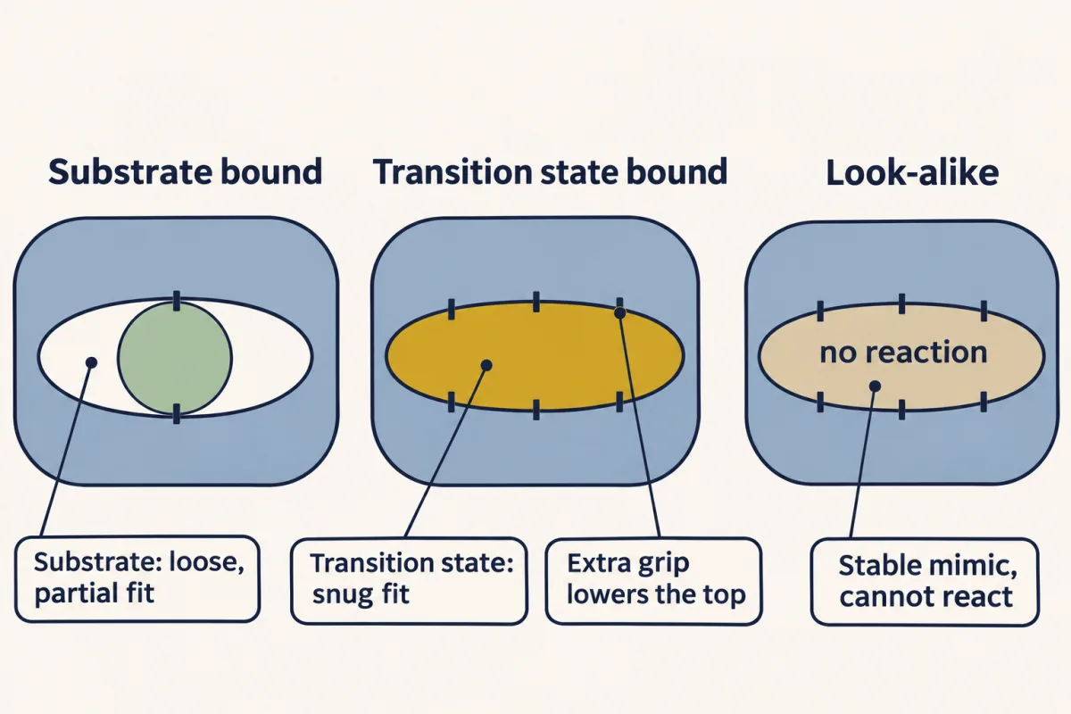
The film shows the grip. The active site, a small cleft built from amino acids brought together from distant parts of the chain, fits the distorted transition state better than the substrate. Binding releases energy (binding energy), and the extra that goes to the transition state comes off the climb from the bound substrate. A thermodynamic cycle (Going deeper) turns that into a rule:
speed-up from the bound substrate = how many times more tightly the enzyme holds the transition state than the substrate
Written with dissociation constants, Kd (the lower the Kd, the tighter the grip): kcat / kuncat = Kd(substrate) / Kd(transition state), where kcat is the rate of the step from the bound substrate and kuncat the rate with no enzyme. The 1.9 × 10⁸ enzyme above holds its transition state about 190 million times more tightly than its substrate, worth the same 47 kJ/mol.
Predict. A team designs an enzyme whose active site holds the substrate exactly as tightly as it holds the transition state. How much faster does the reaction go once the substrate is bound?
Check your prediction
Not at all. The bound substrate and the top drop by the same amount, so the climb from the bound substrate is as high as before. Worse, tightening the substrate’s grip alone shrinks that speed-up: take its Kd from 1 mM to 1 µM, a thousandfold tighter, with the transition state’s grip fixed, and the ratio falls a thousandfold, so a 10⁸ speed-up becomes 10⁵. The loss bites whenever the enzyme is saturated with substrate. Enzymes hold substrates moderately and transition states extremely tightly.
Binding is not the only tool. Enzymes also hold reacting groups close together and lined up, pass protons to and from the substrate (acid-base catalysis), form brief covalent bonds with it (covalent catalysis), and use metal ions. Each makes the transition state easier to reach. On paper any speed-up can still be written as extra grip on the transition state, but covalent catalysis in particular changes the transition state itself, so the simple rule then compares two different transition states.
The evidence: transition-state analogs bind tighter
If the active site is built for the transition state, it should grip a stable look-alike of it harder than the real substrate. Proline racemase, which interconverts the two mirror forms of proline, passes through a planar transition state, its central carbon going flat as it flips, and binds its substrate, L-proline, with a Kd of 3.6 mM. Chemists made pyrrole-2-carboxylate, a stable flat look-alike the enzyme cannot turn over.
Predict. Which will the enzyme hold harder, its own substrate or the flat look-alike?
Check your prediction
The look-alike: Kd 0.022 mM, 160 times tighter than the substrate it actually works on. The enzyme grips the shape it is built to stabilize.
Count it: 160-fold is about 2.2 factors of ten, or 12.6 kJ/mol, small next to the 47 to 97 kJ/mol that enzymes like those above take off their barriers. An analog is an imperfect mimic of so fleeting a structure, so it captures only part of the grip; the tightest designed analogs, made for other enzymes, bind far more tightly than this one. Read 160× as a down payment, not the full account.
A separate question: does the active site change shape?
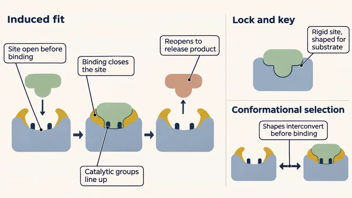
What the active site fits best and whether it changes shape are different questions. Emil Fischer’s 1894 lock-and-key picture explained why an enzyme acts on one sugar and ignores its near-twin. But a rigid lock built for the substrate would bind the substrate most tightly, and enzymes don’t: the site fits the transition state best, an idea from Haldane (1930) and Pauling (1940s), not from the shape models.
Flexibility is the second question. In 1958 Daniel Koshland proposed induced fit: substrate binding changes the enzyme’s shape and brings its catalytic groups into line. His central argument was hexokinase and water (scientist panel). Later work showed that many enzymes also shift between shapes before the substrate arrives, and the substrate picks the one that fits (conformational selection). Real enzymes often use a mix of both.
Tight binding as a drug strategy
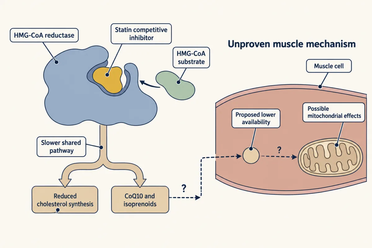
A molecule that binds the active site more tightly than the substrate does, and that the enzyme cannot turn over, can shut the enzyme down. Drugs shaped like a transition state exploit the same logic: they bind the active site far more tightly than the substrate does. Statins get there another way. They block 3-hydroxy-3-methylglutaryl coenzyme A (HMG-CoA) reductase, the rate-limiting enzyme of cholesterol synthesis. They are competitive inhibitors (they sit in the active site and keep the substrate out), not true transition-state analogs: part of each statin mimics the HMG part of the substrate and fills its pocket, and extra contacts nearby let the drug bind with nanomolar affinity, far more tightly than the substrate. Slowing the pathway makes the liver pull more low-density lipoprotein (LDL) cholesterol out of the blood, lowering blood cholesterol as intended. Some patients report muscle symptoms; the cause is unproven (Going deeper). Inhibitors return in enzyme kinetics.
Naming enzymes and their helpers
An enzyme is a reusable catalyst, usually a protein, with one important exception: some enzymes are RNA (ribozymes), and the ribosome that builds every protein in your body is itself one. An enzyme’s activity depends on temperature, pH, and often on a helper molecule, and its name usually ends in -ase.
There are seven official classes, sorted by the chemistry they do. Oxidoreductases move electrons. Transferases move a chemical group from one molecule to another. Hydrolases use water to break a bond. Lyases add or remove groups across a double bond (simplified; Going deeper has the official definition). Isomerases rearrange a molecule into its isomer. Ligases join two molecules and usually spend ATP to do it. Translocases, added in 2018, move ions and molecules across membranes. Each enzyme gets a four-part EC number whose first digit is its class: hexokinase, a transferase, is EC 2.7.1.1.
Predict. Fumarase adds a water molecule across the double bond of fumarate. It uses water. Is it a hydrolase?
Check your prediction
No, a lyase. A hydrolase uses water to cut one molecule into two; fumarase adds water across a double bond and leaves one molecule. Ask what happens to the bonds, not whether water appears.
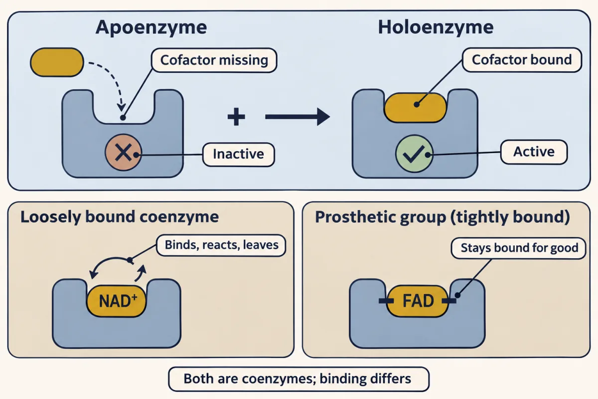
Many enzymes cannot work alone. A cofactor is a small helper required for activity, of two kinds. A coenzyme is an organic molecule, usually derived from a vitamin (niacin becomes NAD⁺; thiamine becomes thiamine pyrophosphate (TPP)). A metal ion is the inorganic kind (Zn²⁺, Mg²⁺). When a cofactor, organic or metal, binds tightly and permanently rather than coming and going, we call it a prosthetic group; the heme buried in catalase, an organic ring holding an iron ion, is the classic example. The pair of terms that trips students up most: an enzyme missing its cofactor is an apoenzyme and it does not work; the assembled, working complex is the holoenzyme. Apo means away; holo means whole.
The classifier below shows one reaction at a time.
Pick the class. The trick is to ignore the enzyme's name and ask what happens to the chemical bond.
EC class is decided by the reaction, not the name. Oxidoreductases move electrons; transferases move a group; hydrolases break a bond with water; lyases add or remove a group across a double bond, even water, as long as no bond is cut by hydrolysis and nothing is oxidized; isomerases rearrange one molecule; ligases join two, usually spending ATP; translocases move things across a membrane.
Going deeper
The bookkeeping behind the rule
The speed-up rule comes from a thermodynamic cycle: going from free substrate to bound transition state costs the same free energy whichever way round you go. It compares kcat with kuncat and assumes the same kind of transition state and the same frequency factor in both. Written for kcat/KM (free enzyme plus free substrate) instead, the same cycle measures how tightly the enzyme holds the transition state outright. That free-end speed-up is the one that is equal forward and reverse: (kcat/KM) forward over (kcat/KM) reverse equals Keq (the Haldane relation), which is why no enzyme can move the equilibrium.
It applies to the highest barrier of a multistep reaction, which has several transition states and intermediates (the 7TIM structure holds an intermediate analog). Once an enzyme is limited by how fast substrate diffuses in, as triosephosphate isomerase nearly is, extra grip on the chemical transition state no longer helps. And it does not cover coupling, which changes the reaction itself (the hexokinase answer above).
Fischer, Haldane, Pauling and shape
Fischer called lock-and-key a hypothesis that could only be tested once someone could hold a pure enzyme and see its shape; when crystallographers finally did, many active sites turned out to flex. Shape changes also continue after the chemistry: in an induced-fit enzyme, the site reopens to help the product leave.
The full class definitions and EC numbers
Hexokinase’s EC 2.7.1.1 reads as transferase, phosphate group, alcohol acceptor, first on the list. Strictly, EC 2.7 is “transferring phosphorus-containing groups”, so “phosphate group” is a fair gloss. The official lyase definition is broader than “add or remove groups across a double bond”: lyases break bonds by means other than hydrolysis or oxidation, often forming a double bond or a ring. The short version is enough for the MCAT.
Statins and muscle symptoms
The classic worry: a patient starts a cholesterol drug and within weeks their muscles ache and a blood marker called creatine kinase (CK) climbs. The pathway statins slow also makes CoQ10 for mitochondrial energy and the isoprenoids muscle cells need. One proposed explanation for statin muscle symptoms is that blocking the pathway lowers CoQ10 and isoprenoids in muscle and harms its mitochondria. It is not proven. Other mechanisms are being studied, CoQ10 supplements have not reliably helped, and in blinded trials most muscle aches reported on statins also occur on placebo. True statin muscle damage with a large CK rise is rare.
Your notes
Select any text in the lesson to highlight it or add a note to it. Your highlights and notes are saved in this browser and collected in your notebook.
This browser is not letting the site save anything (a private window or strict privacy settings), so highlights and notes will be lost when you leave the page.
How we measure it
Measuring rate enhancement
Run the reaction with the enzyme and without it, take each rate, and divide. A catalyzed rate of 540 s⁻¹ over an uncatalyzed 2.9×10⁻⁶ s⁻¹ gives 1.9×10⁸ — the number that says how much work the protein is doing.
Reading ΔG°′ from the equilibrium constant
Let a reaction reach equilibrium, measure [products]/[reactants], and that ratio is Keq. Plug it into ΔG°′ = −RT ln Keq (R = 8.314 J/mol·K, T = 298 K) to convert a concentration you can measure into a free energy you cannot.
Transition-state analog binding
Build a stable molecule shaped like the transition state, then measure how tightly the enzyme holds it versus the real substrate. Proline racemase binds its analog 160× tighter — a classic example of evidence that the active site is complementary to the transition state, not the substrate.
Practice questions on this lesson (5) → MCAT-style, original, each with an explanation
Where this lesson fits
Before thisReview first
- Water, Weak Bonds, and the Generation of OrderThis course · Lesson 3
You need weak, reversible bonds from there to understand how an active site grips a substrate or transition state.
- Protein Three-Dimensional Structure: The Sequence Sets the ShapeThis course · Lesson 5
The active site is a cleft formed by folding, so you need how sequence sets a protein's shape.
After thisStudy next
- Enzyme Kinetics: The Michaelis–Menten ModelThis course · Lesson 8
It measures kcat and KM, the rates this lesson links to transition-state binding, and shows saturation.
- Metabolism: Basic Concepts and DesignThis course · Lesson 15
It uses ΔG, Q and coupling to ask which metabolic reactions can run in cells.
- Protein Binding and Allostery: How Hemoglobin Loads and Delivers OxygenThis course · Lesson 9
It takes binding and Kd further, showing how binding changes with shape and other ligands.
Elsewhere on the siteFilms, simulations and method films in other courses
- Speed up and slow down an enzymeSimulation · Biology
It lets you speed up and slow down an enzyme and see the catalyst come out unchanged.
- Count the Working SitesFilm · Biology
It shows the substrate fitting the active site and the enzyme working again unchanged.
- Osmosis chamberSimulation · Biology
It shows a free-energy downhill direction setting net movement, the same ΔG-decides-direction idea applied to transport.
Suggested by an AI model from the lessons themselves, then checked. How we use AI
Finish line
Test yourself
9 questions
Practice and MCAT-style, with explained answers.
Next · Lesson 8
Enzyme Kinetics: The Michaelis–Menten Model
Why doesn't doubling the substrate keep doubling the rate? Enzymes saturate.
Continue →← Lesson 6 · Techniques in Protein Biochemistry: Purify It, Then Prove It