Biology · Unit 2: Cells
The Cell Theory and Its Evidence
Rewritten on : new text, flashcards and film. Your notes and highlights stay, but the text around them may have moved.
Robert Hooke saw tiny boxes in a slice of cork in 1665, so why did it take about 175 more years to claim that plants and animals alike are made of cells?
Pause when the reading reaches a film, and offer to play it. Saved on this device.
Watch the film now, or keep listening
Builds on:What Am I Made Of?Why these?
Where this lesson sits
- Virginia SOL BIO.3a
- Practices BIO.1d, BIO.1e, BIO.1f
Essential question: How did scientists figure out that everything alive is made of cells, and why did it take better tools?
-
Life Science (grade 7) LS.2 a
Review it at grade 7 level
In grade 7 you learned that all living things are made of one or more cells. You learned that the cell theory was built over time, as microscopes improved and scientists looked at more kinds of living things. You also learned the main ideas: living things are made of cells, cells are the basic unit of life, and cells come from other cells. Biology adds three more ideas, about metabolism, DNA and what cells are made of, and asks why some parts of cells could be seen only with newer tools.
-
Biology BIO.3a You are here
This lesson
-
Biology II Biology II: Advanced Survey of Biology Topics, BIOII.2 a
Preview where it goes next
Virginia's Biology II: Advanced Survey of Biology Topics course (BIOII.2 a) treats the cell as a system in which the nucleus, mitochondria, ribosomes, endoplasmic reticulum, Golgi apparatus and cytoskeleton work together. One topic this lesson left out is how scientists now beat the 0.2 µm limit of ordinary light microscopes. Special fluorescence methods track single labeled molecules. In a 2017 test, a method called MINFLUX separated glowing labels only 6 nanometers apart. That does not give a school microscope this power; it needs special labels and equipment.
-
College
 ▶ Watch What a Microscope Can Show · 3:19
Read the transcript of “What a Microscope Can Show”
Film transcript: What a Microscope Can Show
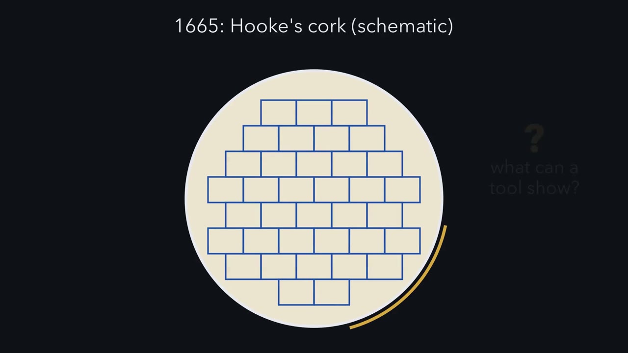
In 1665, Robert Hooke saw rows of tiny boxes in a thin slice of cork. He called the boxes cells. What decides what a microscope can show? And why was seeing those boxes not enough? We start with the tool. Two words matter here. Magnification is how many times larger an image looks than the real object.
Resolution is the smallest distance between two points at which you still see two separate points. Watch two dots on a slide under an ordinary light microscope. A micrometer is one thousandth of a millimeter. The dots start close together and look like one blur.
Slowly they move apart. Near a gap of about 0.2 micrometers, they begin to look like two. Why the blur? Even a well-made ordinary light microscope blurs the finest details. Light spreads out as it passes through the opening of the lens. So each point becomes a small smudge, and close smudges merge into one. Now set the dots 0.15 micrometers apart. They look like one blur. We triple the magnification.
Will you see one dot or two? Still one. The blur got bigger, and it is still one blur. Making the image bigger does not shrink the limit. Magnification makes an image bigger. Resolution decides how much detail it holds. So here is the rule. A tool shows a detail only if the detail is larger than the tool's limit. Enlarging the image adds no detail. A whole cell can be clear while its parts are not. Each tool has its own limit. Picture a ladder of sizes, each rung ten times smaller than the one above. Your eye's limit sits near a tenth of a millimeter. The light microscope's gold line sits far lower. The electron microscope's line sits lower still.
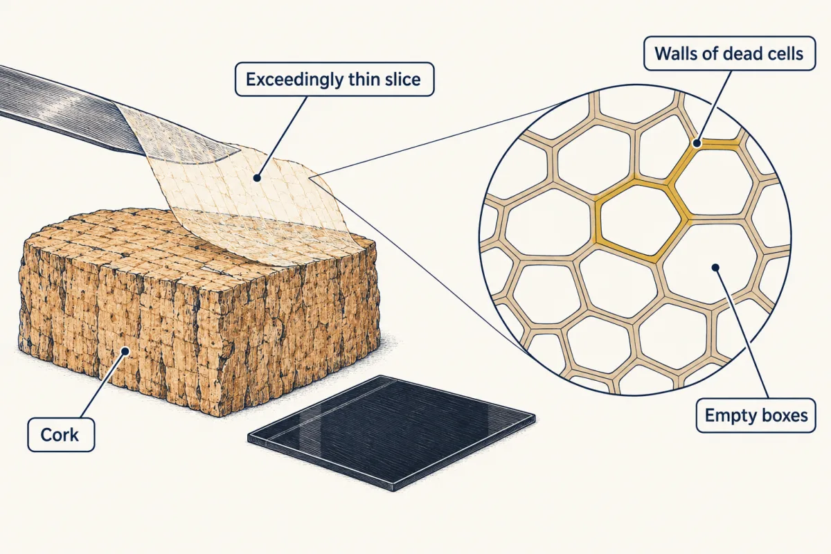
Hooke needed a microscope to make out the outlines and spacing of the cork boxes. Much smaller or low-contrast details inside cells needed better resolution, more contrast and later tools. But look at what he had. Empty walls, left after the cork's cells had died. One material, from one kind of plant. That cannot show that all living things are made of cells. Making out cells needs a tool with fine enough resolution. Building the theory needed more: comparing many kinds of living things, and testing where new cells come from. About 175 years passed before scientists argued that plants and animals alike are built of cells.
From the film
- Magnification makes an image bigger; resolution decides how much detail it holds.
- An ordinary light microscope cannot separate two points closer than about 0.2 micrometers, however much you enlarge the image.
- A tool shows a detail only if the detail is larger than the tool's limit, so a whole cell can be clear while its parts are not.
- Hooke's cork showed the walls of dead cells from one plant, which was not enough to claim that all life is made of cells.
The first film ended with Hooke’s cork boxes: in view, but not enough to build a theory on. Now test its rule on real objects.
What do you think? Decide before you read.
-
Scientists disagree: Measurements under microscopes show huge differences. Many bacteria are about 1 to 2 micrometers long, a human cheek cell is about 50 to 80 micrometers across, and the yolk of an unfertilized chicken egg is a single cell you can hold. Shapes vary too, from flat sheets to long threads.
What scientists think
Scientists disagree: Measurements under microscopes show huge differences. Many bacteria are about 1 to 2 micrometers long, a human cheek cell is about 50 to 80 micrometers across, and the yolk of an unfertilized chicken egg is a single cell you can hold. Shapes vary too, from flat sheets to long threads.
-
Scientists disagree: Bacteria and archaea are living things made of cells, and their cells are neither plant nor animal cells. Fungi and many single-celled organisms in ponds have their own kinds of cells too. Microscopes and, later, other laboratory methods showed this variety.
What scientists think
Scientists disagree: Bacteria and archaea are living things made of cells, and their cells are neither plant nor animal cells. Fungi and many single-celled organisms in ponds have their own kinds of cells too. Microscopes and, later, other laboratory methods showed this variety.
-
Scientists disagree: In science, a theory is a well-tested explanation that ties together many lines of evidence. The cell theory rests on a very large number of observations of many kinds of living things, made with many different tools. It has also been corrected as new evidence came in, which is how scientific theories work.
What scientists think
Scientists disagree: In science, a theory is a well-tested explanation that ties together many lines of evidence. The cell theory rests on a very large number of observations of many kinds of living things, made with many different tools. It has also been corrected as new evidence came in, which is how scientific theories work.
Explore: zoom in and see what appears
The simulation gives you a microscope with a magnification slider (how many times larger the image looks) and a switch for the type of tool: a hand lens, a light microscope or an electron microscope. A set of objects sits on the slide: a frog egg (about 1 mm across), a human cheek cell (about 50 to 80 µm), a bacterium (about 1 to 2 µm long), a virus (smaller than a bacterium) and a ribosome (about 0.02 to 0.03 µm). A micrometer (µm) is one thousandth of a millimeter. Before each step, compare each object’s size with the tool’s limit.
- Smallest detail this tool separates
- about 0.1 mm
- Smallest detail the eye separates
- about 0.1 mm
In this sketch: an object is seen only when (1) the tool separates details finer than the object and (2) the enlarged image is at least 0.1 mm, the eye’s limit (this rule is the sketch’s own; the lesson does not state it). Sizes use the small end of the lesson’s ranges (cheek cell 50 µm, bacterium 1 µm, ribosome 0.02 µm; frog egg is the African clawed frog, 1 mm). The lesson gives no limit for a hand lens, so it gets the eye’s 0.1 mm here. The lesson gives no virus size, so the virus is placed at an assumed 0.1 µm, between the two microscope limits. Electron limit: 0.002 µm (the coarse end of 1 to 2 nm). The limits are typical values; real ones vary with the instrument and the sample. The electron microscope needs a very thin, specially prepared sample. The 1 to 10,000 times range is part of the sketch; real hand lenses and light microscopes do not reach the top of it. The shapes are labelled sketches, not to scale.
Making the image bigger does not change what a tool can resolve: a tool shows a structure only if its resolution is finer than the structure.
Try this.
- Predict which objects you can see with the hand lens. Write your guess down, then test it.
- Switch to the light microscope and turn the magnification all the way up. Predict whether the ribosome will appear, then check.
- Find the lowest magnification at which you can trace the cheek cell’s outline all the way around. Does turning it higher show the virus?
- Switch to the electron microscope. Which objects appear now that did not appear before?
The one idea. A tool shows a detail only if the detail is larger than the tool’s limit, its resolution (the smallest gap at which two points still look separate). So a whole cell can be clear while its parts are not.
Scope: the rule is about making out a shape or a part, not about whether an object shows up at all. The 0.2 µm limit belongs to ordinary light microscopes; special methods beat it (see the Biology II note).
Explain: seeing smaller things
- cell
- cell theory
- metabolism
- DNA
- magnification
- resolution
- stain
- electron microscope
- hypothesis
- theory
- law
Here are all three tools against the simulation’s objects. A nanometer (nm) is one thousandth of a micrometer.
| Tool | Smallest detail it can separate (about) | Can it show (and everything larger)… |
|---|---|---|
| Unaided eye | about 0.1 mm (100 µm) | an African clawed frog egg, about 1 mm across |
| Ordinary light microscope | 0.2 µm | a cheek cell or a bacterium |
| Electron microscope, biological samples | 1 to 2 nm (0.001 to 0.002 µm) | most viruses, or a ribosome |
These are typical values for biological samples; real values vary with the instrument and the sample. An electron microscope forms its image with a beam of electrons instead of light. Electrons travel as waves with a much shorter wavelength (the distance from one crest to the next) than visible light, so electrons can separate much finer details (see Going deeper). In practice, the lenses and the delicate sample, not the wavelength, set the 1 to 2 nm limit. Ribosomes, the cell’s protein builders, are about 0.02 to 0.03 µm across, far below the light microscope’s limit. That is why they were seen clearly only with electron microscopes.
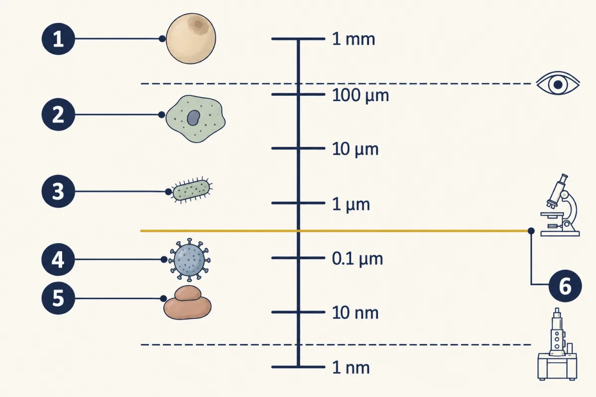
- frog egg
- cheek cell
- bacterium
- virus
- ribosome
- limit of the light microscope
- frog egg
- cheek cell
- blank
- blank
- blank
- blank
Word bank: bacterium, limit of the light microscope, ribosome, virus
- blank
- blank
- blank
- blank
- blank
- blank
Check your labels
- frog egg
- cheek cell
- bacterium
- virus
- ribosome
- limit of the light microscope
Predict. Many bacteria are about 1 to 2 µm long. One of Leeuwenhoek’s surviving microscopes separated details 1.35 µm apart. Could it show a bacterium’s shape and parts? Could it show anything at all?
Check your prediction
Its parts, no: they are far smaller than 1.35 µm. Its shape, only roughly at best. The 1 to 2 µm is the bacterium’s length, and a rod is narrower than it is long. So a long rod might look slightly stretched, and a short one like a round blur. Anything at all, yes: an object near or below the limit can still show up as a blurred speck. Detecting something is not the same as resolving it.
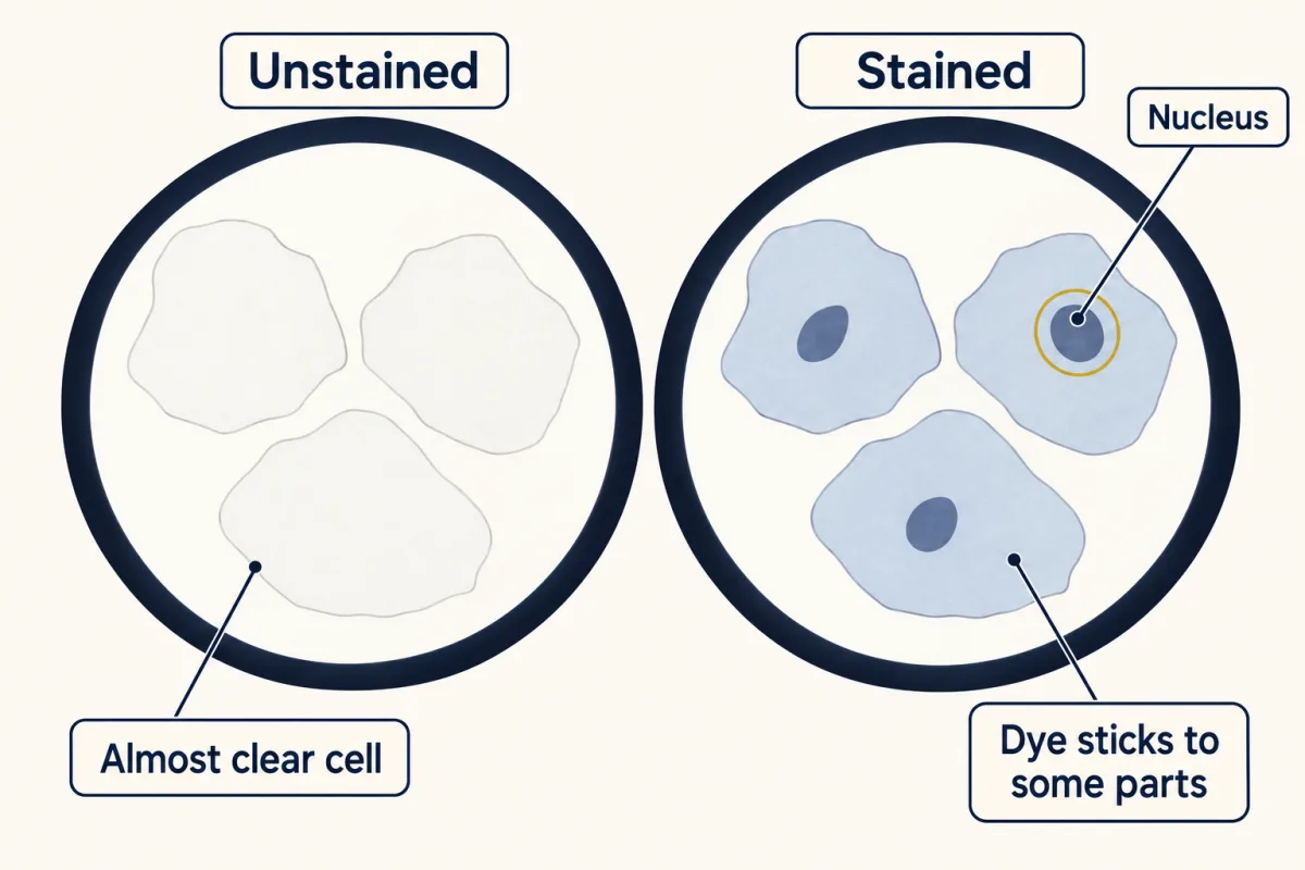
Resolution is not the only problem. Many cells are almost clear inside. A stain is a dye that sticks to some cell parts more than others. It adds contrast, a difference in darkness between a part and its background. Stains made parts inside clear animal cells, such as the nucleus, much easier to see.
Explain: the six ideas of the cell theory
The claim about cells is the cell theory, the scientific explanation of how cells relate to living things. A cell is the smallest unit that can carry out all the processes of life. Virginia lists six ideas:
- All living organisms are composed of cells.
- Cells are the basic unit of life.
- All cells arise from pre-existing (already existing) cells.
- Metabolism occurs within cells.
- Hereditary information (DNA) is passed from cell to cell.
- All cells have the same basic composition.
Metabolism is all the chemical reactions that happen in a living thing, such as breaking down sugar to release energy for the cell to use. Enzymes speed these reactions inside cells (that lesson). DNA is the molecule that carries hereditary information. When a cell divides, each new cell gets a copy (that lesson).
“Same basic composition” means that cells are built from the same main kinds of molecules: proteins, lipids, carbohydrates and nucleic acids (that lesson). Typical cells have a membrane around them, a watery inside and DNA. The details still differ between kinds of cells.
The same basic plan does not mean the same size or shape: compare the simulation’s frog egg and bacterium. Viruses are not made of cells (that lesson).
Idea 3 had to be fought for, and the second film shows how, starting from comparisons of tissues, organized groups of cells that work together.
Read the transcript of “Where Do New Cells Come From?”
Film transcript: Where Do New Cells Come From?
Idea three says every cell comes from a cell. The first scientists to argue for cells everywhere did not think so. So where did they think new cells came from? By the 1830s, improved microscopes were in use. The argument was not only about seeing. In 1838, Matthias Schleiden described plant tissues as made of cells. In 1839, Theodor Schwann compared plant and animal tissues, such as tadpole cartilage. He found the same cellular plan. Comparison was the key step. Many kinds of tissue, plant and animal, showed one shared plan. That was evidence that plants and animals alike are built of cells. But Schleiden and Schwann also thought new cells could form inside a structureless fluid. Picture a crystal growing in a solution.
No cell divides to make it. Schleiden proposed this for plants. Schwann extended it to animals. So there were two models. In the fluid model, a new cell forms inside a structureless fluid, and no cell divides. In the division model, one existing cell splits into two. Suppose you could watch a growing tissue for a while. What would each model lead you to expect to find?
The fluid model predicts new cells taking shape out of fluid, with no cell dividing. The division model predicts a series of stages. One cell pinches in two, then its daughters split again. Watching over time can tell the two models apart. In 1852, Robert Remak published evidence that new cells form when existing cells divide. In 1855, Rudolf Virchow spread the idea widely in a short article. Every cell comes from a cell. Remak's evidence came first; Virchow made it widely known. The fluid idea was wrong, so it was dropped. The core stayed: plants and animals are made of cells. A corrected idea took the fluid idea's place: cells come from existing cells.
That is how a scientific theory works. It is a well-tested explanation, tied to many lines of evidence. When strong new evidence appears, it can be revised. Being revised does not make it a guess. One limit on the rule. Every cell comes from a cell is about cells living today. How the very first cells arose is a separate question that scientists still study.
From the film
- Schleiden and Schwann compared many plant and animal tissues and found the same cellular plan.
- They also thought new cells could form inside a structureless fluid, like a crystal, with no cell dividing, and that part was wrong.
- Remak published evidence in 1852 that new cells form when existing cells divide, and Virchow spread the idea in 1855: every cell comes from a cell. That rule is about cells living today; how the very first cells arose is a separate question.
- The cell theory kept its core and replaced the wrong part with a corrected idea, which is how a well-tested scientific theory changes.
Explain: a theory that grew and changed
The other ideas arrived differently. Schwann already treated cells as the places where nutrition and growth happen, which points toward idea 4. The idea that DNA carries hereditary information (idea 5) came much later.
Three words people often mix up:
| Word | What it is | Example |
|---|---|---|
| Hypothesis | A testable statement about nature | Hypothesis: all plant tissues are made of cells. Prediction: thin slices of other plants will show cells too. |
| Theory | A well-tested explanation of some part of nature, supported by many lines of evidence | The cell theory |
| Law | A description of how something behaves under stated conditions | The law of conservation of mass (below) |
A prediction is what you should observe if a hypothesis is true.
Predict. The cell theory rests on a huge amount of evidence. With enough more, will it become a law?
Check your prediction
No. A theory and a law do different jobs. The law of conservation of mass says that when a chemical reaction runs in a closed container, the total mass stays the same; it does not say why. The atomic theory, the explanation that matter is made of atoms, says why: a reaction rearranges atoms but does not create or destroy them. No amount of evidence for atoms will turn the atomic theory into the law of conservation of mass.
Elaborate: tools and discoveries over time
In the 1670s Antonie van Leeuwenhoek already saw living single-celled organisms, which he called animalcules. Compound microscopes use two or more lenses in a row. They did not clearly beat his best single lenses until improved microscopes came into use in the 1830s, the decade of Schleiden and Schwann. So tools were part of the wait, but not all of it: animalcules had been known for more than 150 years, and the theory still needed the work in the Ideas table.
Tools
| Year | Tool | Magnification (times) | Smallest detail separated (µm) | Note |
|---|---|---|---|---|
| Before 1723 | Leeuwenhoek microscope (one of two surviving ones studied in 2021) | 266 | 1.35 | A single bead-shaped lens |
| 1933 | Ruska’s electron microscope | 12,000 | not reported | Electrons can form a magnified image |
| 1937 | Electron image of a diatom shell | 3,500 | 0.13 | A diatom is a single-celled alga with a glassy shell; the caption gives 130 nm, finer than the light limit |
| Today | Ordinary light microscope | – | 0.2 | The typical limit for biological samples |
Ideas
| Year | Who | What they did | Kind of work |
|---|---|---|---|
| 1665 | Hooke | Saw small chambers in cork and called them cells | Looking |
| 1838 | Schleiden | Described plant tissues as made of cells | Comparing |
| 1839 | Schwann | Found the same cellular plan in animal tissues, such as tadpole cartilage | Comparing |
| 1852 | Remak | Published evidence that new cells form by division | Observing cell division, which tested the fluid idea |
| 1855 | Virchow | Spread the idea: every cell comes from a cell | Stating the corrected idea |
Hooke’s magnification and smallest separated detail are not reported in these sources. The tables are a compilation, not one measured data set (sources in Going deeper).
- How many times greater was the magnification of Ruska’s 1933 microscope than that of the Leeuwenhoek microscope in the table? Round to the nearest whole number.
- The Leeuwenhoek microscope in the table separated details 1.35 µm apart. An ordinary light microscope today separates about 0.2 µm. About how many times finer is the modern limit?
- The Tools table lists no tool between the Leeuwenhoek microscope (before 1723) and Ruska’s 1933 microscope, although microscopes had improved in the 1830s. Look at the Ideas table: besides better microscopes, what kind of work moved the theory forward from 1838 to 1855?
Claim, evidence, reasoning
Make a claim that answers the essential question: why did it take better tools, and more than tools, to build the cell theory? Use the two tables.
Start with: The cell theory needed blank and also blank.
Start with: The Tools table shows that blank, and the Ideas table shows that the steps from 1838 to 1855 came from blank.
Start with: This matters because a tool can show a detail only when the detail is blank than the tool's limit, and a theory needs blank.
Compare with a model answer
The cell theory needed better tools and also many comparisons and a corrected idea. Hooke saw cork chambers in 1665, but the claim that animals share the cellular plan came only in 1839, after many plant and animal tissues were compared. Microscopes had improved in the 1830s, but the steps from 1838 to 1855 came mainly from comparing tissues and testing how new cells form with those improved microscopes, not from a new kind of tool. Tools mattered too: the caption of a 1937 electron image of a diatom gives 130 nm (0.13 µm), finer than the 0.2 µm light limit.
Saved in this browser only.
Design problem
Hooke cut his cork slice exceedingly thin. Does the thickness of a slice change how many separate cells you can count in one view of a light microscope? Plan a fair test using an onion or a potato, a light microscope and slides.
Saved in this browser only.
One way to do it
- Hypothesis: If the slice is thinner, then more separate cells will be countable in one view, because fewer layers of cells overlap.
- Independent variable: slice thickness, for example very thin, medium and thick.
- Dependent variable: the number of cells with walls you can trace all the way around in one field of view.
- Constants: the same onion or potato, microscope, magnification, light setting and stain.
- Comparison: the thickest slice serves as the baseline.
- Repeated trials: make five slides at each thickness, count three views on each, and calculate the mean for each thickness.
- Safety: cutting thin slices needs a sharp blade. Use a guarded blade or a peeler, cut away from your hand, and work only with an adult’s permission.
Evaluate: check your thinking
Hooke’s cork put cells in view. Claiming them for all life took better light microscopes, many comparisons and a corrected idea about new cells. Stains showed pale parts more clearly, and electron microscopes later showed much finer details inside cells. A theory built that way is a well-tested explanation, not a guess.
Science practices you used: predicting before testing, reading data tables, telling a hypothesis, a prediction, a theory and a law apart, modeling resolution and its limits, and planning a fair test.
Going deeper
Why even a good lens blurs
Physicists call the first film’s spreading of light through a lens opening diffraction. The 0.2 µm limit is typical, not exact for every microscope or viewing direction. Shorter wavelengths make smaller smudges, which is why electrons help. A stretched, blurry phone photo is only a loose comparison: photos also blur from poor focus, motion or too few pixels.
Archaeal membranes
Archaea build their membranes from lipids that differ from those of bacteria, plants and animals in both their chains and the chemical links that join them. This is one of the main features that sets archaea apart.
Where the table’s numbers come from
Two of the few surviving microscopes made by Antonie van Leeuwenhoek, both made before 1723, are in museums in Leiden and Utrecht. A 2021 study (Cocquyt and colleagues, Science Advances) scanned their lenses and reported their magnifications. One, labelled V7017, magnified 118 times, and no resolution was reported for it; its single lens was ground and polished. The other, UM-1, magnified 266 times and separated details 1.35 µm apart, and its lens was a single bead shape; the 1.35 µm value is an earlier measurement that the study cites. The 1933 magnification and the 1937 diatom image come from Ernst Ruska’s 1986 Nobel Lecture. Dates for Schleiden, Schwann, Remak and Virchow come from the OpenStax Microbiology textbook (2016).
Viruses near the limit
The table says most viruses because a few unusually large viruses come close to the light microscope’s limit or pass it.
Metabolism outside cells?
“Metabolism occurs within cells” is the Virginia wording. Some reactions do run outside cells, such as digestive enzymes working in the gut. But those enzymes are made by cells, and most of metabolism happens inside cells.
Your notes
Select any text in the lesson to highlight it or add a note to it. Your highlights and notes are saved in this browser and collected in your notebook.
This browser is not letting the site save anything (a private window or strict privacy settings), so highlights and notes will be lost when you leave the page.
How we measure it
Thin-section light microscopy
A very thin slice of a sample is cut so that light can pass through it, and a light microscope enlarges it. Thin slices show the walls and spaces of separate cells instead of a thick, dark block.
Staining
Many cells are almost clear, so a dye is added that sticks to some parts more than others. The added contrast lets you see parts inside cells, such as the nucleus, that are hard to see in an unstained sample.
Transmission electron microscopy
A beam of electrons passes through a very thin, specially prepared sample, and the electrons that get through form the image. Because electrons have much shorter wavelengths than visible light, this microscope can separate details about a hundred times finer than a light microscope can. In practice the lenses and the delicate sample, not the wavelength, set that limit.
Where this lesson fits
Before thisReview first
- What Am I Made Of?This course · Lesson 2
The sixth idea says cells share the same main molecules, which this lesson names and explains.
After thisStudy next
- The Cell Cycle and MitosisThis course · Lesson 10
It shows how an existing cell divides into two cells, the process behind the idea that all cells come from cells.
- Living or Not? Viruses and BacteriaThis course · Lesson 12
It tests the cell theory on viruses, which are not made of cells.
- Organelles as a SystemThis course · Lesson 7
It uses the finer cell parts that electron microscopes later revealed.
Elsewhere on the siteFilms, simulations and method films in other courses
- Zoom into a dropSimulation · Life Science
Zooming into pond water shows which things sit below the eye's size limit and need a microscope.
- Electron microscopyMethod film
It shows how electron beams reveal cell parts that light microscopes blur.
Suggested by an AI model from the lessons themselves, then checked. How we use AI
Finish line
Test yourself
8 questions
Practice and MCAT-style, with explained answers.
Next · Lesson 7
Organelles as a System
The insulin gene never leaves a beta cell's nucleus, yet insulin leaves the cell after a meal. What carries it out, and in what order?
Continue →