Biology · Skim version
The Cell Cycle and Mitosis
Read the full lesson → Skim the whole course →
The guiding idea
When one cell becomes two, what keeps either new cell from receiving only half the chromosomes?
Copy each chromosome first; separate the copies so each nucleus gets one.
From the film: How One Set Becomes Two
- Copy each chromosome first; separate the copies so each nucleus gets one.
- Interphase includes growth, DNA copying and preparation; it is not a rest.
- After S phase, two joined sister chromatids still count as one copied chromosome.
- Mitosis divides the nucleus; cytokinesis divides the cytoplasm, often starting in late anaphase or telophase.
From the film: A Snapshot Becomes a Clock
- In a fair snapshot, a longer phase holds more cells.
- A stained root tip is a snapshot of many cells caught at different moments.
- A phase-time estimate is the fraction of cells in that phase times an assumed or measured total cycle time.
- Hand-picked pictures, cells that stopped cycling, or a changing pace can break the estimate.
-
Explore: step a cell through division
-
Explain: the cell cycle
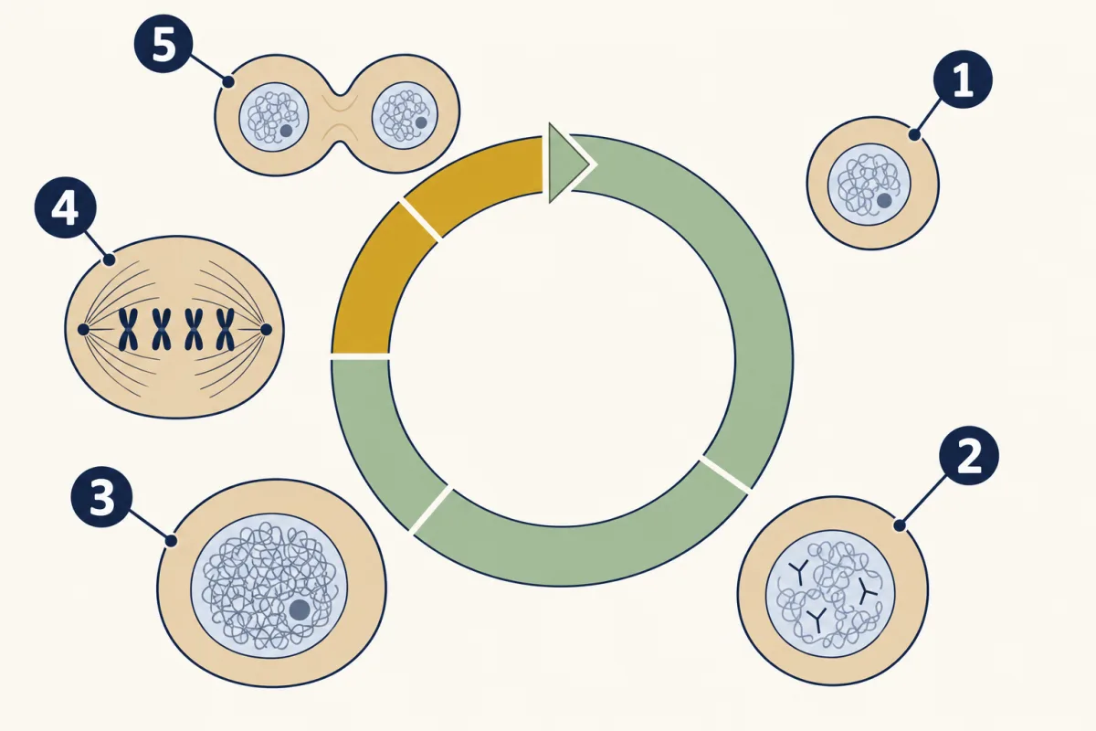
- G1
- S
- G2
- mitosis
- cytokinesis
-
Explain: the stages of mitosis
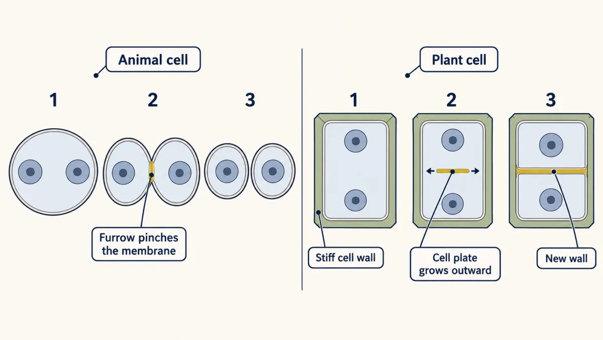
Cytokinesis in two kinds of cell: a furrow pinches an animal cell in two, while a plant cell, held by its stiff wall, builds a cell plate that grows outward into a new wall. AI-generated illustration -
Explain: why cells divide and stay small
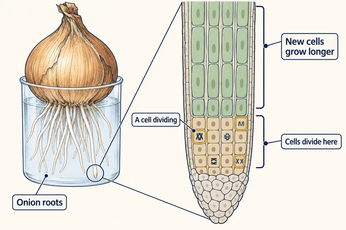
Near the tip of an onion root, cells divide; just behind them, the new cells stretch. Both push the root deeper. AI-generated illustration -
Elaborate: count onion root-tip cells
-
Evaluate: check your thinking
-
Going deeper
Where this lesson fits
Before thisReview first
- The Cell Theory and Its EvidenceThis course · Lesson 6
You need the idea that all cells come from earlier cells to see why division matters here.
- Organelles as a SystemThis course · Lesson 7
You need to know the nucleus holds the DNA that is copied and split here.
- Crossing the MembraneThis course · Lesson 8
The surface-area-to-volume argument here relies on food and wastes crossing the membrane.
After thisStudy next
- Meiosis and VariationThis course · Lesson 19
It reuses the copy-then-separate rule but separates chromosomes twice, making cells that differ from each other.
- DNA: Structure and CopyingThis course · Lesson 16
It explains how the DNA copying done in S phase actually works.
- One Cell or ManyThis course · Lesson 11
It builds on why cells stay small and how many cells share the jobs of an organism.
Elsewhere on the siteFilms, simulations and method films in other courses
- Heal the scrapeSimulation · Life Science
Heal a scrape to see cell division at work in repairing skin.
- Copy First, Then ShareFilm · Life Science
It shows how a cell copies first and then shares, as in interphase and mitosis.
Suggested by an AI model from the lessons themselves, then checked. How we use AI