Sensory Systems · Skim version
Hot, Warm, Cool and Cold
Read the full lesson → Skim the whole course →
The guiding idea
Mice lacking the cold-and-menthol receptor TRPM8 failed completely to learn to report forepaw warming in a 10-day task. Why would a cold receptor matter for warmth?
Skin temperature is read from a few afferent lines at once, and a cool line that fires at rest can report warming by slowing down.
From the film: A Fibre That Fires at Rest
- Skin temperature is read from a few afferent lines at once, not one channel per feeling.
- A cool line that fires at rest can report warming by slowing down.
- In the mouse forepaw, losing a channel that supplies a line's firing can cost warmth, though the circuit that reads the lines has not been identified.
From the film: Take Away the Cool Line
- Without TRPM8, mice failed to report forepaw warming in this task, even after 10 days of training.
- TRPM8 loss removes the cool fibres' resting firing in isolated skin, so warming has no ongoing firing left to silence.
- Loss of heat channels blunted warmth detection, whereas TRPM8 loss abolished it in this task.
- Whether gentle warmth rests mainly on warm fibres or on quieter cool fibres is not settled.
From the film: Three Channels, One Burn
- In mice, acute withdrawal from noxious heat survives the loss of any one of TRPV1, TRPM3 or TRPA1.
- The three channels act in parallel: only mice lacking all three lacked acute withdrawal in the reported assays.
- The same mice still preferred moderate temperatures, so withdrawal and preference are different behaviours.
-
The map: ranges, fibres and channels
-
Lines that fire at rest
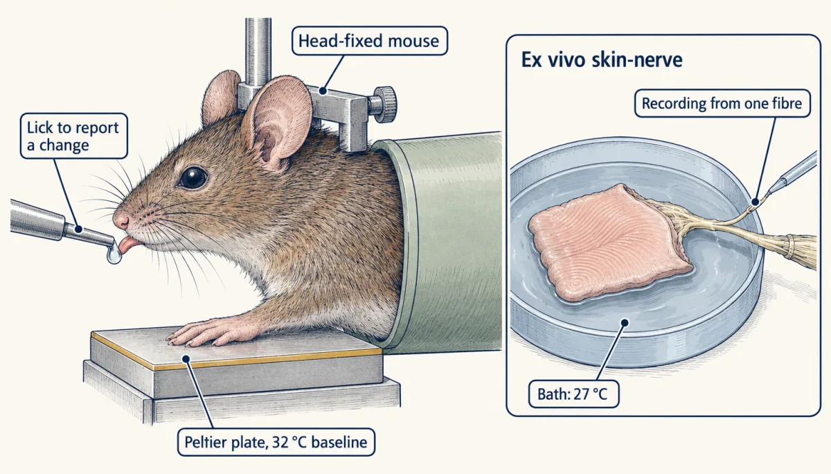
Two preparations in the 2020 mouse forepaw study: behavioural reporting used an 8 × 8 mm Peltier contact surface at a 32 °C baseline. Ex vivo recordings included a 27 °C skin bath and a separate mineral-oil compartment for recording from teased nerve filaments. AI-generated illustration -
Measuring detection: hits, false alarms and d′
-
Take away the cool line
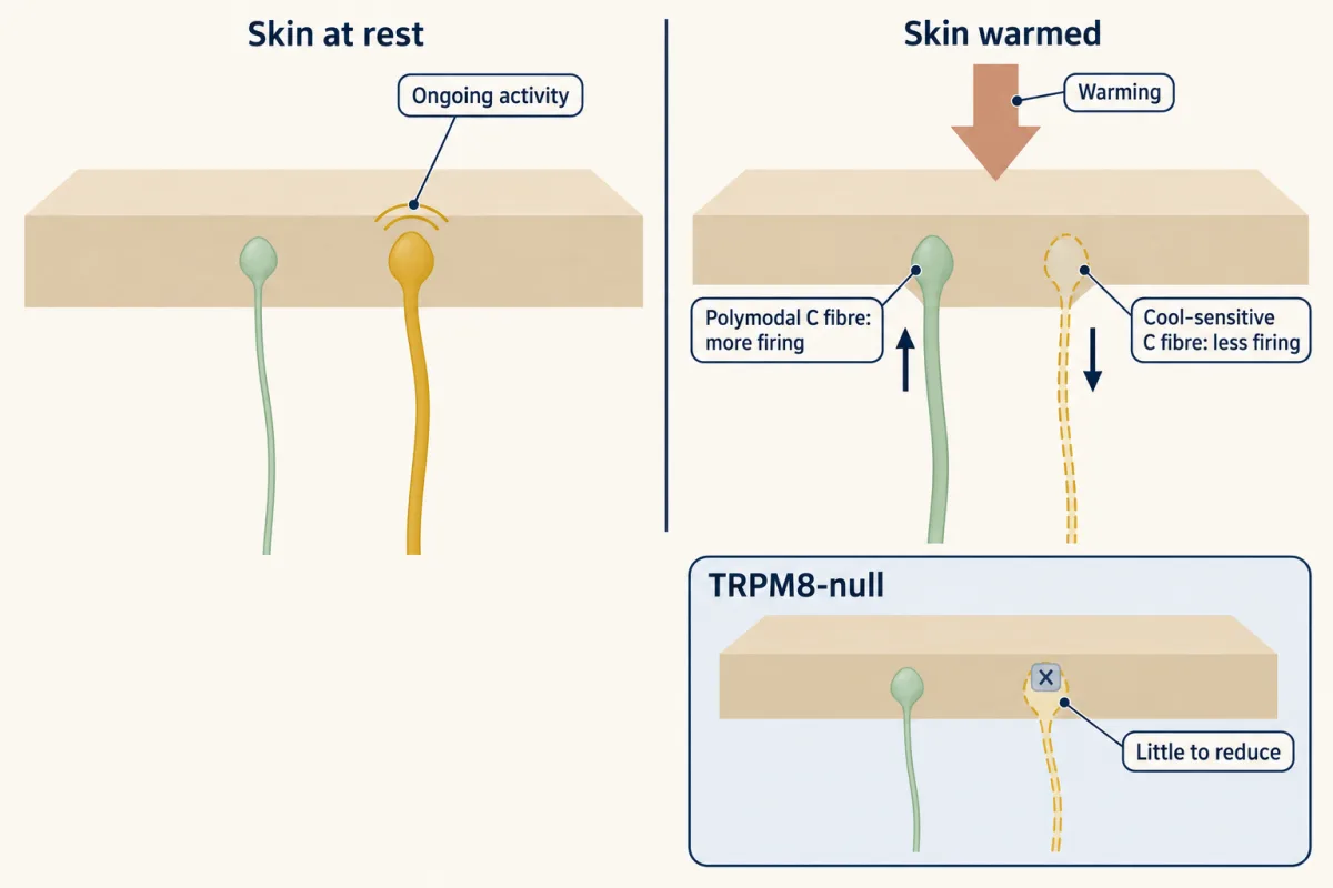
In the mouse forepaw, warming raised firing in one population of polymodal C fibres and reduced the ongoing firing of cool-sensitive C fibres, so warmth is signalled by excitation and by reduced cool input. Mice lacking TRPM8 lose the cool-fibre activity whose reduction is one of the two parts of that signal; the study did not identify the central circuit that integrates the two signals. AI-generated illustration -
Three channels for one burn: redundancy, not a chain
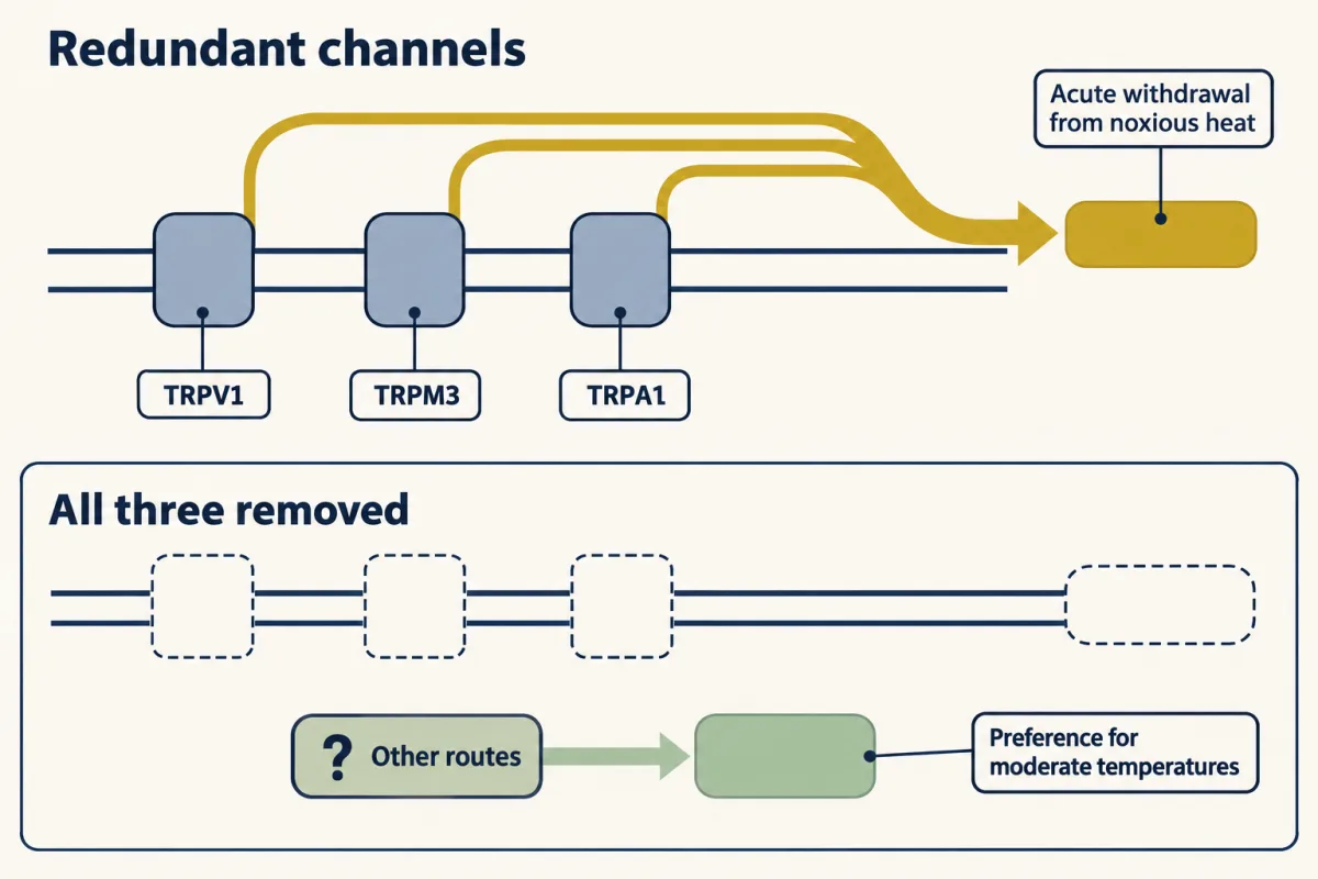
In mice, the three channels act in parallel for acute noxious-heat withdrawal: single knockouts do not abolish it, but losing TRPV1, TRPM3 and TRPA1 together does. The same animals still preferred moderate temperatures, so whatever supports that preference is not exhausted by these three channels. AI-generated illustration -
Reading the lines: spinal cord, ganglia and insular cortex
-
A different question: where is the sensor inside the channel?
-
Going deeper
Where this lesson fits
Before thisReview first
- Five Ways to Turn a Stimulus into a CurrentThis course · Lesson 1
It sets out how a stimulus is turned into a current, which this lesson assumes when it discusses temperature-gated channels.
- Sensory Neurons by Their Genes: the Atlas EraThis course · Lesson 3
It defines the molecular classes of dorsal root ganglion neurons whose cool, warm and heat lines are read here.
After thisStudy next
- Nociceptors and the Sodium Channels of PainThis course · Lesson 11
It takes up what happens after heat transduction, when sodium channels turn nociceptor signals into spikes.
- Spinal Circuits That Gate PainThis course · Lesson 12
It follows the heat and cool lines into the spinal cord, where gating decides what reaches the brain.
- When Pain PersistsThis course · Lesson 14
It shows how these heat and cold lines change after injury.
Elsewhere on the siteFilms, simulations and method films in other courses
- The Signal Opens a HoleFilm · Cell Signaling
It shows how a chilli pepper and a hot stove open the same heat-gated channel, the TRPV1 story used here.
- Why Capsaicin Feels HotFilm · Neuroscience
It shows capsaicin opening the heat channel TRPV1 above about 43 degrees Celsius, the cloning result this lesson revisits.
Suggested by an AI model from the lessons themselves, then checked. How we use AI