Biochemistry · Molecular Recognition & Allostery
Protein Binding and Allostery: How Hemoglobin Loads and Delivers Oxygen
Rewritten on : new text, figures, flashcards and film. Your notes and highlights stay, but the text around them may have moved.
Your blood has to grab oxygen in the lungs and let go of about two-thirds of it in hard-working muscle. A tighter grip than hemoglobin's would make it worse. What does hemoglobin do instead?
Pause when the reading reaches a film, and offer to play it. Saved on this device.
Watch the film now, or keep listening
Builds on:Protein Three-Dimensional Structure: The Sequence Sets the ShapeWater, Weak Bonds, and the Generation of OrderAmino Acids: How One Side Chain Decides EverythingWhy these?
 ▶ Watch The Delivery Problem · 2:30
Read the transcript of “The Delivery Problem”
Film transcript: The Delivery Problem
Your blood must grab oxygen in the lungs and let it go in working muscle. How can one protein do both? In the lungs, the oxygen pressure is about 100 torr. Oxygen is plentiful there. Blood leaving resting tissue sits near 40 torr. Blood leaving hard-working muscle sits near 20. So blood has two opposite jobs. Load oxygen where it is plentiful. Unload it where it is scarce. A protein that binds tightly is good at the first job. It is bad at the second. Predict. Would a protein that binds oxygen more tightly make a better carrier?
No. A tight binder fills in the lungs, but it stays nearly full in the muscle. Oxygen that stays aboard is not delivered. Myoglobin, the muscle's storage protein, is that kind of tight binder. Across the same gap it would unload only about seven percent. Hemoglobin gives up about two-thirds of its oxygen between the lungs and hard-working muscle. At resting tissue it gives up much less, about a fifth. Blood leaving resting tissue is still mostly full. Hard work drains far more. The harder the muscle works, the more oxygen the blood hands over. Both proteins use the same heme and the same iron. So what is hemoglobin doing differently? Myoglobin has one binding site.
Hemoglobin has four that cooperate, and its grip is set for the pressures between lung and muscle. Here is the idea for this whole series. Hemoglobin is always a mix of two shapes. One shape, called T, holds oxygen weakly. The other, called R, holds it tightly. How full hemoglobin is depends on how full each shape would be, and how much of each is present.
Anything that binds one shape better shifts the mix toward that shape. Oxygen itself favors R. So oxygen pushes hemoglobin toward the shape that grabs oxygen best. That is cooperativity, and it is why blood delivers.
Your blood has to grab oxygen in the lungs and let go of it in working muscle, and a protein that gripped oxygen harder than hemoglobin does would deliver less, not more.
Hemoglobin delivers about two-thirds of its oxygen to hard-working muscle. Myoglobin, which uses the same heme (the iron-holding ring that binds oxygen) and the same iron, would deliver about 7%. The difference is not simply that one grips harder. Hemoglobin can change its own grip, oxygen is what changes it, and its grip is tuned so the change happens between the lungs and the muscle.
The delivery problem: load in the lungs, unload in the tissues
Start with the pressures. In the lungs the partial pressure of oxygen, pO₂ (the share of the gas pressure due to oxygen), is near 100 torr (torr is a unit of pressure; one atmosphere is 760 torr). Blood leaving resting tissue is near 40 torr, and blood leaving hard-working muscle is near 20 torr.
So blood has two opposite jobs. It must load oxygen where oxygen is plentiful and unload it where oxygen is scarce.
Predict. Would a protein that binds oxygen more tightly make a better carrier?
Check your prediction
No. A tight binder fills up in the lungs, but it is still nearly full when it reaches the muscle, and oxygen that stays aboard is not delivered. Myoglobin, the muscle’s high-affinity storage protein, is that kind of binder: across the gap from 100 to 20 torr it would unload only about 7%.
Hemoglobin does both jobs: it gives up about 66% of its oxygen between the lungs and hard-working muscle, but only about a fifth at resting tissue pressures, so blood leaving resting tissue is still mostly full.
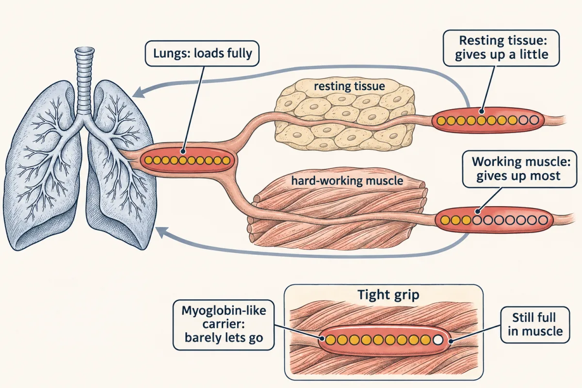
The difference: myoglobin has one binding site, while hemoglobin has four that cooperate, and its grip is set for the pressures between the lungs and the muscle. This lesson is about what that cooperation is.
The one idea to carry through this lesson. Hemoglobin is always a mix of two shapes: T, which holds oxygen weakly, and R, which holds it tightly. How full hemoglobin is at any pO₂ is the average of how full each shape would be, weighted by how much of each shape is present. Anything that binds one shape better than the other shifts the mix toward that shape. Oxygen and CO favor R; H⁺, CO₂ and 2,3-BPG favor T. Cooperativity is oxygen shifting the mix toward the shape that binds oxygen best.
This is the two-state picture. It explains the S-shaped curve, the regulators and CO poisoning. It does not explain sickle-cell disease on its own (there the T shape itself turns sticky), and real hemoglobin is a little more complicated than two shapes (Going deeper, “The two-state fit used in this lesson”).
To use the rule you need one tool first: a number for how tightly a single site holds its ligand.
One site: Kd and the hyperbola
Read the transcript of “One Site, One Curve”
Film transcript: One Site, One Curve
How do you put a number on how tightly a protein holds its partner? Binding is never permanent. A protein and its ligand keep joining and coming apart. Write the reaction in the direction it comes apart. The bound complex splits into free receptor and free ligand. The dissociation constant, Kd, is the ratio of what is loose to what is bound. Two concentrations on top and one below leave one. So Kd has units of concentration. Now pick the ligand level where exactly half the receptors are bound. Free and bound receptor are then equal. They cancel. So Kd is simply the free ligand concentration at half occupancy. That makes the scale run backwards.
A small Kd means tight binding, because little ligand fills half the sites. The most common error is flipping it. Its reciprocal, the association constant runs the other way. For Kd, lower is tighter. Plot fraction bound against free ligand. You get a hyperbola. It rises steeply, then bends over and creeps toward full. Oxygen is a gas, so we use its pressure. The dissolved concentration is proportional to the partial pressure. So for a single site, P50, the pressure at half full, is just Kd measured in torr. Myoglobin is the test case. One chain, one heme, one site. Its P50 is about 2 torr. Predict. Myoglobin is about 98% % full in the lungs.
How full is it at 20 torr, in working muscle? Still about 91% % full. 20 torr is far above its P50, so it barely lets go. So myoglobin gives up only about seven percent. It is a sponge that grips oxygen and lets go only when oxygen gets truly scarce. Hemoglobin's curve is not a hyperbola at all. It is an S. The next chapter builds that S. One site gives a hyperbola, and Kd, or P50, sets where it sits.
Most of life is molecules bumping into each other, and once in a while two of them agree to stick. The sticking is never permanent. Binding is an equilibrium, a partnership that keeps forming and breaking. A receptor is a protein that binds a specific ligand far more tightly than molecules that look almost right, and that discrimination is what lets a signal be a signal.
To measure how tightly a protein holds a ligand, write the binding reaction in the direction it comes apart:
RL ⇌ R + L
The dissociation constant is the ratio of what is loose to what is bound:
Kd = [R][L] / [RL]
Kd has units of concentration: in [R][L] / [RL], two concentrations on top and one below leave one. Read it as a stickiness rating with the scale running backwards: a small Kd means tight binding, and a large Kd means loose. The most common student error is flipping it. Kd is not Ka, the association constant, which is its reciprocal (Ka = 1/Kd, so a bigger Ka means tighter binding).
Why Kd is the concentration at half-saturation
Set the free ligand concentration to the value where exactly half the receptor is bound, and call it L½. At that point the bound and free receptor populations are equal, [R] = [RL], and they cancel:
Kd = [R][L] / [RL] = [L] when [R] = [RL], so Kd = L½
So Kd is just the ligand concentration that gets you to half occupancy. That is why a small Kd means tight binding: very little ligand fills half the sites. The same definition gives the whole curve.
In words The fraction of sites occupied equals the free ligand concentration, divided by Kd plus the free ligand concentration.
- Y“Y”
- fractional saturation: the fraction of sites with ligand bound0 to 1
- L“L”
- the free ligand concentrationM
- Kd“K d”
- the dissociation constant: the free ligand concentration at half occupancyM
Sanity check When L equals Kd, Y is exactly one half. When L is far below Kd, Y is near zero. When L is far above Kd, Y creeps toward 1 but never quite reaches it.
Try one number
Take any single site and measure the ligand in units of its own Kd.
At L = Kd: Y = 1 / (1 + 1) = 0.5, half full.
At L = 3 × Kd: Y = 3 / (1 + 3) = 0.75.
At L = 9 × Kd: Y = 9 / (1 + 9) = 0.9.
Going from half full to 90% full takes nine times as much ligand. That slow climb is the bend of the hyperbola.
Where it comes from optional
Step 1, saturation is bound sites over all sites: Y = [RL] / ([R] + [RL]).
Step 2, rearrange the definition of Kd to get [R] = Kd × [RL] / [L], and substitute it: Y = [RL] / (Kd × [RL] / [L] + [RL]).
Step 3, divide the top and bottom by [RL], then multiply both by [L]: Y = [L] / (Kd + [L]).
Need a refresher? rates and “per” · units and prefixes
Plotted against L, this is a hyperbola. The clean result holds for a single binding site, with no cooperativity and ligand in excess.
From Kd to P50
For oxygen there is one more step. Oxygen is a gas, and the concentration of a dissolved gas is proportional to its partial pressure. So for a single site, P50, the pO₂ at half-saturation, is just Kd measured in torr, and the curve is Y = pO₂ / (P50 + pO₂).
Myoglobin is the test case. It is the storage protein in muscle, one polypeptide chain wrapped around one heme. Its binding curve is a plain hyperbola, the same shape as the Kd plot, with a P50 of about 2 torr. There is no cooperativity, because there is nothing for a lone site to cooperate with.
Predict. Myoglobin is about 98% full at 100 torr, in the lungs. How full is it at 20 torr, in hard-working muscle?
Check your prediction
Still about 91% full: 20 / (2 + 20) ≈ 0.91. Twenty torr is far above its P50, so the site is still nearly always occupied. Myoglobin hands over only about 7% (98 − 91), a sponge that grips oxygen and barely lets go. That suits a storage protein, which holds its oxygen and releases it only when oxygen gets truly scarce.
Two shapes: building the S
Read the transcript of “Two Shapes Make the S”
Film transcript: Two Shapes Make the S
Myoglobin draws a hyperbola. Hemoglobin draws an S. Where does the S come from? Hemoglobin has four sites. And the whole molecule can take either of two overall shapes. T, for tense, is held by extra salt bridges between the subunits. Each heme in T holds oxygen weakly. R, for relaxed, has fewer of those bridges. Each heme in R holds oxygen tightly. In one illustrative fit, a T site is half full only near 110 torr. An R site is half full below 1 torr. A molecule does not wait for oxygen to change shape. Even empty, it flickers between T and R. But with no oxygen, T is far more stable. R molecules are almost absent. So a sample is a mix. Its fullness is the average of each shape's fullness, weighted by how much of each is present.
At 20 torr, about a fifth of the molecules are R. Together the mix is about one-third full. At 100 torr, nearly all the molecules are R. The mix is about 98% % full. Between those two points, about two-thirds is delivered. But notice what changed. The fraction of R climbed with oxygen. Why? R holds oxygen far better than T. So each oxygen bound makes R a little more favorable. Oxygen is a reactant that only R uses well. Adding oxygen pulls the balance from T toward R. Follow one molecule as it fills. Each oxygen multiplies the odds of R by the same large factor. That factor is how much more tightly R holds oxygen than T does. At low pressure, nearly everything is T. The curve follows T's weak curve and barely rises.
Through the middle, each oxygen tips more molecules into R. Their empty sites fill easily. This is the steep part. At high pressure, nearly everything is R. The curve flattens onto R's curve near full. Predict. Suppose T and R held oxygen equally well. What shape would the curve have?
A plain hyperbola. Oxygen would favor neither shape, so the mix would never shift. So cooperativity is oxygen shifting the mix toward the shape that binds oxygen best. The four sites share one shape, so a shift changes all four at once. The Hill coefficient scores how steep the S is at half full. Identical, independent sites score 1. Hemoglobin scores about 2.8. The score can be no larger than the number of sites. It measures steepness. It does not count anything. Predict. A carrier as steep as hemoglobin, but with myoglobin's P50. How much would it deliver?
Almost none. Its steep part sits far below 20 torr, so it is still full in the muscle. A hyperbola with hemoglobin's P50 delivers only about a third. Hemoglobin needs both: an S, and an S in the right place. The S comes from two shapes that differ in grip, and oxygen shifting the mix between them.
Myoglobin has one site and one shape, so its curve is the hyperbola you just built. Hemoglobin has four sites, and the molecule can take either of two overall shapes.
- T (tense) is held together by extra salt bridges (attractions between oppositely charged side chains) between the subunits. In this shape each heme holds oxygen weakly. In one illustrative fit of hemoglobin’s curve, a T site is half full only at about 110 torr.
- R (relaxed) has fewer of those bridges, and each heme holds oxygen tightly. In the same fit, an R site is half full at about 0.37 torr.
A molecule does not have to wait for oxygen to change shape. Even with no oxygen at all, it flickers between T and R, but T is so much more stable that R molecules are almost absent.
So at any moment, a sample of hemoglobin is a mix of the two shapes. How full is the mix? Ask how full each shape would be on its own, then average, counting each shape in proportion to how much of it there is.
In words Overall fullness is the fullness of the R shape times the fraction that is R, plus the fullness of the T shape times the fraction that is T.
- Y“Y”
- fractional saturation of all the hemoglobin: the fraction of hemes carrying oxygen0 to 1
- fR“f sub R”
- the fraction of molecules in the R shape at this pO₂0 to 1
- YR“Y sub R”
- how full an R site would be: pO₂ / (pO₂ + KR), the single-site curve with R's small half-full pressure0 to 1
- YT“Y sub T”
- how full a T site would be: pO₂ / (pO₂ + KT), with T's much larger half-full pressure0 to 1
Sanity check If every molecule were R (fR = 1), Y would be R's hyperbola. If every molecule were T (fR = 0), it would be T's hyperbola. Hemoglobin's curve is neither: it starts on the T curve and ends on the R curve.
Try one number
At 20 torr, in blood leaving hard-working muscle: an R site would be about 98% full and a T site about 15% full. About a fifth of the molecules are R.
R’s share: 0.21 × 0.98 ≈ 0.21. T’s share: 0.79 × 0.15 ≈ 0.12. Total ≈ 0.33, so hemoglobin is about one-third full.
At 100 torr, in the lungs: about 96% of molecules are R, so Y ≈ 0.96 × 1.00 + 0.04 × 0.48 ≈ 0.98.
Unloaded between the lungs and the muscle: 0.98 − 0.33 ≈ 0.65, about two-thirds, the opening’s 66% within rounding. (Computed without rounding, this two-state fit gives about 64%, against 65–66% from the Hill curve used in the opening, the table and the sandbox. The gap is not a discrepancy in hemoglobin: the two are different idealized curves fitted to the same P50 and steepness.)
Need a refresher? rates and “per”
That example hides the key move. In the same fit, fR is about 0.2% at 5 torr; by 20 and 100 torr it has climbed to a fifth and 96%. Oxygen changed the mix. Why?
Compare a T molecule and an R molecule that each pick up one oxygen. R holds oxygen far more tightly, so every oxygen bound makes R a little more favorable compared with T. It is Le Chatelier’s principle applied to shape: oxygen is a reactant that R uses far better, so adding oxygen pulls the T ⇌ R balance toward R.
Follow one molecule as it fills. The numbers are from the same illustrative fit; the pattern, not the exact values, is the point.
| Oxygens bound | Odds that the molecule is R |
|---|---|
| 0 | T wins by millions to one |
| 1 | T still wins by tens of thousands to one |
| 2 | T wins by a few hundred to one |
| 3 | roughly even: R slightly ahead |
| 4 | R wins by hundreds to one |
Each oxygen multiplies the odds of R by the same large factor, about 300 in this fit (110 / 0.37), and that factor is how much more tightly R holds oxygen than T does. That is where the S comes from.
- At low pO₂ almost every molecule is T, so the curve follows T’s weak hyperbola and barely rises. The first oxygens are hard to load.
- Through the middle each bound oxygen tips more molecules into R, and an R molecule’s empty sites are suddenly easy to fill. Fullness climbs faster than either shape alone would allow. This is the steep part.
- At high pO₂ nearly everything is R, so the curve flattens onto R’s hyperbola near full.
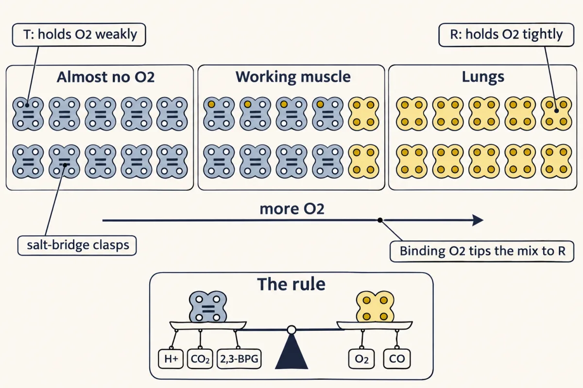
So binding gets easier as the molecule fills, and most of the gain comes late, as the mix tips to R (Going deeper, “Step by step”).
Predict. Suppose T and R held oxygen equally well. What shape would the curve have?
Check your prediction
A plain hyperbola. If both shapes bind equally, oxygen gives neither an advantage, so the mix never shifts, and the curve is just one single-site curve. The S needs two shapes that differ in affinity, plus oxygen that shifts the mix between them. In the model, setting the two affinities equal gives a Hill coefficient of exactly 1.
This is cooperativity. In the two-state picture no separate message has to pass from subunit to subunit: the four sites are coupled because they share one overall shape, carried across the contacts between the subunits, so whatever shifts the shape changes all four at once. That shared shape is how the subunits talk.
The Hill coefficient, nH, scores how steep the S is at half-saturation: it is the slope there of a Hill plot, log[Y/(1 − Y)] against log pO₂. For identical, independent sites it is 1, as for myoglobin. It can be no larger than the number of sites, so 4 for hemoglobin. Human hemoglobin’s is about 2.8: strong cooperation that stops short of the limit. It is a measure of steepness, not a count of anything.
Steepness alone does not make a good carrier. Where the steep part sits matters just as much, and P50 sets that. Hemoglobin’s P50 is about 26 torr, against myoglobin’s 2. Compare four curves between 100 and 20 torr:
| Carrier | P50 | nH | Oxygen delivered, 100 → 20 torr |
|---|---|---|---|
| Myoglobin | 2 torr | 1 | about 7% |
| A hyperbola with hemoglobin’s P50 | 26 torr | 1 | about 36% |
| A cooperative carrier with myoglobin’s P50 | 2 torr | 2.8 | almost none |
| Hemoglobin | 26 torr | 2.8 | about two-thirds |
The third row is the surprise: a steep curve in the wrong place delivers almost nothing, because it is still nearly full at 20 torr. Hemoglobin needs both: an S, and an S whose steep part sits between the lungs and the muscle.
In the sandbox below, which draws curves from the Hill equation, start on Normal HbA, then switch to Myoglobin. Watch the S flatten into a hyperbola and the delivered fraction fall from about two-thirds to about 7%. Then set the Hill coefficient back to 2.8 and drag P50 down to 2 torr to see the third row of the table, and drag the Hill coefficient to 1 at a P50 of 26 to see the second.
- Saturation at lungs (100 torr)
- —
- Saturation at working muscle (20 torr)
- —
- O₂ delivered (lungs − tissue)
- —
At nH 2.8 and P50 26, hemoglobin delivers about two-thirds of its O₂ between lungs (100 torr) and hard-working muscle (20 torr). Hit "Myoglobin" to see a non-cooperative, high-affinity carrier give up only ~7%.
Hemoglobin's S starts near the T-alone curve at low pO₂ and finishes on the R-alone curve: oxygen shifts the mix from T to R. (Dashed: single-site curves for a T site, half full at about 110 torr, and an R site, half full at about 0.37 torr, from the lesson's illustrative fit.)
The switch inside the molecule
Read the transcript of “The Switch Inside the Molecule”
Film transcript: The Switch Inside the Molecule
Why does the R shape hold oxygen better than T? The answer starts with a movement smaller than an atom. Hemoglobin is four chains, two α and two β. They pair up as two αβ dimers. Each chain cradles one heme. Heme is a ring, protoporphyrin IX, with an iron at its center. The iron makes four bonds to the ring's nitrogens. A fifth bond goes to a histidine below, the proximal histidine. The sixth site is saved for oxygen. Across the pocket sits the distal histidine, which hydrogen-bonds to bound oxygen. The iron must stay in its plus-two form. Oxidized, it becomes methemoglobin, which cannot carry oxygen at all. Step one. In T, the iron is high-spin and slightly too big for the ring's hole.
It sits about 0.4 Å below the ring, toward the histidine. That is about a seventh of the width of a water molecule. When oxygen binds, the iron turns low-spin, shrinks, and rises into the plane. Step two. The iron is bonded to the proximal histidine, so the histidine follows it. Step three. That histidine sits on a helix, the F helix. The helix shifts and presses on the neighboring subunit. Step four. In Max Perutz's classic account, this is levered into a rotation of one dimer against the other, about 15°. degrees. The salt bridges that held T break, and the molecule moves toward R. In T, those salt bridges resist this chain of movements. That is why heme in T binds oxygen weakly.
Later work shows the coupling is looser than a gear train. Each iron shift raises the odds of the switch rather than forcing it. That fits the two-shape picture. Each oxygen bound makes R more likely, not certain. Perutz saw the two end points by solving deoxy and oxy hemoglobin side by side. Do all four subunits switch together, or one at a time? Real hemoglobin fits neither textbook model perfectly. Its overall switch looks concerted. But each subunit's own shape also shifts as it binds. A tiny shift at one iron, levered through a helix, raises the odds that the whole molecule turns to R.
Why does R hold oxygen better than T? The answer starts with a movement smaller than an atom.
Hemoglobin is four chains, two α and two β, arranged as a pair of αβ dimers (α₂β₂). Each chain cradles one heme with a single iron atom. Heme is protoporphyrin IX with an iron(II) at its center. The iron makes four bonds to the porphyrin’s nitrogen atoms, holds a fifth bond to a histidine reaching up from the protein (the proximal histidine), and saves its sixth coordination site for oxygen. The iron must stay Fe²⁺; oxidize it to Fe³⁺ and you get methemoglobin, which cannot carry oxygen at all. Across the pocket sits the distal histidine, which hydrogen-bonds to bound oxygen and steadies it.
T and R are hemoglobin’s two classic quaternary states (arrangements of the four chains): T is the deoxy form, R the oxy form, and the molecule also visits other relaxed shapes. In Max Perutz’s classic account, four linked movements lead from T to R.
- The iron moves into the ring. In the deoxy T state the iron is high-spin and slightly too big to fit the porphyrin hole, so it sits about 0.4 Å out of the ring plane, on the side of the proximal histidine. That is about a seventh the width of a water molecule. (High-spin means some of the iron’s electrons sit in orbitals that point at the ring’s nitrogens, which makes the ion effectively bigger.) When oxygen binds on the other face, the iron flips to low-spin, pulls those electrons in, shrinks and moves into the plane.
- The histidine follows. The iron is bonded to the proximal histidine, so the histidine is pulled along.
- The helix shifts. The histidine sits on a helix, the F helix, and that helix presses against the neighboring subunit.
- The dimers rotate. The twitch at the iron is levered into roughly a 15° rotation of one αβ dimer against the other, and the salt bridges that held the T state break.
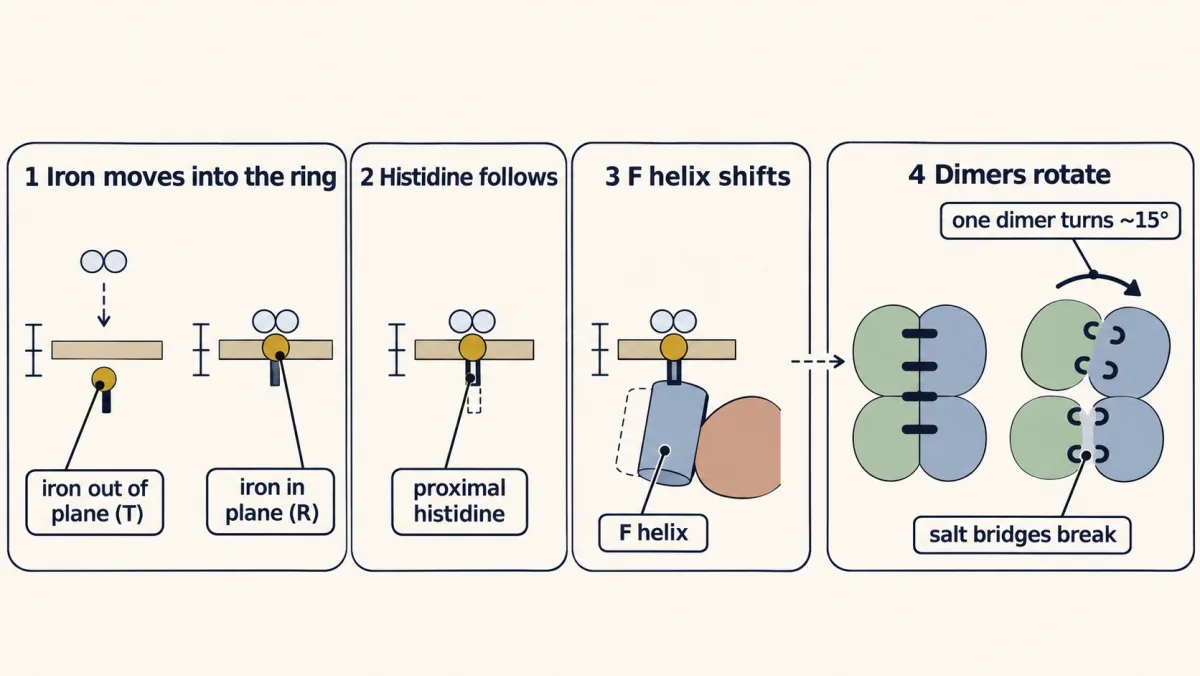
Read backwards, the same chain says why T is the weak binder: in T the salt bridges resist these movements, so binding oxygen is less favorable there. Structure talks to structure. A protein takes a sub-ångström event at one iron and broadcasts it across the whole molecule.
One caveat matters for the rule. Later work shows the coupling is looser than a gear train: each iron shift raises the odds of the switch rather than forcing it. That is just what the two-state picture needs, since each oxygen bound makes R more likely, not certain. Perutz saw the two end points by solving deoxy- and oxyhemoglobin side by side; open 2HHB and 1HHO in the structures panel to compare them.
Whether the four subunits switch all together (the concerted model) or one at a time (the sequential model) is a long-running question, and real hemoglobin fits neither perfectly.
The simulator below runs the concerted, two-state picture with round, illustrative settings, not the hemoglobin fit above, so read its pattern rather than its numbers. Raise the O₂ concentration and watch the population tip from T to R; then add an inhibitor (BPG or low pH) and notice that it takes more oxygen to make the same tip.
fraction R × how full R is + fraction T × how full T is
Cooperativity needs no new physics, just a population. Each O₂ bound multiplies the odds of R by the same factor, how much more tightly R holds O₂ than T does (100 in this simulator, about 300 in the lesson's hemoglobin fit), so the more O₂, the more R, the more easily the next O₂ binds. An inhibitor like 2,3-BPG stabilizes T and shifts the whole curve right (releasing more O₂ to hungry tissue); an activator stabilizes R and shifts it left. The single-subunit reference, myoglobin, has no T⇌R conversation at all, so its curve is a plain hyperbola.
Tuning the mix: allosteric regulators
Read the transcript of “Tuning the Mix”
Film transcript: Tuning the Mix
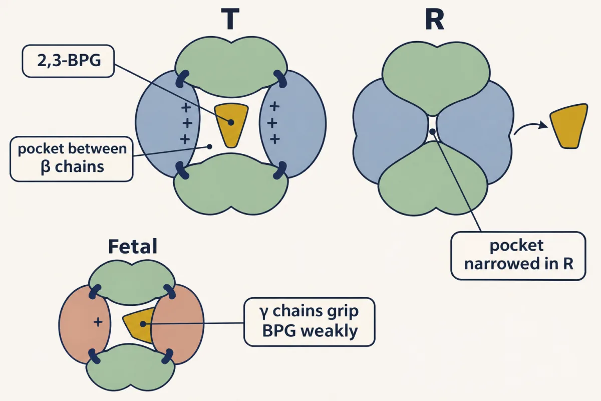
Working muscle needs more oxygen. How does the blood know to let go of more there? Use the rule. Anything that binds one shape better shifts the mix toward that shape. Three regulators prefer T: acid carbon dioxide and a small molecule called BPG. Its full name is 2,3-bisphosphoglycerate. It is a small anion with about four to five negative charges. Red cells hold nearly one BPG for every hemoglobin. It wedges into a central pocket between the β chains. That pocket is wide open only in T. Predict. Add BPG to hemoglobin. Which way does P50 move?
BPG holds the molecule in T, like a doorstop. It takes more oxygen to tip the mix, so the curve shifts right. Acid works through the Bohr effect. Protons add to certain groups, chiefly a histidine near the end of each β chain. Those groups can then form salt bridges that steady T. So low pH lowers oxygen affinity. Working muscle makes acid. The acid tips hemoglobin toward T exactly where a straining muscle needs the oxygen. Carbon dioxide binds the ends of hemoglobin's chains as carbamate. Those groups form their own salt bridges that favor T. In exercising muscle, three fast signals arrive at once: falling oxygen, falling pH, and rising carbon dioxide.
Does a right shift spoil loading in the lungs? Acid and carbon dioxide are high in tissue, not in the lungs. So their shift happens where unloading happens. BPG shifts the curve everywhere. But the lung end sits on the flat top of the curve, so a modest shift costs little loading. A very large shift would start to cost loading, so more is not always better. BPG is the slower dial. It rises over hours to days at high altitude, part of how you acclimatize. Fetal hemoglobin has two γ chains in place of the two β chains. At position 143, it has a serine instead of a histidine. So it binds BPG weakly and stays at higher oxygen affinity than the mother's hemoglobin.
That lets fetal blood pull oxygen across the placenta from the mother's blood. Acid, carbon dioxide and BPG all favor T. So they shift the mix toward T where oxygen must let go.
Cooperativity sets the baseline. The rule then predicts what any other molecule will do. If it binds T better than R, it shifts the mix toward T, so it takes more oxygen to tip the population to R and the curve moves right, toward higher pO₂ on the x-axis: a higher P50 and lower affinity. Three regulators work this way.
2,3-bisphosphoglycerate (2,3-BPG) is a small, ferociously charged anion (around four to five negative charges), present in red cells at nearly 1:1 with hemoglobin. It wedges into a central pocket between the β chains that is wide open only in the T state (relaxed forms narrow it and bind BPG far more weakly).
Predict. Add 2,3-BPG to purified hemoglobin. Which way does P50 move?
Check your prediction
Up: the curve shifts right. BPG binds T and acts as a mechanical doorstop that holds the molecule tense, so more oxygen is needed to tip the mix to R. At a given tissue pO₂, more of the oxygen is let go.
BPG is the slower dial, compared with the fast H⁺ and CO₂ signals below. It rises in your red cells over hours to days at high altitude, which is part of how you acclimatize, and its response to exercise varies.
Hydrogen ions act through the Bohr effect. Protonating certain groups on hemoglobin, chiefly a histidine near the end of each β chain, lets them form salt bridges that steady the T state, so low pH lowers oxygen affinity. Lung blood runs slightly alkaline (pH ~7.4) and favors loading; respiring tissue is more acidic (venous blood ~7.36, lower in working muscle) and favors unloading. At rest that gap is small. The effect is large where it counts: working muscle makes acid, and the acid tips hemoglobin toward tense exactly where a straining muscle needs the oxygen.
Carbon dioxide binds as carbamate groups (CO₂ bonded to an amino group) on hemoglobin’s N-termini, which form their own T-stabilizing salt bridges. Carbonic anhydrase, the enzyme that turns most CO₂ into bicarbonate, also supplies the Bohr protons.
During exercise, three fast signals arrive at once in working muscle: falling pO₂, falling pH and rising CO₂.
Does a right shift spoil loading in the lungs? H⁺ and CO₂ are high in tissue, not in the lungs, so their shift happens where unloading happens. BPG shifts the curve everywhere, but the lung end sits on the flat top of the curve, so a modest shift costs little loading. A very large shift would start to cost loading (Going deeper, “How big a shift helps”).
Fetal hemoglobin runs the rule the other way. Fetal hemoglobin (α₂γ₂) has a serine where the adult β chain has a histidine at position 143, so it binds 2,3-BPG weakly. With less BPG holding it in T, it stays at higher oxygen affinity and pulls oxygen across the placenta from the mother’s blood.
Evolution can also tune the shapes themselves. The bar-headed goose, which flies over the Himalayas, carries a hemoglobin with higher oxygen affinity that loads oxygen better from thin air.
When the shapes betray you: hemoglobin pathology
Read the transcript of “When the Shapes Betray You”
Film transcript: When the Shapes Betray You
What happens when something holds hemoglobin in the wrong shape, or makes one shape dangerous? Carbon monoxide binds the heme about 250 times more tightly than oxygen does. Like oxygen, it favors R. So it shifts the mix toward R. Predict. One subunit carries carbon monoxide. Is the oxygen on the other three held more or less tightly?
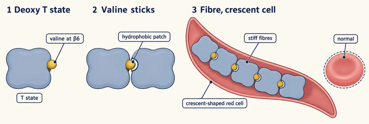
More tightly. The molecule is held in R, so its oxygen is released far less readily. The curve shifts left. So carbon monoxide takes sites away from oxygen, and it makes the oxygen still aboard stay put. Carbon monoxide hemoglobin is bright red. But the classic cherry-red skin is rarely seen in living patients. A pulse oximeter cannot tell it from oxygen hemoglobin. It reads falsely normal while tissues suffocate. The fix is high-concentration oxygen, to compete the carbon monoxide back off. Sickle-cell disease is a different failure. Here the T shape itself turns sticky. One amino acid changes. Glutamate becomes valine at position 6 of the β chain.
That valine sits on the surface in every state. But in T, a neighbor's hydrophobic pocket opens, and the valine plugs in. That neighbor sticks to the next. The molecules polymerize into stiff fibers that deform the red cell into a crescent. So the trigger is losing oxygen. Exertion raises oxygen use. Cold slows blood through small vessels. Both leave more hemoglobin in T for longer. Dehydration concentrates sickle hemoglobin, so fibers form faster. Carriers with one copy are usually healthy. They are partly protected against severe falciparum malaria. One newer treatment uses gene editing to switch fetal hemoglobin back on. Fetal hemoglobin does not join the sickle polymer.
It dilutes the sickle hemoglobin around it, so cells sickle far less. Almost everything in this story follows one rule. Hemoglobin is a mix of a weak shape and a tight shape. Oxygen shifting that mix is how blood delivers. Locking the mix, or making T sticky, is how it fails.
The rule explains one failure and sets up the other: in CO poisoning the mix is locked toward R, and in sickle-cell disease the rule’s T shape is the one that turns dangerous.
Carbon monoxide is the R-state lock. It binds the heme about 250-fold more tightly than oxygen does, and like oxygen it favors R.
Predict. One subunit of a hemoglobin carries CO, and the other three carry oxygen. Is the oxygen on those three held more or less tightly than usual?
Check your prediction
More tightly. CO holds hemoglobin in the high-affinity R state, so the oxygen still aboard the other subunits is released far less readily, and the curve shifts left. That is double damage: fewer sites carry oxygen, and the oxygen that is carried stays put in the tissues.
CO-hemoglobin is bright red, though the classic cherry-red skin is rarely seen in living patients. A pulse oximeter, which cannot tell O₂-hemoglobin from CO-hemoglobin, reads falsely normal while tissues suffocate. The fix is to flood the system with high-concentration oxygen to compete the CO back off; hyperbaric oxygen is reserved for selected severe cases.
Sickle-cell disease is the textbook molecular disease, and here the T shape itself turns sticky. One amino acid. Glutamate to valine, at position 6 of the β chain. That valine sits on the surface in every state, but the hydrophobic pocket it docks into (Phe85, Leu88) on a neighboring hemoglobin is open only in the deoxy T shape. So in T, the valine of one molecule plugs into the pocket of the next, which plugs into the next. The molecules polymerize into stiff fibers that deform the red cell into a crescent.
So the trigger is deoxygenation. Crises come with exertion, which raises oxygen use, and with cold, which slows blood through small vessels. Exertion and cold leave more hemoglobin in T for longer. Dehydration concentrates HbS, so fibers form faster. Carriers (one copy) are usually healthy and are partly protected against severe falciparum malaria, which is why the mutation persists.
One of the newest treatments, Casgevy, uses CRISPR to switch fetal hemoglobin back on in red-cell precursors. HbF does not join the sickle polymer and dilutes the HbS around it, so cells sickle far less.
The thalassemias fail differently again: the problem is mostly not a broken chain but the wrong amounts of chain.
That is the lesson in one line. Hemoglobin is a mix of a weak shape and a tight shape, and oxygen shifting that mix is why blood loads in the lungs and gives up about two-thirds of its oxygen in hard-working muscle. A 0.4 Å iron twitch, levered into a ~15° dimer rotation, makes the switch; acid, CO₂ and BPG tune the mix; CO locks it; and a single wrong amino acid makes the T shape stick. Sickle-cell anemia is what happens when structure talks, and structure lies.
Going deeper
The main line above does not depend on these details.
The two-state fit used in this lesson
The numbers marked “one illustrative fit” come from fitting a two-state (concerted) model to human hemoglobin’s curve: P50 = 26 torr, a Hill coefficient of 2.8 at P50, and 97.5% full at 100 torr. One fit puts an R site half full at about 0.37 torr and a T site half full at about 110 torr, with T outnumbering R about 1.7 × 10⁷ to one when no oxygen is bound. Fitted two-state parameters are not unique, so other fits give other numbers with the same pattern.
The picture has limits. Real hemoglobin also changes shape within each quaternary state (tertiary T and R), so its Hill coefficient of about 2.8 is not a clean two-state parameter.
One trap: the rule averages how full each shape is, not their Kd values. Averaging the two shapes’ half-full pressures by population at P50 gives about 67 torr, far from the true 26.
Step by step: the measured binding constants
The old summary, “the second oxygen comes a little easier, the third easier still, the fourth easiest of all”, is right as a trend, but measured step-by-step (Adair) constants do not rise evenly: the last oxygen binds on the order of a hundred times more tightly than the first.
How big a shift helps
In the two-state fit, tripling T’s advantage, as a T-favoring effector would, moves P50 from 26 to about 35 torr and raises delivery between 100 and 20 torr from about 64% to about 71%. A tenfold increase moves P50 to about 48 torr, and delivery falls back from 71% to about 66%, because the curve no longer fills fully at 100 torr. A tenfold shift toward R moves P50 to about 14 torr and cuts delivery to about 24%, the direction CO pushes. (For normal hemoglobin the two-state fit gives about 64% and the Hill curve used in the opening and the sandbox about 65%, only because they are different idealized curves fitted to the same P50 and steepness; the lesson rounds both to two-thirds.) The sandbox’s BPG preset uses the Hill equation with the Hill coefficient held at 2.8, so it shows a somewhat larger gain than the two-state model.
Inside the red blood cell
The hemoglobin in this lesson does its work packed inside red blood cells, which also hold 2,3-BPG at nearly 1:1 with it. About 5 million red blood cells sit in a microliter of your blood.
Pressure bookkeeping
Where does the lesson’s lung value of about 100 torr come from? Dry sea-level air is 21% oxygen, so about 159 torr of the 760, and by the time air reaches the alveoli, humidified and mixed with CO₂, pO₂ has fallen to roughly 100 torr.
How picky binding gets
A receptor earns the name by binding its specific ligand far more tightly than molecules that look almost right, and Kd is how that pickiness is scored. Inside the cell, the estrogen receptor binds estradiol far more tightly than testosterone, a steroid with the same four-ring core that differs in only a few chemical groups.
You will mostly interpret Kd values, not compute them. Three ranges are worth memorizing. Enzymes often hold their metabolites loosely, in the micromolar to millimolar range, which suits molecules that must be released after each reaction. Hormones and their receptors bind in the nanomolar range. Antibodies, refined by your immune system, can reach picomolar, and that refinement is not metaphorical: booster shots can drive further rounds of selection for B cells that make lower-Kd antibodies, so antibody binding often gets stickier over time.
Myoglobin in the clinic
Myoglobin, the single-site example from earlier in this lesson, is the oxygen-storage protein of muscle. When myoglobin leaks into the blood after a heart attack or crush injury, clinicians read that escaped storage protein as a marker of muscle damage.
Magnetic hemoglobin and functional MRI
The iron switch has a clinical echo. Deoxyhemoglobin is paramagnetic (its iron carries unpaired electrons) and oxyhemoglobin is diamagnetic. Functional MRI uses that difference: active brain regions pull in more oxygenated blood, which lowers the local amount of paramagnetic deoxyhemoglobin, and the magnetic signal shifts. The same change in the iron’s electrons that lets it slide into the ring is what the scanner picks up, summed over vast numbers of molecules.
CO and the distal histidine
Oxygen binds bent, carbon monoxide prefers to bind straight, and the distal histidine favors oxygen, mostly by hydrogen-bonding to it, cutting CO’s advantage roughly 100-fold. That discrimination is a big part of why we tolerate the trace CO our own cells produce. CO’s 250-fold edge in hemoglobin is what is left after that cut; on bare heme the gap is far larger.
Concerted or sequential?
There are two textbook models for how four subunits switch. In the concerted (MWC) model all four flip together, and ligand binding only nudges the T↔R balance. In the sequential (KNF) model each subunit changes one at a time. Hemoglobin’s quaternary switch looks concerted, but each subunit’s own shape also shifts as it binds, and how the two levels combine is still studied.
Which groups carry the Bohr effect
Protonating histidine β146 (favored at low pH) lets it form a salt bridge with aspartate β94 that steadies the T state; this is the biggest single part of the Bohr effect, and other groups, such as the α-chain N-termini, add the rest.
How CO₂ travels
Carbon dioxide travels three ways: under a tenth dissolved, about 70% as bicarbonate (made by carbonic anhydrase), and the rest as carbamate groups bound to hemoglobin’s N-termini. Together with more blood flow to working muscle, the three exercise signals help push whole-body oxygen extraction from about 25% at rest to about 70% in heavy exercise.
The thalassemias
The thalassemias are the quantitative cousins of sickle-cell disease, and they can be just as severe. Too few β chains and the leftover α chains precipitate and wreck the cell; too few α chains and the surplus β chains assemble into β₄ tetramers (HbH; before birth, surplus γ chains form γ₄, Hb Bart’s) that bind oxygen tightly but with no cooperativity, a delivery truck that forgot how to open its doors. Matched α-to-β synthesis matters because imbalance is toxic either way.
How the sickle-cell gene edit works
Casgevy, approved in late 2023, uses CRISPR to disrupt the erythroid enhancer of BCL11A, a repressor that silences the fetal γ-globin genes after birth. Less BCL11A in red-cell precursors switches fetal hemoglobin back on.
Your notes
Select any text in the lesson to highlight it or add a note to it. Your highlights and notes are saved in this browser and collected in your notebook.
This browser is not letting the site save anything (a private window or strict privacy settings), so highlights and notes will be lost when you leave the page.
How we measure it
The oxygen-binding (saturation) curve
Fractional saturation Y plotted against the partial pressure of O₂. A hyperbola signals independent, non-interacting sites (myoglobin has just one); an S-shaped sigmoid signals cooperative sites whose affinity rises as the molecule fills (hemoglobin). P50, the pO₂ at half-saturation, is read straight off the x-axis.
The Hill coefficient (n_H)
A number for cooperativity, taken from the slope of a linearized binding plot: n_H = 1 means identical, independent sites, n_H > 1 means positive cooperativity, n_H < 1 means negative cooperativity or a mix of sites with different affinities. Hemoglobin's n_H ≈ 2.8 (out of a maximum of 4). The Hill equation is an empirical curve fit; the two-state picture explains the cooperativity it measures.
X-ray crystallography of T and R states
Solving deoxy- and oxyhemoglobin side by side reveals the tense (T, low-affinity) and relaxed (R, high-affinity) quaternary states and the sub-ångström iron movement that triggers the switch. The classic Perutz structures made allostery a thing you could see.
Protein electrophoresis
Separating proteins by charge in an electric field. Pauling used it in 1949 to show that sickle-cell and normal hemoglobin migrate differently, pinning a human disease to a charge change in one molecule.
BOLD / functional MRI
Imaging that reads the magnetic difference between paramagnetic deoxyhemoglobin and diamagnetic oxyhemoglobin. Active brain regions draw more oxygenated blood, changing the signal — the same iron chemistry that carries oxygen also lets a scanner track blood oxygenation, an indirect readout of brain activity.
Practice questions on this lesson (8) → MCAT-style, original, each with an explanation
Where this lesson fits
Before thisReview first
- Protein Three-Dimensional Structure: The Sequence Sets the ShapeThis course · Lesson 5
You need how a sequence folds into a shape, and how one residue change alters it, to follow the T and R shapes.
- Water, Weak Bonds, and the Generation of OrderThis course · Lesson 3
The salt bridges that hold the T shape together are weak bonds, so you need their strength and reversibility from there.
- Amino Acids: How One Side Chain Decides EverythingThis course · Lesson 4
Histidine's charge and pKa from there explain why protons stabilize the T shape in the Bohr effect.
After thisStudy next
- Oxidative Phosphorylation: The Electron-Transport ChainThis course · Lesson 20
It uses the oxygen that hemoglobin delivers, as the final electron acceptor of the respiratory chain.
- Signal-Transduction Pathways: How Cells Compute With Molecular SwitchesThis course · Lesson 13
It reuses the idea that a ligand shifts a protein's balance between active and inactive shapes.
- Integration of Metabolism: How the Body Switches FuelsThis course · Lesson 30
It follows how tissues' oxygen and energy demands change with fed and fasting states.
Elsewhere on the siteFilms, simulations and method films in other courses
- Why Blood Needs an S-Shaped CarrierFilm · Organismal Biology
It shows why plasma alone carries little oxygen and hemoglobin's S-shaped curve raises the load and release.
- Osmosis chamberSimulation · Biology
Its simulation is not about this idea, so it should be skipped.
- Dose–response explorerSimulation · Cell Signaling
It lets you explore occupancy against ligand concentration, the same Kd and half-saturation idea.
Suggested by an AI model from the lessons themselves, then checked. How we use AI
Finish line
Test yourself
8 questions
Practice and MCAT-style, with explained answers.
Next · Lesson 10
Carbohydrates: One Bond Decides Fuel or Fiber
Closing the glucose ring makes one special carbon, and the direction of the bond it forms decides whether a polymer feeds you or holds a plant up.
Continue →