Biochemistry · Glucose Homeostasis
Gluconeogenesis: Pushing Glucose Back Uphill
Rewritten on : new text, figures and flashcards. Your notes and highlights stay, but the text around them may have moved.
When food runs out, your liver (with help from the kidney) builds the glucose your brain and red blood cells still need, and it does it with four new enzymes, three compartments, and real ATP and GTP spent to make the climb.
Pause when the reading reaches a film, and offer to play it. Saved on this device.
Watch the film now, or keep listening
Builds on:Glycolysis: The Investment-and-Payoff PathwayMetabolism: Basic Concepts and DesignBasic Concepts of Enzyme ActionWhy these?
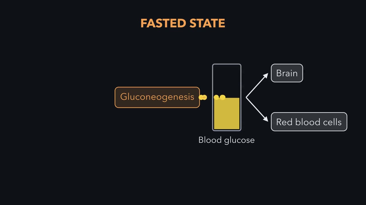 ▶ Watch Not Glycolysis in Reverse · 3:05
Read the transcript of “Not Glycolysis in Reverse”
Film transcript: Not Glycolysis in Reverse
Think of your body as running two completely different supply chains depending on whether you have just eaten. In the fed state, blood glucose is abundant, so cells simply burn it. Glycolysis feeds the Krebs cycle, which makes ATP. But hours after a meal, glucose runs scarce, and the brain and red blood cells still demand it constantly. Now the body switches to gluconeogenesis, manufacturing glucose from non-sugar raw materials. Three streams feed that factory. Lactate from muscle and red cells, alanine and glutamine from muscle, and glycerol from fat breakdown. Hold onto this fed versus fasted framing because it explains every regulatory decision we make today.
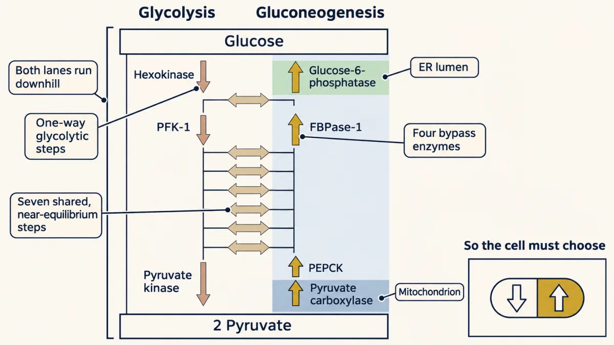
Most of glycolysis is freely reversible, and gluconeogenesis simply runs those steps backward. The problem lives in exactly three places where glycolysis releases so much free energy that the reaction is effectively one-way. Look at the numbers—hexokinase, phosphofructokinase, and pyruvate kinase—each have large negative delta ΔG values, so reversing them directly is impossible. Gluconeogenesis solves this with four dedicated bypass enzymes, shown in orange, glucose 6 -phosphatase, fructose 1, 6 -bisphosphatase, and the pyruvate carboxylase, plus PEP carboxykinase pair. Notice that pyruvate to PEP is split into two reactions, whose combined energetics make the climb feasible.
Same map, different roads around the cliffs. Here is a logistics problem the cell must solve. Pyruvate carboxylase lives inside the mitochondrial matrix, so oxaloacetate is made there. But the next enzyme, PEP carboxykinase, works in the cytoplasm in this route, and the membrane has no oxaloacetate transporter. The workaround is elegant. Reduce oxaloacetate to malate, which can cross, then reoxidize malate back to oxaloacetate on the other side. From pyruvate or alanine, that second oxidation also regenerates cytoplasmic NADH, which gluconeogenesis needs later. So one shuttle accomplishes two jobs at once, moving carbon out and delivering reducing power exactly where the downstream reactions will consume it.
Why does gluconeogenesis need its own enzymes instead of just reversing glycolysis? Think of it as a bargain the cell strikes with thermodynamics. Glycolysis releases energy, about 85 kilojoules per mole, so it is downhill and spontaneous. Gluconeogenesis consumes energy, and that extra cost, the six nucleotide triphosphate equivalents, is precisely what tips the synthetic direction to also be favorable. That means both pathways could run at once in the same cell, a futile cycle that burns ATP and dumps it as heat with no net product. Regulation, not the energy cost, is what holds that in check.
Skip breakfast and lunch, and by the early afternoon your liver is building much of your glucose out of spare parts. It has to. Your brain runs almost entirely on glucose until a fast lasts several days and ketones take over part of the load, and your red blood cells, which have no mitochondria, run on nothing else. Glycogen breakdown supplies most of the glucose in the first hours of a fast, with gluconeogenesis already contributing; gluconeogenesis rises as glycogen falls, and the two share the load until glycogen runs low within a day or two. After that, blood sugar stays in its narrow lane (about 70–100 mg/dL fasting) only because the liver, with help from the kidney cortex, is manufacturing fresh glucose. (When the film at the top says the body “switches”, it means this gradual handover.) Fasting is not running out of energy but switching fuels: gluconeogenesis supplies the glucose-dependent tissues while most others switch to fat and ketones.
On a map, gluconeogenesis looks like glycolysis run backward. That raises two puzzles. If glycolysis runs downhill, its steps run backward should run uphill, so how does the liver make glucose from pyruvate at all? And once it can, what stops a liver cell from making glucose and burning it at the same time?
Where the carbons come from
There are three precursors to know. Lactate comes back from hard-working muscle and red cells; lactate dehydrogenase converts it to pyruvate. Glucogenic amino acids, alanine chief among them, plus glutamine, the kidney’s main gluconeogenic amino acid, lose their nitrogen and enter at pyruvate or at citric acid cycle intermediates that become oxaloacetate. Glycerol, released when fat is mobilized, enters as dihydroxyacetone phosphate (DHAP) by way of glycerol kinase and glycerol-3-phosphate dehydrogenase.
Notice what is missing: the fatty acids that make up most of fat. Even-chain fatty acids cannot become glucose. This trips up almost everyone; the reason comes after Bypass 1.
Three cliffs: why the pathway can’t just run backward
An enzyme speeds a reaction both ways; it does not pick a direction. ΔG, the free-energy change under the conditions inside the cell, does. When ΔG is close to zero, the step is near equilibrium: a two- or threefold change in concentrations tips it either way, so one enzyme can serve both pathways. When ΔG is large and negative, the step is far from equilibrium and in practice runs one way only. That is all “irreversible” means here.
Seven of glycolysis’s ten steps are near equilibrium, and gluconeogenesis shares those seven enzymes. Three are far from it. These are classic estimates of their ΔG inside a cell (calculated from metabolite levels measured in red blood cells; textbooks differ by a few kJ/mol):
| Glycolytic step | What it does | ΔG in the cell |
|---|---|---|
| Hexokinase | ATP’s phosphate onto glucose | about −33 kJ/mol |
| Phosphofructokinase-1 (PFK-1) | ATP’s phosphate onto fructose-6-phosphate | about −22 kJ/mol |
| Pyruvate kinase | PEP’s phosphate onto ADP, making ATP | about −17 kJ/mol |
Predict. Could the cell run pyruvate kinase backward just by piling up pyruvate and ATP?
Check your prediction
Only in principle. At body temperature, every 6 kJ/mol or so of ΔG is a tenfold change in the ratio of products to reactants. Just to bring pyruvate kinase to equilibrium the cell would have to shift that ratio about 650-fold, and for hexokinase about 400,000-fold; running either one backward would take more still. No cell can hold its ATP, ADP and sugar phosphates that far from normal.
So at each cliff, gluconeogenesis uses a different enzyme:
- Where glycolysis put a phosphate on with ATP (hexokinase, called glucokinase in liver, and PFK-1), gluconeogenesis does not hand the phosphate back to ADP; it cuts it off with water. These hydrolyses, by glucose-6-phosphatase and fructose-1,6-bisphosphatase (FBPase-1), are downhill (about −14 and −16 kJ/mol; these are standard values, unlike the table’s in-cell ones).
- Where glycolysis made ATP (pyruvate kinase), there is no cheap trick: even with one ATP spent, turning pyruvate back into phosphoenolpyruvate (PEP) is uphill by about 31 kJ/mol under standard conditions (about 17 in the cell, the table’s number with its sign flipped). The cell spends two high-energy phosphates, an ATP at pyruvate carboxylase and a GTP at PEP carboxykinase (PEPCK). The second phosphate is worth about 30.5, so with both the step is about even under standard conditions (+31 − 30.5 ≈ +1) and comfortably downhill in the liver.
The one idea to carry through this lesson. Every step has to run downhill in the direction it is used, the near-equilibrium ones only slightly. Gluconeogenesis keeps glycolysis’s seven near-equilibrium steps and swaps each of the three one-way steps for a different reaction that runs downhill toward glucose. With both pathways downhill, thermodynamics no longer picks the direction; the cell has to choose, by regulation.
This holds in the liver and kidney cortex, which carry all four bypass enzymes.
Bypass 1: pyruvate to PEP, across two compartments
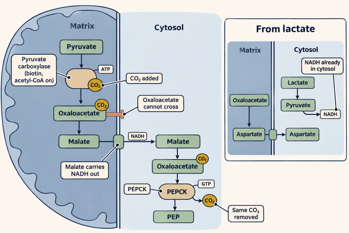
Inside the mitochondrion, pyruvate carboxylase adds CO₂ to pyruvate to make oxaloacetate, spending one ATP. It uses biotin, a vitamin that carries the CO₂ (see Going deeper), and is switched on by acetyl-CoA, the cell’s way of saying plenty of fuel here, make glucose rather than burn more pyruvate.
Next, a shipping problem. Phosphoenolpyruvate carboxykinase (PEPCK), the next enzyme, works in the cytosol in the route followed here (human liver also has a mitochondrial form), and oxaloacetate has no transporter across the inner mitochondrial membrane. The fix is the malate shuttle: oxaloacetate is reduced to malate, malate rides out on the malate–α-ketoglutarate carrier, and in the cytosol it is oxidized back to oxaloacetate.
The trip also moves reducing power, and where the NADH must come from decides whether oxaloacetate leaves as malate or as aspartate. Later, GAPDH runs in reverse and needs a cytosolic NADH for each three-carbon unit. From pyruvate or alanine, that NADH must come out of the mitochondrion, and malate brings it. From lactate, lactate dehydrogenase already made the NADH in the cytosol, so oxaloacetate can leave as aspartate instead.
Back in the cytosol, PEPCK spends a GTP and removes the very CO₂ that pyruvate carboxylase added, producing phosphoenolpyruvate.
Predict. Why would a cell add a CO₂ only to remove it one step later?
Check your prediction
PEP needs about two high-energy phosphates’ worth of energy, and in animals no single enzyme turns pyruvate into PEP. The CO₂ is the handle that lets two enzymes, in two compartments, share the job. Pyruvate carboxylase spends ATP to attach it, storing energy in oxaloacetate. PEPCK pulls the same CO₂ off; that decarboxylation is downhill, and with a GTP it pays for PEP. That is what “CO₂ as a thermodynamic lever” means.
Why fat can’t become sugar
Every phosphoenolpyruvate (PEP) made from pyruvate uses up one oxaloacetate, so gluconeogenesis from anything but glycerol (which enters at dihydroxyacetone phosphate (DHAP), above PEP) needs a net source of oxaloacetate or of something that becomes it, such as pyruvate or citric acid cycle intermediates. Even-chain fatty acids supply none. Their carbons enter the citric acid cycle as acetyl-CoA, which joins one oxaloacetate to get in; over a turn, two carbons leave as CO₂ and exactly one oxaloacetate comes back. Net oxaloacetate gain is zero, so net glucose from fatty acids is zero. (Carbon label from a fatty acid can still turn up in glucose, because the cycle shuffles atoms; see the isotope-tracing technique above.)
Why not turn acetyl-CoA back into pyruvate? Pyruvate dehydrogenase releases CO₂ in a step so far downhill that it runs one way only (next chapter). Plants and bacteria have a way around: two glyoxylate-cycle enzymes let acetyl-CoA skip the cycle’s two CO₂-releasing steps, giving them a net gain of oxaloacetate. Humans and other vertebrates lack them. The one exception is odd-chain fatty acids, whose final three-carbon propionyl-CoA can be routed through succinyl-CoA to oxaloacetate. Of the fat you actually carry, only the glycerol backbone can become sugar.
Bypasses 2 and 3, and why muscle can’t share
From phosphoenolpyruvate (PEP), the shared enzymes run in reverse up to fructose-1,6-bisphosphate. There FBPase-1 hydrolyzes off a phosphate, a clean snip past the phosphofructokinase-1 (PFK-1) cliff. It is a major regulated step, inhibited by AMP and by fructose-2,6-bisphosphate and activated by citrate.
Predict. FBPase-1 makes no ATP and spends none. How can it run downhill, and what did it cost?
Check your prediction
Cutting the phosphate off with water is downhill on its own (the −16 above). Handing it back to ADP, the reverse of PFK-1, would be uphill by about 14, since PFK-1 is downhill by about 14 under standard conditions. The gap, about 30 kJ/mol, is one ATP’s worth: the ATP that PFK-1 spent, which gluconeogenesis never gets back. The cost is a refund given up, not a payment.
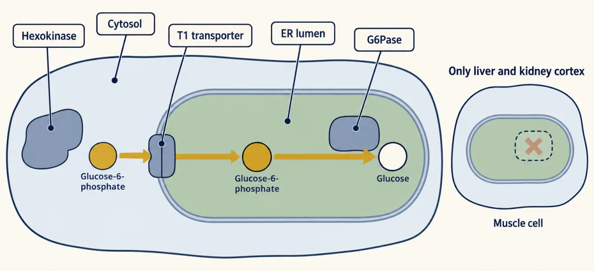
The last cliff, glucose-6-phosphatase, works the same way, and it hides in a revealing place: the lumen of the endoplasmic reticulum. Glucose-6-phosphate is carried in by the T1 transporter to be dephosphorylated (why there is unsettled; Going deeper). Only liver, kidney cortex and, to a lesser degree, the small intestine express the glucose-releasing form, G6Pase-α. Muscle does not, so it cannot send its glycogen into the blood as glucose; it uses that glycogen locally and, in hard work, ships out the lactate it makes faster than it can use.
The bill
Count what one glucose costs from two pyruvates. Every step from pyruvate up to the trioses happens twice, once for each three-carbon half.
Predict. Before you open the map, count: how many high-energy phosphates and how many NADH does one glucose cost?
Check your prediction
- Pyruvate carboxylase: 1 ATP per pyruvate, so 2.
- PEPCK: 1 GTP per pyruvate, so 2.
- Phosphoglycerate kinase, a shared step run in reverse: 1 ATP per pyruvate, so 2.
- GAPDH, run in reverse: 1 NADH per pyruvate, so 2.
- FBPase-1 and glucose-6-phosphatase: nothing.
That is 4 ATP + 2 GTP + 2 NADH per glucose. Two of the six high-energy phosphates are spent at a shared step, which a count of only the bypass enzymes misses.
Check your count: tap each step to reveal it and watch the tally.
regulated step − used up
Mitochondrial. A biotin cofactor on a flexible lysine tether, carried on a mobile domain, ferries the added CO₂. The enzyme is strongly activated by acetyl-CoA — the cell's signal that fuel is plentiful and it should build glucose, not burn more pyruvate.
Regulation. Allosterically activated by acetyl-CoA; this is the anaplerotic gate into gluconeogenesis.
Clinical. Pyruvate carboxylase deficiency causes lactic acidosis (pyruvate shunted to lactate); the severe newborn form often adds hypoglycemia (no OAA to start) and high ammonia.
OAA can't cross the inner membrane, so it is reduced to malate, ferried out on the malate–α-ketoglutarate carrier, and re-oxidized to OAA in the cytosol. With pyruvate or alanine precursors this relocation also supplies the NADH GAPDH will need; with lactate, LDH already made that NADH, so OAA can instead leave as aspartate, which is transaminated back to OAA in the cytosol. The reducing power was spent in the matrix making malate, so it is moved, not minted — no free NADH credit here.
Cytosolic. Removes the very CO₂ pyruvate carboxylase just added. That carboxylation-then-decarboxylation pair is a thermodynamic lever that makes forming high-energy PEP favorable. Spends a GTP, tracked here as a high-energy phosphate.
Regulation. Set mainly by how much enzyme the cell makes (insulin and glucagon control it over hours); its level alone controls surprisingly little of the flux.
Clinical. In insulin resistance, insulin no longer holds hepatic glucose output down, largely because more substrate arrives from fat; the excess gluconeogenesis feeds fasting hyperglycemia.
A near-equilibrium glycolytic enzyme run in reverse, a hydration this direction. No new enzyme needed.
Shared and reversible. Moves the phosphate from C2 back to C3.
Runs the substrate-level step in reverse, so here it COSTS an ATP rather than making one. This is one of the places gluconeogenesis pays its energy bill: a shared step, so counting only the bypass enzymes misses it.
Consumes the cytosolic NADH, reducing the acyl-phosphate back to an aldehyde. This is the reductive step the malate shuttle was feeding.
Two trioses condense back into one six-carbon sugar, the mirror of the aldolase cleavage in glycolysis. Glycerol enters here, as DHAP.
A simple hydrolysis that lops off the C1 phosphate, bypassing irreversible PFK-1. This is a major regulated step: inhibited by AMP and by fructose-2,6-bisphosphate, activated by citrate.
Regulation. Fructose-2,6-bisphosphate is the key inhibitor at this pair — high when fed, low when fasting.
Clinical. FBPase-1 deficiency causes fasting hypoglycemia and lactic acidosis in infants, especially when sick.
Shared and reversible. Ketose back to aldose.
Hydrolysis in the ER lumen; the T1 transporter carries G6P in. Only liver, kidney cortex and small intestine express this form (G6Pase-α), which is why muscle cannot export its glycogen as blood glucose.
Regulation. The glucose pair is set by glucokinase's regulatory protein, which pulls glucokinase into the nucleus when glucose is low, and by enzyme amounts over hours.
Clinical. Von Gierke disease (glycogen storage disease type I): loss of G6Pase or its transporter gives severe fasting hypoglycemia, lactic acidosis, and a glycogen-laden liver.
4 ATP + 2 GTP + 2 NADH per glucose. Glycolysis returns only 2 ATP, so the extra 4 high-energy phosphates are what make the climb run downhill toward glucose. With both pathways downhill, the cell has to choose by regulation, not thermodynamics.
Glycolysis nets only 2 ATP, so building a glucose costs four more high-energy phosphates than breaking one down returns. (The film calls the six “the extra cost”; six is the total, and four is the extra beyond what glycolysis returns.) Those four make the pathway run. Under standard conditions glycolysis is downhill by about 85 kJ/mol, so running it literally backward would be uphill by 85. Four more high-energy phosphates, at about 30.5 kJ/mol each, turn that into about −37 kJ/mol. Glucose does sit uphill of two pyruvates; the ATP and GTP are the push that makes the coupled climb run downhill.
In the Cori cycle, muscle and red cells break glucose to lactate and bank 2 ATP; the liver rebuilds the glucose for 6. Each loop costs the body 4 high-energy phosphates, paid by the liver (in a fast, fueled largely by fat oxidation). Drag the effort slider to send more lactate to the liver and watch the ledger.
The Cori cycle makes no new energy — it relocates the bill. Muscle makes lactate faster than it can oxidize it, and the liver, fueled largely by fat oxidation, spends 6 high-energy phosphates (4 ATP + 2 GTP) to rebuild glucose. That spend is what makes the climb from lactate back to glucose run downhill.
Downhill both ways: the switch at each pair
The catch: if both pathways ran at full speed in one cell, glucose would be broken down and rebuilt with no product: a futile cycle that burns ATP as heat. Each turn wastes four high-energy phosphates: one at the glucose pair, one at the middle pair, and two at the pyruvate pair (lifting each pyruvate to phosphoenolpyruvate (PEP) costs an ATP and a GTP, and pyruvate kinase returns one ATP). That matches the bill: six spent, two made.
The one idea, in short: downhill both ways, so the cell has to choose. As the film puts it, regulation, not the energy cost, holds the cycle in check, and each of the three pairs has its own controls.
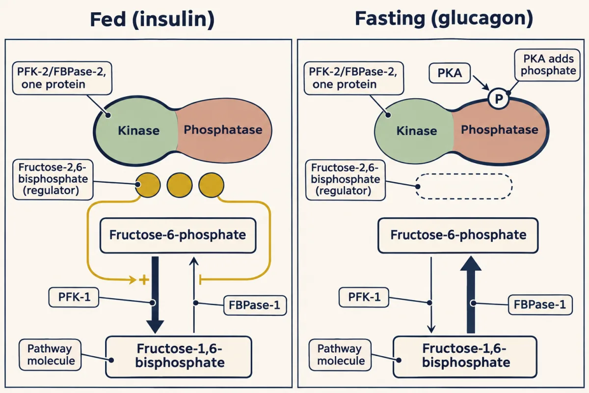
The middle pair, phosphofructokinase-1 (PFK-1) and FBPase-1. The best-known switch, fructose-2,6-bisphosphate (F-2,6-BP), works here, activating PFK-1 and inhibiting FBPase-1. 1,6 is the pathway; 2,6 is the regulator.
| Enzyme | What it acts on | Wins in the liver when you are |
|---|---|---|
| PFK-1 | makes fructose-1,6-bisphosphate (glycolysis) | fed |
| FBPase-1 | breaks fructose-1,6-bisphosphate (gluconeogenesis) | fasting |
| PFK-2/FBPase-2 | one protein; its two domains make (PFK-2) or break (FBPase-2) fructose-2,6-bisphosphate | fed: PFK-2; fasting: FBPase-2 |
In the fed liver, insulin keeps this bifunctional enzyme dephosphorylated, the kinase wins, F-2,6-BP rises, and glycolysis runs.
The pyruvate pair. Liver pyruvate kinase is shut off when protein kinase A (PKA) phosphorylates it; fructose-1,6-bisphosphate activates it, and ATP and alanine inhibit it. Pyruvate carboxylase is switched on by acetyl-CoA.
The glucose pair is set by glucokinase’s regulatory protein, which pulls glucokinase into the nucleus when glucose is low, and by enzyme amounts that insulin and glucagon control over hours.
Predict. A fast begins. Glucagon raises cAMP and activates PKA, and the liver burns fat, so its acetyl-CoA rises. Predict the middle and pyruvate pairs.
Check your prediction
- Middle pair: PKA phosphorylates the bifunctional enzyme, the phosphatase wins, and F-2,6-BP falls. PFK-1 loses its activator and FBPase-1 is released.
- Pyruvate pair: PKA shuts off liver pyruvate kinase, so new PEP is not turned back into pyruvate, and acetyl-CoA turns on pyruvate carboxylase.
- Glucose pair: glucokinase is sequestered as glucose falls, and over hours enzyme amounts shift toward glucose-6-phosphatase.
So in a healthy liver cell one direction dominates and the waste stays small. Notice that “fasting” and “energy high” point the same way: burning fat supplies the liver’s ATP and acetyl-CoA, and while its energy state stays high, AMP stays low, which also leaves FBPase-1 free.
When a bypass breaks
Pyruvate carboxylase deficiency breaks Bypass 1: pyruvate cannot be carboxylated to start gluconeogenesis, so it piles up and is converted to lactate, and lactic acidosis is the constant finding. In the severe newborn form hypoglycemia often joins it, with high ammonia, because the same oxaloacetate also feeds the citric acid cycle and, as aspartate, the urea cycle.
Von Gierke disease (glycogen storage disease type I) breaks Bypass 3. Without glucose-6-phosphatase or its T1 transporter, the liver can build glucose-6-phosphate but cannot release free glucose through its usual route: severe fasting hypoglycemia, lactic acidosis, and a liver swollen with glycogen it cannot cash out.
In type 2 diabetes no enzyme is missing. Insulin no longer holds the liver’s glucose output down, and fasting blood sugar rises; much of the cause is extra fuel arriving from fat.
The alanine cycle, beside the Cori cycle, carries muscle nitrogen to the liver packaged on alanine, because muscle lacks the urea-cycle machinery to dispose of ammonia. A rising serum alanine aminotransferase (ALT), the enzyme that runs that cycle, flags liver damage.
In the next chapter we follow pyruvate the other way, into the mitochondrion, where the pyruvate dehydrogenase complex turns it irreversibly into acetyl-CoA, which the citric acid cycle can burn completely.
Going deeper
The swinging biotin arm of pyruvate carboxylase
Pyruvate carboxylase uses a biotin cofactor on a flexible lysine tether, carried on a mobile domain that swings the carboxyl group between two active sites. At the first site the enzyme spends its ATP to load CO₂ onto biotin; at the second, biotin hands the carboxyl to pyruvate, making oxaloacetate.
Why glucose-6-phosphatase sits in the ER lumen
The reason is not settled. Hexokinase (glucokinase, in liver) sits in the cytosol running the opposite reaction, but both enzymes draw on the same pool of glucose-6-phosphate, so the separate room does not by itself stop a futile cycle; regulation of the two enzymes does most of that work.
Type 2 diabetes, PEPCK and metformin
Much of the liver’s lost insulin response is indirect. Insulin-resistant fat releases more glycerol and fatty acids, the fatty acids raise liver acetyl-CoA and so pyruvate carboxylase flux, and glucagon stays high. Phosphoenolpyruvate carboxykinase (PEPCK) levels alone control surprisingly little of the flux. Metformin lowers glucose output, by mechanisms still debated (AMPK activation is one candidate, a drop in liver energy state another).
Fasting across cultures
Fasting itself is old and human. The Teton Sioux fasted to invite a spiritual awakening, and cultures across the world still fast for clarity or devotion. Every such fast runs on the handover this lesson opened with: glycogen and gluconeogenesis first, then, after several days, ketones taking over part of the brain’s load.
Your notes
Select any text in the lesson to highlight it or add a note to it. Your highlights and notes are saved in this browser and collected in your notebook.
This browser is not letting the site save anything (a private window or strict privacy settings), so highlights and notes will be lost when you leave the page.
How we measure it
Isotopic carbon tracing of precursors
Feed a precursor labeled at a known carbon — ¹⁴C-lactate, ¹³C-alanine, or ¹³C-glycerol — and track where the atoms land in newly made glucose. The carbon skeleton is what becomes glucose (alanine's nitrogen is stripped by transamination first), so the label has to sit on carbon. Tracing also exposed a subtlety at the heart of the chapter: label from an even-chain fatty acid can turn up in glucose, because the cycle shuffles acetyl carbons into oxaloacetate, yet there is never any net glucose, because for every two carbons that enter as acetyl-CoA, two leave as CO₂.
Stable-isotope tracing of glucose production
To measure how much glucose the body is making, infuse a labeled glucose tracer in an overnight-fasted (basal) subject and read the dilution: total glucose appearance minus any infused glucose is endogenous glucose production, from liver and kidney. After an overnight fast roughly half of it still comes from glycogen breakdown, so splitting out gluconeogenesis needs a second method, such as heavy water or ¹³C NMR. A hyperinsulinemic-euglycemic clamp answers a different question — it raises insulin while holding blood glucose constant to see how well insulin SUPPRESSES that output, so failure to suppress is the read-out of hepatic insulin resistance (insulin's action on fat, which sets substrate supply to the liver, feeds into it too), not a measure of fasting gluconeogenesis itself.
Allosteric enzyme assay with single effectors
To map a control point you purify the enzyme and change one effector at a time while watching velocity. Adding acetyl-CoA to pyruvate carboxylase, AMP or fructose-2,6-bisphosphate to FBPase-1, or running the bifunctional PFK-2/FBPase-2 enzyme with and without PKA phosphorylation is how the reciprocal regulators of this pathway were assigned to their enzymes, one molecule at a time.
Practice questions on this lesson (3) → MCAT-style, original, each with an explanation
Where this lesson fits
Before thisReview first
- Glycolysis: The Investment-and-Payoff PathwayThis course · Lesson 16
You need the ten glycolytic steps and the three one-way ones from there to see which steps get bypassed.
- Metabolism: Basic Concepts and DesignThis course · Lesson 15
The free-energy and coupling ideas from there explain why ATP and GTP spent make the climb run downhill.
- Basic Concepts of Enzyme ActionThis course · Lesson 7
You need the idea that an enzyme speeds a reaction both ways but does not set its direction.
After thisStudy next
- The Pyruvate Dehydrogenase Complex: The One-Way Gate to the CycleThis course · Lesson 18
It follows pyruvate the other way, through a one-way step that is why fat cannot become glucose.
- Integration of Metabolism: How the Body Switches FuelsThis course · Lesson 30
It shows how hormones switch the liver between glycolysis and gluconeogenesis as fed turns to fasting.
- Fatty Acid Degradation: The β-Oxidation SpiralThis course · Lesson 25
It shows the fat burning that fuels the liver during a fast and supplies acetyl-CoA to switch on pyruvate carboxylase.
Elsewhere on the siteFilms, simulations and method films in other courses
- What the pump rate measuresSimulation · Physiology
It shows how insulin and glucagon steer liver glucose output, which gluconeogenesis supplies during fasting.
- A Beta Cell Burns Glucose to Read ItFilm · Physiology
It shows how beta cells sense glucose, the signal that decides whether the liver makes or stores glucose.
Suggested by an AI model from the lessons themselves, then checked. How we use AI
Finish line
Test yourself
8 questions
Practice and MCAT-style, with explained answers.
Play it · about 4 min
Mixing Board
Set insulin, glucagon, F-2,6-BP and the energy signals so the liver runs the right pathway, and never both at once.
Play →Next · Lesson 18
The Pyruvate Dehydrogenase Complex: The One-Way Gate to the Cycle
Pyruvate meets a one-way gate: one machine turns it into acetyl-CoA, CO₂ and NADH, and there is no way back.
Continue →Also in the arcade for this lesson: Wire the Cell: Cori-cycle liver