Biochemistry · Skim version
Signal-Transduction Pathways: How Cells Compute With Molecular Switches
Read the full lesson → Skim the whole course →
The guiding idea
One epinephrine molecule can mobilize millions of glucose units, and the same relay has to stop on time.
One epinephrine molecule can mobilize millions of glucose units.
-
The parts: messengers, receptors, effectors
-
One switch first: the G-protein cycle
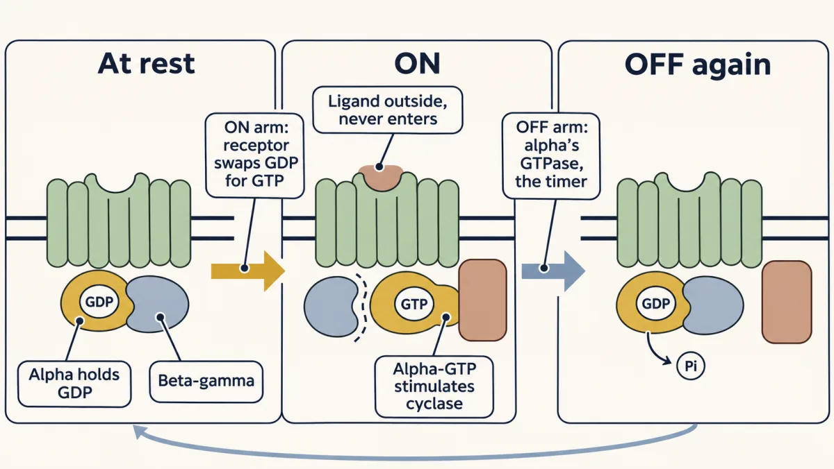
The G-protein switch, ON and OFF. The activated receptor is alpha's GEF: it swaps GDP for GTP, and alpha-GTP stimulates adenylate cyclase. Alpha's own GTPase is the timer: it cuts GTP back to GDP, and alpha rejoins beta-gamma. Subunit separation is schematic; activation can involve separation or rearrangement. AI-generated illustration -
The cAMP relay: a chain of switches
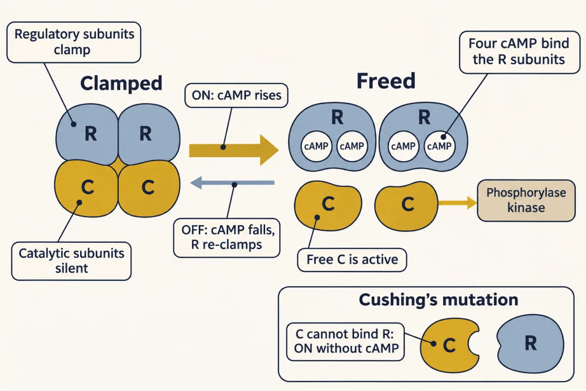
PKA as a switch. Two regulatory subunits clamp two catalytic subunits silent. Four cAMP bind the regulatory subunits and release the catalytic subunits, which go on to phosphorylate their targets. As PDE clears cAMP, the regulatory subunits clamp again. Inset: in some cases of Cushing's syndrome the catalytic subunit cannot bind its regulatory subunit, so PKA is ON with no cAMP at all. AI-generated illustration -
How big: speed times lifetime, multiplied down the chain
-
How long: the slowest OFF step
-
Broken switches: four ways to break the contest
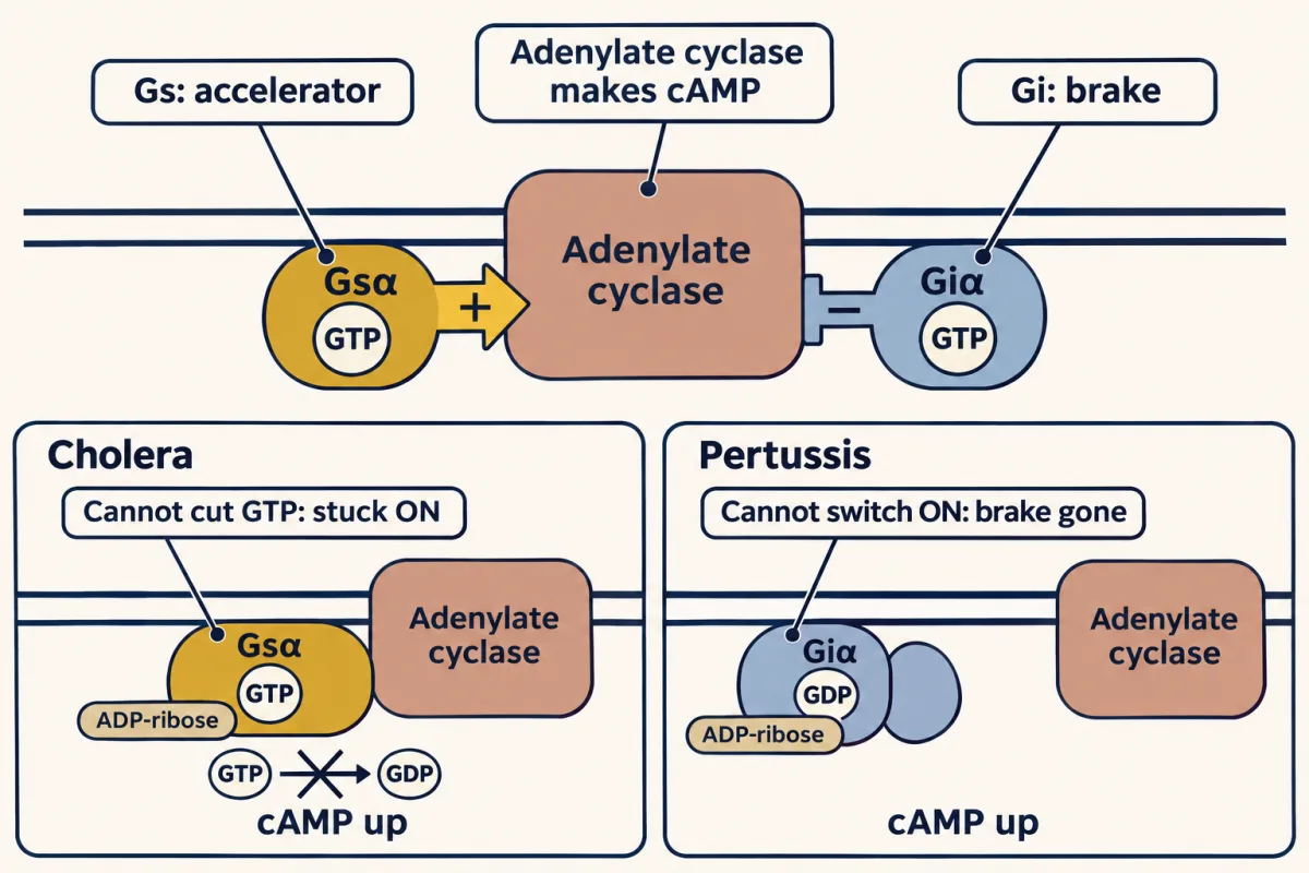
One chemical trick, two jammed switches. Cyclase sits between an accelerator (Gs) and a brake (Gi). Cholera toxin ADP-ribosylates Gs so it cannot cut its GTP and stays ON. Pertussis toxin ADP-ribosylates Gi so it cannot be switched ON and the brake is lost. Both raise cAMP. AI-generated illustration -
Same template, new parts
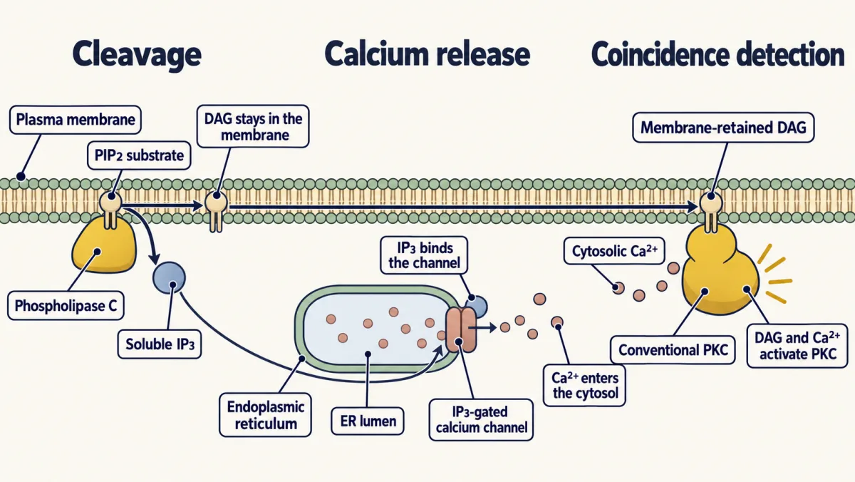
Phospholipase C makes a single cut in PIP2. DAG stays in the membrane; soluble IP3 floats to the endoplasmic reticulum and opens a calcium channel. Together, DAG and Ca²⁺ activate protein kinase C. AI-generated illustration 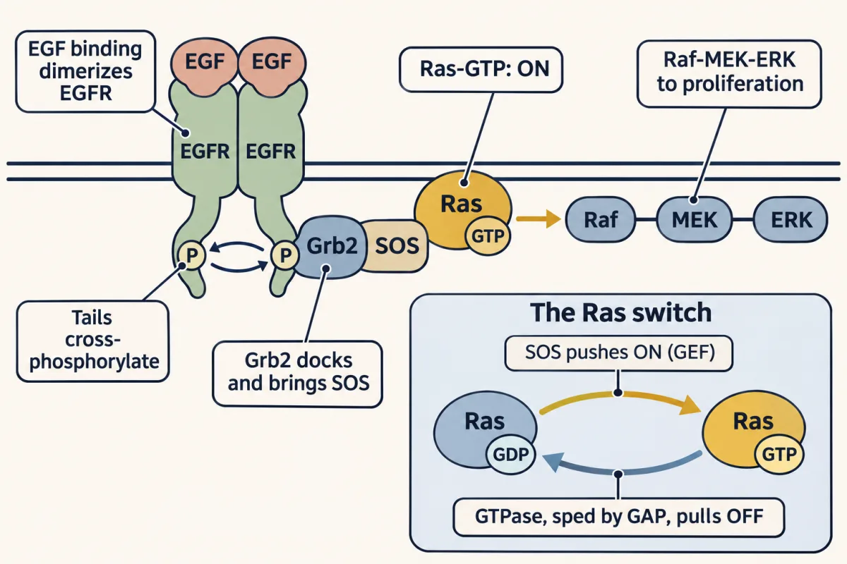
A receptor that is a kinase. EGF dimerizes EGFR, the halves cross-phosphorylate, and Grb2 brings SOS to the membrane. SOS is Ras's GEF and pushes it ON; active Ras drives Raf-MEK-ERK. Inset: Ras is a small version of G-alpha, pushed ON by SOS and pulled OFF by its own GTPase, which GAPs speed up. AI-generated illustration 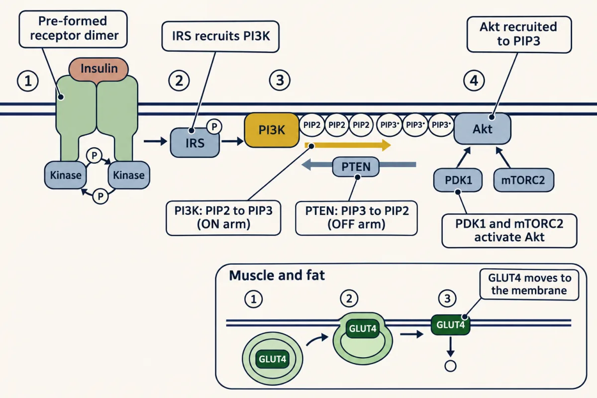
Insulin's lipid switch. The pre-formed receptor dimer cross-phosphorylates and phosphorylates IRS, which recruits PI3K. PI3K turns PIP2 into PIP3 on the cytosolic leaflet (the ON arm); PTEN turns PIP3 back into PIP2 (the OFF arm). PIP3 recruits Akt, which PDK1 and mTORC2 activate. Inset: in muscle and fat, Akt drives GLUT4 to the membrane for glucose uptake. AI-generated illustration -
Drugs on the wiring
-
Going deeper
Where this lesson fits
Before thisReview first
- Enzyme Kinetics: The Michaelis–Menten ModelThis course · Lesson 8
You need saturation of enzymes by substrate from there to follow why the product rule stops working at large signals.
- Protein Binding and Allostery: How Hemoglobin Loads and Delivers OxygenThis course · Lesson 9
Ligand binding that changes a protein's shape is the idea behind a receptor switching on a G protein here.
- Membranes: The Impermeable Sheet That Builds ItselfThis course · Lesson 12
Why large polar signals cannot cross the membrane and need a spanning receptor comes from there.
After thisStudy next
- Integration of Metabolism: How the Body Switches FuelsThis course · Lesson 30
Insulin, glucagon and epinephrine signals turn pathways up or down here, using the switches built in this lesson.
- Gluconeogenesis: Pushing Glucose Back UphillThis course · Lesson 17
Glucagon's cAMP and PKA signal is what switches liver glucose building on during fasting.
Elsewhere on the siteFilms, simulations and method films in other courses
- GTPase timerSimulation · Cell Signaling
This simulation's cousin shows the G-protein timer that turns the signal off.
- The Messenger Level: Made and ErasedFilm · Cell Signaling
It follows the cAMP second messenger and PKA step by step, the core relay used here.
- A Membrane Address for AktFilm · Cell Signaling
It covers the PI3K, PIP3 and Akt steps that carry insulin to GLUT4 in this lesson.
- One Cut, Two DestinationsFilm · Cell Signaling
It shows PLC cutting PIP2 into IP3 and DAG, the Gq branch described here.
Suggested by an AI model from the lessons themselves, then checked. How we use AI