Biochemistry · The Citric Acid Cycle
The Pyruvate Dehydrogenase Complex: The One-Way Gate to the Cycle
Rewritten on : new text, figures and flashcards. Your notes and highlights stay, but the text around them may have moved.
Pyruvate meets a one-way gate: one machine turns it into acetyl-CoA, CO₂ and NADH, and there is no way back.
Pause when the reading reaches a film, and offer to play it. Saved on this device.
Watch the film now, or keep listening
Builds on:Glycolysis: The Investment-and-Payoff PathwayMetabolism: Basic Concepts and DesignBasic Concepts of Enzyme ActionWhy these?
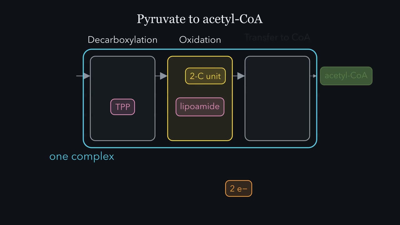 ▶ Watch The gate to the cycle · 3:29
Read the transcript of “The gate to the cycle”
Film transcript: The gate to the cycle
Converting pyruvate to acetyl-CoA is not one reaction but three, carried out in sequence by a single complex. First comes decarboxylation, where pyruvate loses carbon dioxide, and the remaining two-carbon fragment is captured on thiamine pyrophosphate. Second is oxidation, where two electrons leave and the fragment is handed to the lipoamide arm. Third is transfer to coenzyme A, producing acetyl-CoA. Follow the carbon across the three boxes and follow the electrons separately, because those electrons end up reducing NAD⁺ to NADH. Notice that each step uses its own coenzyme, and several of those coenzymes trace back to B vitamins. Watch the lipoamide arm make its rounds.
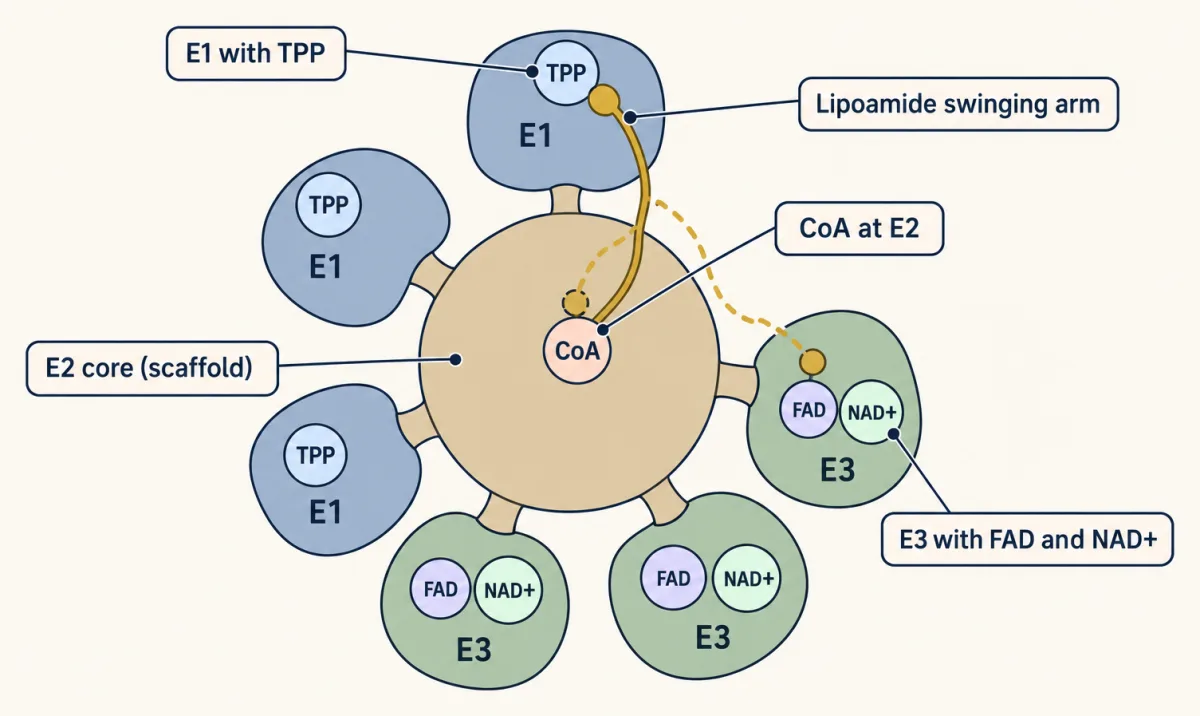
Tethered to the E2 core, it first swings to E1 and picks up the acetyl group, becoming acetyl-lipoamide. Next, it pivots back to E2 and delivers that acetyl unit to coenzyme A, releasing acetyl-CoA and leaving the arm reduced as dihydrolipoamide. Finally, it reaches E3 to be reoxidized, regenerating the disulfide so the cycle can repeat. Because the arm rides on a mobile lipoyl domain and the E2 core has many copies, 24 or 60 depending on the organism, all three active sites stay within reach. The intermediates never escape. They ride the tethered arm from station to station. Let me put the whole mechanism on one timeline so you know exactly where NADH is born. Step one, decarboxylation on E1, releases carbon dioxide and makes no NADH.
Step two, oxidative transfer, hands the hydroxyethyl group to lipoamide as it is oxidized to an acetyl group, still no NADH. Step three, acetyl transfer on E2, produces acetyl-CoA and leaves dihydrolipoamide, again, no NADH. Only in step four, regeneration on E3, is the reduced lipoamide reoxidized by FAD and the electrons flow onward to make NADH. So three steps build the product and exactly one step, the last, generates the reducing power. Enzyme E1 is the key regulatory site of the entire complex, and it is controlled by a simple on-off switch built from a kinase and a phosphatase that travel with the complex. A kinase phosphorylates E1 and inactivates it.
A phosphatase removes that phosphate and reactivates it. The switch reads the cell's energy state. When ATP, acetyl-CoA and NADH are abundant, signaling plenty of fuel and product, the complex is inhibited. When ADP and pyruvate accumulate, signaling demand and available substrate, the complex is stimulated. So the gateway from glycolysis to the citric acid cycle opens only when the cell actually needs to burn pyruvate.
Eat more sugar than you burn and your body stores the surplus as fat. Burn that fat later, and almost none of it can come back as sugar. Why does the road run only one way?
The answer starts with one reaction. The pyruvate dehydrogenase complex (PDC) turns pyruvate, the three-carbon end of glycolysis, into acetyl-CoA, releasing a carbon as CO₂ and banking one NADH. Picture an airport: glycolysis in the cytosol is Terminal 1, the citric acid cycle in the mitochondrial matrix is Terminal 2, and PDC is a security gate between them that opens one way. Fat breaks down mostly to acetyl-CoA, which is already on the far side of that gate.
The net reaction is worth memorizing exactly:
Pyruvate + CoA + NAD⁺ → Acetyl-CoA + CO₂ + NADH + H⁺
It is an oxidative decarboxylation: pyruvate (3 carbons) loses its carboxyl carbon as CO₂ and is oxidized to a 2-carbon acetyl group carried by coenzyme A. The two halves of that name happen at different moments. It runs in the mitochondrial matrix, which pyruvate enters through the mitochondrial pyruvate carrier.
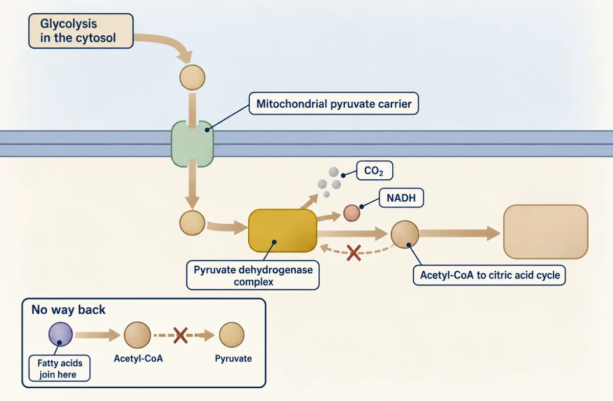
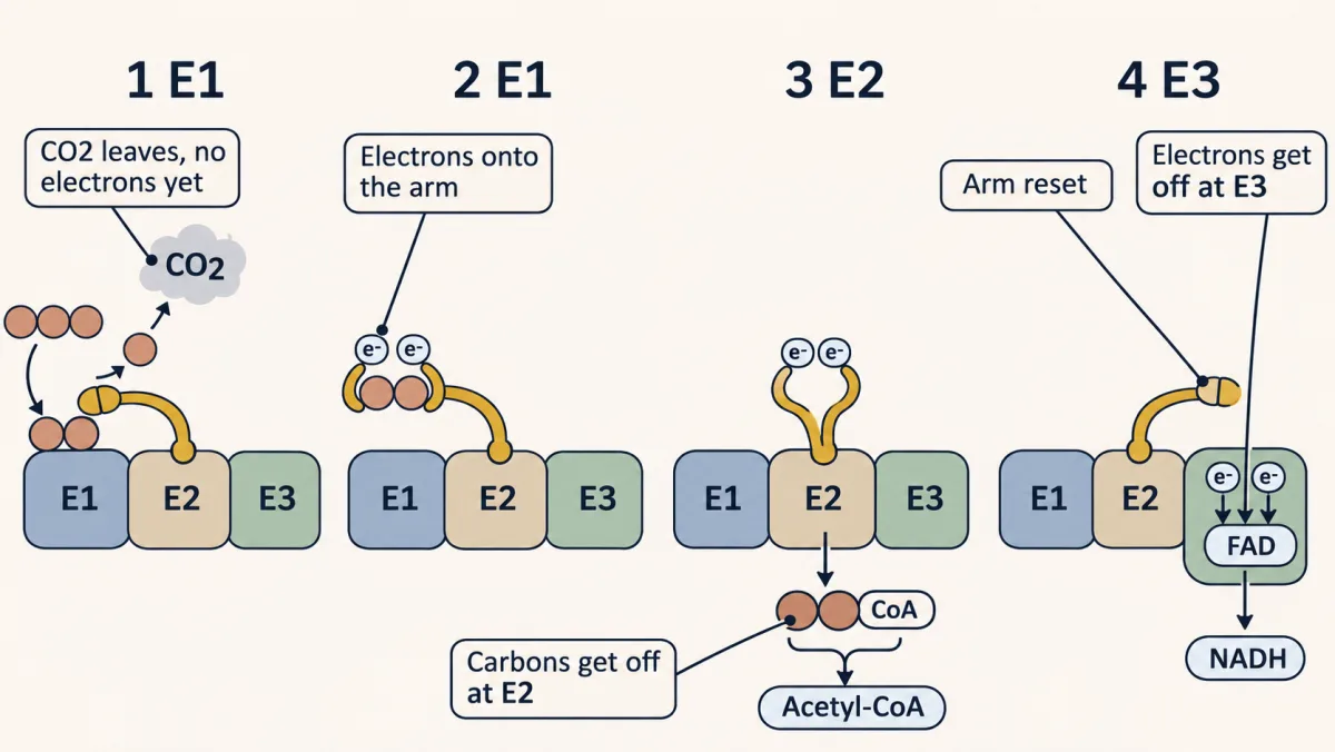
The one idea to carry through this lesson. Follow two things through the machine: the carbons and the electrons. Pyruvate’s carboxyl carbon leaves as CO₂ at E1 before any oxidation happens. The two acetyl carbons that stay and the two electrons removed from them then ride the same swinging arm, and they get off at different stops: the carbons at E2 (onto CoA), the electrons at E3 (through FAD to NAD⁺).
This two-track picture explains the net reaction, the product brakes and the poisons. The phosphorylation switch at E1 is a separate layer on top.
About the film at the top of the page: the “three reactions” it names are steps 1 to 3 below, and its “step four” is our step 4. One correction to its wording: Thiamine pyrophosphate (TPP) grabs pyruvate first, and the CO₂ leaves from that grip.
Following one pyruvate: two tracks on one arm
Keep the two tallies, carbons and electrons, as you go. Oxidation just means losing electrons; for a carbon, that shows up as trading a bond to hydrogen for a bond to oxygen (or, here, sulfur). Chemists rank a carbon’s oxidation level by those bonds: alcohol, then aldehyde, then acid, each with one more.
TPP, the grabber on E1. Thiamine pyrophosphate (made from vitamin B1) has an unusual carbon between a nitrogen and a sulfur in a small ring. It can drop its hydrogen, become negatively charged and grab pyruvate’s middle carbon. The ring then soaks up electrons, which lets the carboxyl group break away as CO₂.
Lipoamide, the arm. Lipoic acid is a short chain ending in a ring that contains two sulfur atoms bonded to each other (an S–S bond, a disulfide). Bolted to a lysine on E2 (by an amide bond), it becomes lipoamide, riding on a floppy lipoyl domain that jiggles at random among the three enzymes, all within reach. Give the S–S two electrons (with two protons) and it breaks into two –SH groups. So the arm can carry two things at once: an acetyl group on one sulfur, and two electrons in the broken S–S.
Predict. Step 1 removes pyruvate’s carboxyl carbon as CO₂. Does that step remove any electrons from the molecule?
Check your prediction
No. CO₂ leaves its bonding electron pair behind (TPP’s ring soaks it up), so no electrons leave the molecule overall, and the two carbons left behind sit at the oxidation level of an aldehyde (acetaldehyde, in effect). Yeast’s pyruvate decarboxylase uses the same TPP chemistry but releases acetaldehyde for fermentation, with no NAD⁺ involved. The carbon is lost for good (it is gone as gas), but the oxidation, the main energy source, is still to come.
Now the walk. Three steps build the product, and one more resets the machine.
| Step | Where | Carbon track | Electron track |
|---|---|---|---|
| 1 · Decarboxylation | E1, with TPP | Pyruvate’s carboxyl carbon (C1) leaves as CO₂. C2 and C3 stay on TPP as a two-carbon (hydroxyethyl) group. | Nothing removed yet. |
| 2 · Oxidative transfer | E1, handing to the arm | The two carbons are oxidized to an acetyl group and hooked onto one sulfur of the arm (a thioester). TPP is free again. | Two electrons removed, and they break the arm’s S–S. The arm now carries both tracks. |
| 3 · Acetyl transfer | E2, with CoA | The arm swings to E2, which moves the acetyl group onto CoA. Acetyl-CoA leaves: the carbon track is finished. | The two electrons are still on the arm, as two –SH groups. |
| 4 · Regeneration | E3, with FAD, then NAD⁺ | — | The arm swings to E3. The electrons pass through E3’s bound FAD (FADH₂ for an instant) to NAD⁺, making NADH. The S–S is restored. The electron track is finished. |
Predict. The oxidation happens at E1, in step 2. So why is the NADH not made there?
Check your prediction
Because E1 has no NAD⁺ site. The electrons it removes go onto the nearest acceptor, the arm’s disulfide, and they ride the arm until it reaches E3, the only enzyme that can hand them to NAD⁺. That is the exam trap in one line: the oxidation is at E1, but the NADH is made at E3. The FADH₂ along the way never leaves E3, which is why the net reaction lists NADH, not FADH₂.
Read down each column and the net reaction writes itself: three carbons in, one CO₂ and one acetyl-CoA out; two electrons out of the carbons, into one NADH. Step 4 makes no product carbon, but it has to happen: an arm still holding electrons cannot accept the next acetyl group.
regulated step
- Carbons
- 3 (pyruvate), crossing into the matrix
- Electrons
- none removed
Pyruvate made by glycolysis in the cytosol is carried across the inner mitochondrial membrane by the mitochondrial pyruvate carrier. No chemistry happens here (a proton comes in along with each pyruvate); this is the geographic move from glycolysis into the matrix, where the complex waits at the one-way gate.
- Carbons
- C1 leaves as CO₂; C2 and C3 stay on TPP
- Electrons
- none removed yet
TPP's reactive carbon (the C2 carbanion, or ylide, between the ring's nitrogen and sulfur) adds to pyruvate's carbonyl carbon, and the carboxyl carbon leaves as CO₂. No electrons are removed yet: CO₂ leaves its bonding pair behind on the TPP ring, and the two carbons left on TPP are at the oxidation level of an aldehyde. Losing CO₂ is where the carbon is lost for good, as gas. What makes the whole reaction one-way is its overall free-energy drop, which comes mainly from the oxidation in the next step, with the CO₂ loss contributing the rest. E1 is also the principal control point.
Regulation. E1 is switched off when PDH kinase (PDK), bound to the complex, phosphorylates it; the kinase is stimulated by a high ATP/ADP ratio and by high acetyl-CoA and NADH, and inhibited by pyruvate and ADP. PDH phosphatase (its PDP1 isoform is stimulated by Ca²⁺) removes the phosphate and switches E1 back on.
Clinical. Thiamine (B1) deficiency starves E1 of TPP; the carbon track stops before it starts, and pyruvate and lactate accumulate. Because α-ketoglutarate dehydrogenase and transketolase also need TPP, they fail too, and beriberi or Wernicke's follows. Give thiamine before or with glucose in malnourished patients, or a glucose load can worsen lactic acidosis.
- Carbons
- 2, now an acetyl group on the arm
- Electrons
- 2 removed, now on the arm (S–S broken)
Still on E1: the two-carbon hydroxyethyl unit is oxidized to an acetyl group and transferred onto lipoamide's disulfide in the same motion. The two electrons removed break the S–S of lipoamide: one sulfur now carries the acetyl group as a thioester and the other is a free thiol. TPP is regenerated. The electrons are now held on the lipoamide arm, NOT yet on NAD⁺, so no NADH is made here, a common trap.
- Carbons
- acetyl-CoA leaves: the carbon track is finished
- Electrons
- still on the arm, as two –SH groups
The swinging lipoamide arm carries the acetyl group to the E2 core, which transfers it to coenzyme A, forming the thioester acetyl-CoA (its hydrolysis releases about 31 kJ/mol). Acetyl-CoA is released, and the carbon track is finished. The lipoamide is left reduced, still holding the two electrons, and must be reset before the complex can run again.
Clinical. Arsenite (As³⁺) bridges the two closely spaced –SH groups of the dithiol lipoamide, locking the arm, and the complex freezes. Mercury (Hg²⁺) also binds thiols, among many other targets.
- Carbons
- none
- Electrons
- arm → FAD → NAD⁺, making NADH: the electron track is finished
First half: E3 reoxidizes the dithiol back to the lipoamide disulfide, resetting the arm, and the electrons pass to E3's tightly bound FAD, making FADH₂ for a moment. Second half: the electrons move from FADH₂ to NAD⁺, producing NADH and regenerating FAD. The FADH₂ stays on the enzyme, an internal relay and not a released product, which is why PDC's net carriers list NADH and not FADH₂. This is the only NADH-producing step of the whole complex. No molecular O₂ is used directly; the complex needs aerobic conditions only because the electron-transport chain must regenerate NAD⁺ downstream.
Card 0 is the trip into the matrix; cards 1 to 4 are the four steps.
Count the coenzymes you just used: TPP (step 1), lipoamide (steps 2 to 4), CoA (step 3), FAD and NAD⁺ (step 4). That is the mnemonic “Tender Loving Care For Nancy,” in the order the pyruvate meets them. Four come from vitamins you must eat: thiamine B1 (TPP, at E1), pantothenate B5 (CoA, at E2), riboflavin B2 (FAD, at E3) and niacin B3 (NAD⁺, at E3). Lipoic acid is the exception; the body makes it, so it is not a true vitamin.
No step uses molecular O₂. Pyruvate dehydrogenase complex (PDC) needs oxygen only indirectly: the electron transport chain must turn NADH back into NAD⁺, or step 4 runs out of acceptor and the relay stops.
One machine, so nothing floats free
PDC is one of the largest enzyme assemblies in the cell, larger than a ribosome. E2 forms the core, and E1 and E3 dock onto it, close enough for the arm to reach every active site. So the intermediates never diffuse into solution, where they could drift away or be lost to side reactions. That is substrate channeling.
Where the energy goes
In animals this reaction is irreversible: no enzyme runs it backward, and acetyl-CoA cannot become pyruvate again. The oxidation is the main energy source: it pays for the NADH and for the thioester, and what is left over, together with the loss of CO₂, makes the gate one-way. To see it, keep the free-energy books (standard values, pH 7).
| Piece of the reaction | ΔG°′ (kJ/mol) |
|---|---|
| The oxidation: the two carbons go from aldehyde level to acid level, and the electrons end on NAD⁺ | about −50 |
| Storing the acetyl group as a thioester, instead of letting it go as free acetate | about +31 (a cost) |
| Losing CO₂ (found by difference, so approximate) | about −14 |
| Overall | about −33 |
Building the thioester spends most of the oxidation’s drop, about 31 of the 50. What is left (about −19), plus about −14 from the CO₂ loss, gives about −33 kJ/mol: an equilibrium constant (K = e−ΔG°′/RT) of about 4 × 10⁵ at body temperature, so far toward the products that in practice the reaction runs only forward.
The +31 line is the same number you get back when you hydrolyze acetyl-CoA: about −31 kJ/mol, roughly ATP’s −30.5. Hydrolyzing an ordinary oxygen ester gives only about −21. This is not energy stored in the bond; breaking a bond always costs energy. Hydrolysis releases energy because the products, acetate and CoA-SH, are much more stable than the thioester, which gets little resonance stabilization: sulfur is larger than oxygen, so its orbitals overlap the carbonyl carbon poorly, whereas an oxygen ester’s lone pairs stabilize its C=O. CoA is the handle, not the valuable part. So acetyl-CoA is a wound spring, and the oxidation wound it. That stored potential lets acetyl-CoA drive the first step of the citric acid cycle and seed fatty-acid synthesis. Acetyl-CoA has two main fates: complete oxidation for ATP, or storage as fat. The liver can also turn it into ketone bodies, and it is the starting point for cholesterol.
Predict. Your body burns fat to acetyl-CoA. Can that acetyl-CoA make new glucose?
Check your prediction
Not a net amount. It is on the far side of the gate and can never become pyruvate, and animals lack the glyoxylate cycle, the separate route plants and bacteria use to make new oxaloacetate (and so glucose) from acetyl-CoA. Gluconeogenesis must start on the near side: from pyruvate (made from lactate or amino acids such as alanine), with pyruvate carboxylase and PEPCK carrying it back to PEP, or from other entry points. That answers the opening puzzle. Two parts of a fat are the exceptions: its glycerol, and the propionyl-CoA made at the end of odd-chain fatty-acid breakdown.
Two layers of regulation
A gate this important is heavily controlled, and pyruvate dehydrogenase complex (PDC) reads the cell’s energy charge (how much ATP it has relative to ADP and AMP) with the same logic as phosphofructokinase-1 (PFK-1) from the glycolysis chapter. The first layer is product inhibition.
Predict. Acetyl-CoA and NADH, the two products, both slow the complex, each by competing with the substrate it resembles. Which enzyme does each one slow?
Check your prediction
Acetyl-CoA competes with CoA at E2, backing up the carbon track. NADH competes with NAD⁺ at E3, backing up the electron track. Either way the relay slows: “we have plenty, slow down.”
The second layer is covalent, and it acts at E1. A dedicated kinase, PDK (pyruvate dehydrogenase kinase), phosphorylates a serine on E1 and switches the complex OFF. A phosphatase, PDP, removes that phosphate and switches it back ON. Both are bound to the complex itself. Note the direction: here phosphorylation inactivates, the same convention as glycogen synthase. Much of the energy-charge sensing at this gate runs through PDK: ATP is its substrate and ADP competes with it, so PDK reads the ATP/ADP ratio.
| The cell signals | What happens | E1 | The gate |
|---|---|---|---|
| Plenty: high ATP/ADP ratio, high NADH, high acetyl-CoA | PDK is stimulated | phosphorylated | closed |
| Need: ADP and pyruvate; in working muscle, Ca²⁺ | ADP and pyruvate inhibit PDK, so PDP wins; Ca²⁺ activates PDP | dephosphorylated | open |
The plenty signals reinforce the product brake.
Try it below: pick a state (fed, sprinting, fasting) or set the effectors, and watch which way PDK and PDP flip the gate.
Pick a physiological state, or tick the signals yourself, and watch which way the switch flips.
Turns PDH OFF
(activates PDK; NADH and acetyl-CoA also brake E3/E2 directly)
Turns PDH ON
(activates PDP / blocks PDK)
E1–Ser–OH With no effectors set, the phosphatase keeps the complex dephosphorylated and active by default.
Two layers. Directly, the products brake the stop where they leave: acetyl-CoA competes with CoA at E2 (carbon track) and NADH with NAD⁺ at E3 (electron track). On top of that, PDK phosphorylates E1 to close the gate when the cell is fuel-rich, and PDP removes the phosphate to reopen it. This switch models the second layer.
The cleanest illustration is muscle. A contracting muscle fills with Ca²⁺; some is taken up into the mitochondrial matrix, where it switches on the phosphatase (the PDP1 isoform). PDC switches on, and acetyl-CoA surges into the citric acid cycle exactly when the muscle needs ATP. The calcium that triggers contraction also unlocks the fuel.
When the gate fails
Each failure below breaks one of the two tracks, and a stall anywhere stops the relay.
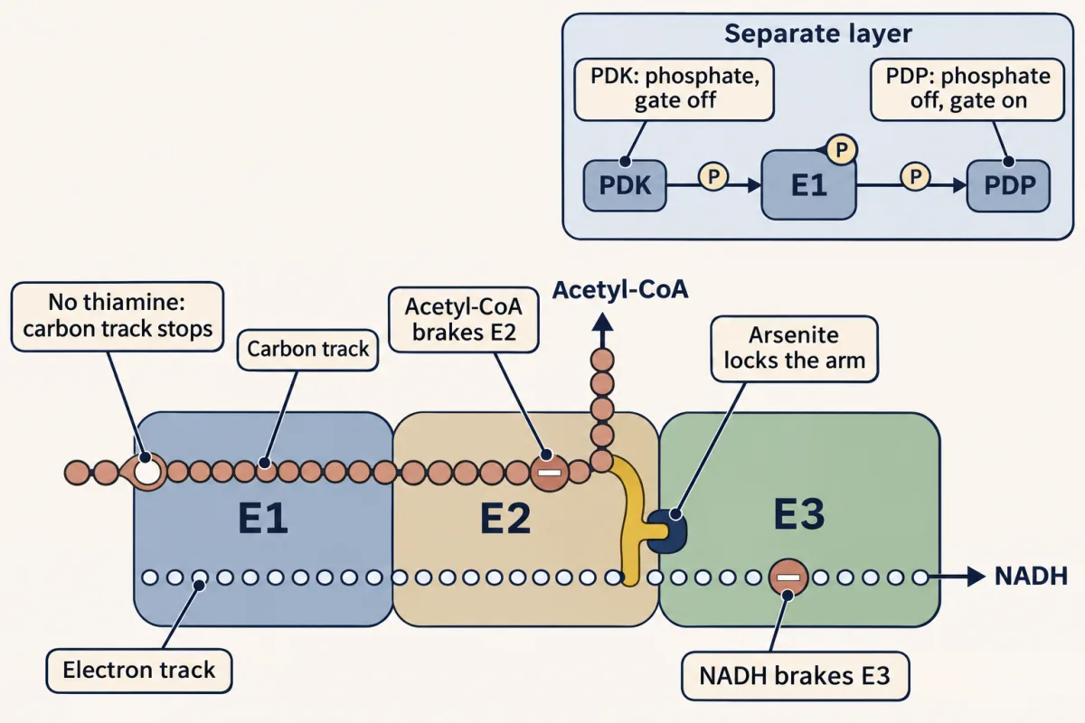
Beriberi is B1 deficiency: no thiamine, no thiamine pyrophosphate (TPP), so the carbon track stops before it starts. Pyruvate and lactate pile up, and the nervous and cardiovascular systems suffer. TPP-dependent α-ketoglutarate dehydrogenase and transketolase fail too, and in the brain they may matter as much as pyruvate dehydrogenase complex (PDC). This is why a malnourished or alcohol-dependent patient should get thiamine before or with a glucose drip: in severe deficiency the sugar load can worsen lactic acidosis, and it has been reported to precipitate Wernicke’s encephalopathy. (Glucose for dangerously low blood sugar is not delayed to wait for thiamine.) Thiamine deficiency is also one part of refeeding syndrome, which is defined by falling blood phosphate, potassium and magnesium after feeding restarts.
Predict. Arsenite grips the arm’s two –SH groups, the state the arm is in after step 3. Which track stops, and why does the other stop too?
Check your prediction
The electron track stops: the locked arm cannot hand its electrons to E3, so no NADH is made and the S–S is never restored. Then the carbon track stops too, because an arm that cannot be reset cannot accept the next acetyl group. The whole complex freezes. Arsenite binds closely spaced pairs of thiols; in lipoamide these are the sulfurs on carbons 6 and 8.
Mercury (Hg²⁺) also binds thiols such as lipoamide’s, though its toxicity involves many other targets.
E3 deficiency stops the electron track in at least three complexes at once, because PDC, α-ketoglutarate dehydrogenase and the branched-chain α-keto acid dehydrogenase share one E3 (it also serves the glycine cleavage system). Patients show lactic acidosis, often with raised branched-chain amino acids and α-ketoglutarate.
The Warburg effect in cancer is partly PDC regulation hijacked: in many tumors HIF-1α (a low-oxygen transcription factor that many tumors keep switched on) drives up PDK, E1 is held largely phosphorylated and off, pyruvate is shunted to lactate even when oxygen is abundant, and the tumor gets fast ATP plus glycolytic intermediates for biosynthesis. Other tumors keep substantial PDC activity and mitochondrial oxidation.
Next: the same relay, again
With acetyl-CoA in hand, we are at the door of Terminal 2, where you will meet this machine again: α-ketoglutarate dehydrogenase runs the same E1 → arm → E2 → E3 relay with the same E3; its own E1 and E2, the group carried (succinyl instead of acetyl) and the build of its core differ. The phosphorylation switch is not shared: α-ketoglutarate dehydrogenase is not controlled by phosphorylation. The next chapter runs that acetyl group through the citric acid cycle, where each turn squeezes out CO₂ and GTP plus the NADH and FADH₂ that feed the electron transport chain.
Going deeper
The energy ledger in numbers
The −50 kJ/mol comes from the reduction potentials of the acetate/acetaldehyde couple (−0.581 V) and NAD⁺ (−0.320 V). The +31.4 thioester cost is 62% of that. The overall −33.4 kJ/mol agrees, within a few kJ, with a cross-check from the acetyl-CoA + CO₂/pyruvate couple (E°′ about −0.48 to −0.50 V, giving −31 to −35 kJ/mol). The −14 for losing CO₂ is found by difference and mixes tables, so it is approximate; the conclusion that the drop comes mainly from the oxidation, with the CO₂ loss contributing the rest, does not hang on it.
Size and docking of the complex
Pyruvate dehydrogenase complex (PDC) is roughly 10 megadaltons in eukaryotes, against about 4 for a eukaryotic ribosome. E2 forms a core of 60 subunits arranged as a pentagonal dodecahedron; in mammals some of those positions are taken by a related protein, E3BP. Some bacteria, such as E. coli, build a 24-subunit cube instead, which is the film’s “24 or 60.” E1 docks onto subunit-binding domains of E2, and E3 onto those of E3BP (in bacteria, E2 binds both). The lipoamide arm, carried on a mobile lipoyl domain, is about 14 Ångströms long.
Hyperglycemia and superoxide
This concerns the electron transport chain, not PDC. In hyperglycemia, one proposed model holds that excess fuel overloads the chain so that more electrons leak out as superoxide. Oxidative stress is thought to contribute to diabetic neuropathy, but its sources are still debated and other pathways are involved too.
Your notes
Select any text in the lesson to highlight it or add a note to it. Your highlights and notes are saved in this browser and collected in your notebook.
This browser is not letting the site save anything (a private window or strict privacy settings), so highlights and notes will be lost when you leave the page.
How we measure it
Substrate channeling in multienzyme complexes
When several active sites are tethered into one physical complex, an intermediate made at one site is handed directly to the next without diffusing into the cytosol. PDC is the textbook case: the lipoamide arm of E2 carries the acetyl unit from E1 to E2 and then visits E3 to be reoxidized, so reactive intermediates do not escape, side reactions are suppressed, and local concentrations stay high. Cryo-EM of the assembled core shows the architecture; kinetic and biochemical experiments test how the intermediates move between active sites.
Tracing the released CO₂ with labeled carbon
Label pyruvate at C1 (the carboxyl) or at C2/C3, and the CO2 released by PDC tells you which carbon left (in whole cells, later turns of the TCA cycle release the other carbons too, so timing matters). This kind of tracing shows that PDC strips the carboxyl carbon (C1) as CO2 and that the surviving two carbons (C2, C3) become the acetyl group. Scaled up, the same logic underlies research ¹³C-pyruvate breath tests and hyperpolarized ¹³C-pyruvate MRI of metabolism in tumors and the failing heart.
Phospho-state assays for covalent regulation
To see whether PDC is on or off you measure the phosphorylation state of E1's regulatory serines (human E1α has three), using phospho-specific antibodies or activity assays before and after adding PDK, PDP, ATP, or Ca2+. Watching activity fall when PDK phosphorylates E1 and rise when PDP (Ca2+-activated) removes the phosphate is how the kinase/phosphatase switch was mapped, and it is the same readout used to test PDK inhibitors like dichloroacetate.
Practice questions on this lesson (3) → MCAT-style, original, each with an explanation
Where this lesson fits
Before thisReview first
- Glycolysis: The Investment-and-Payoff PathwayThis course · Lesson 16
Glycolysis makes the pyruvate this gate receives, and its PFK-1 energy-charge logic is reused here.
- Metabolism: Basic Concepts and DesignThis course · Lesson 15
You need free energy, coupling and activated carriers like NADH and thioesters to follow the energy ledger.
- Basic Concepts of Enzyme ActionThis course · Lesson 7
Coenzymes, intermediates and catalysis of an irreversible step are used here without being re-taught.
After thisStudy next
- The Citric Acid Cycle: Harvesting ElectronsThis course · Lesson 19
Acetyl-CoA from this gate is the fuel the cycle burns, and it uses the NADH relay again.
- Fatty Acid Synthesis: Building the Chain Two Carbons at a TimeThis course · Lesson 26
Acetyl-CoA made at this gate is the starting material for building fat.
Suggested by an AI model from the lessons themselves, then checked. How we use AI
Finish line
Test yourself
9 questions
Practice and MCAT-style, with explained answers.
Play it · about 4 min
Metabolic Autopsy: PDH
Diagnose a broken step at the bridge from the metabolite pile-up.
Play →Next · Lesson 19
The Citric Acid Cycle: Harvesting Electrons
The citric acid cycle makes almost no ATP. Its real product is high-energy electrons.
Continue →Also in the arcade for this lesson: Ledger Rush: PDH