Biochemistry · Skim version
Protein Binding and Allostery: How Hemoglobin Loads and Delivers Oxygen
Read the full lesson → Skim the whole course →
The guiding idea
Your blood has to grab oxygen in the lungs and let go of about two-thirds of it in hard-working muscle. A tighter grip than hemoglobin's would make it worse. What does hemoglobin do instead?
Blood has two opposite jobs: grab oxygen in the lungs (~100 torr) and let go of it in hard-working muscle (~20 torr).
-
The delivery problem: load in the lungs, unload in the tissues
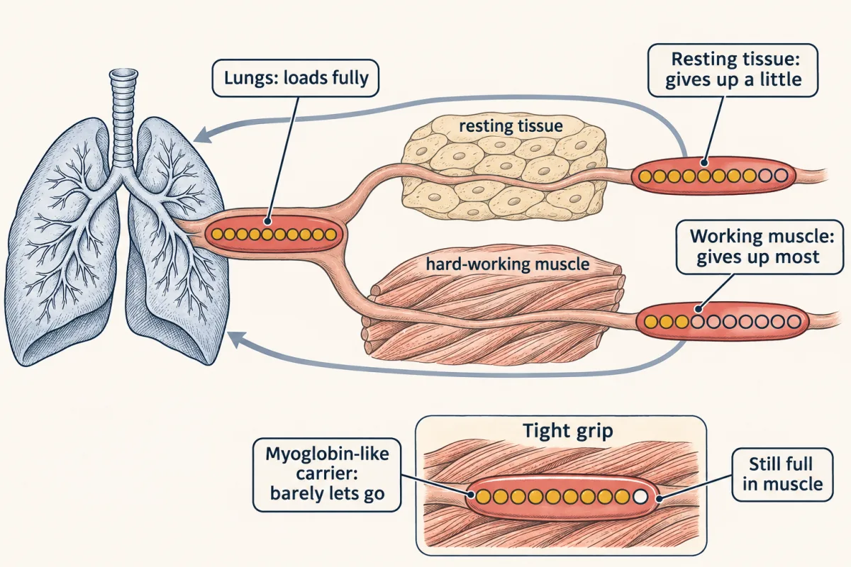
Hemoglobin loads fully in the lungs (pO2 near 100 torr), gives up only about a fifth in resting tissue (near 40 torr) and about two-thirds in hard-working muscle (near 20 torr). A tight-gripping, non-cooperative carrier like myoglobin would still be about nine-tenths full in the muscle, delivering about 7%. Dots are schematic: each stands for a tenth of the cell's oxygen. AI-generated illustration -
One site: Kd and the hyperbola
-
Two shapes: building the S
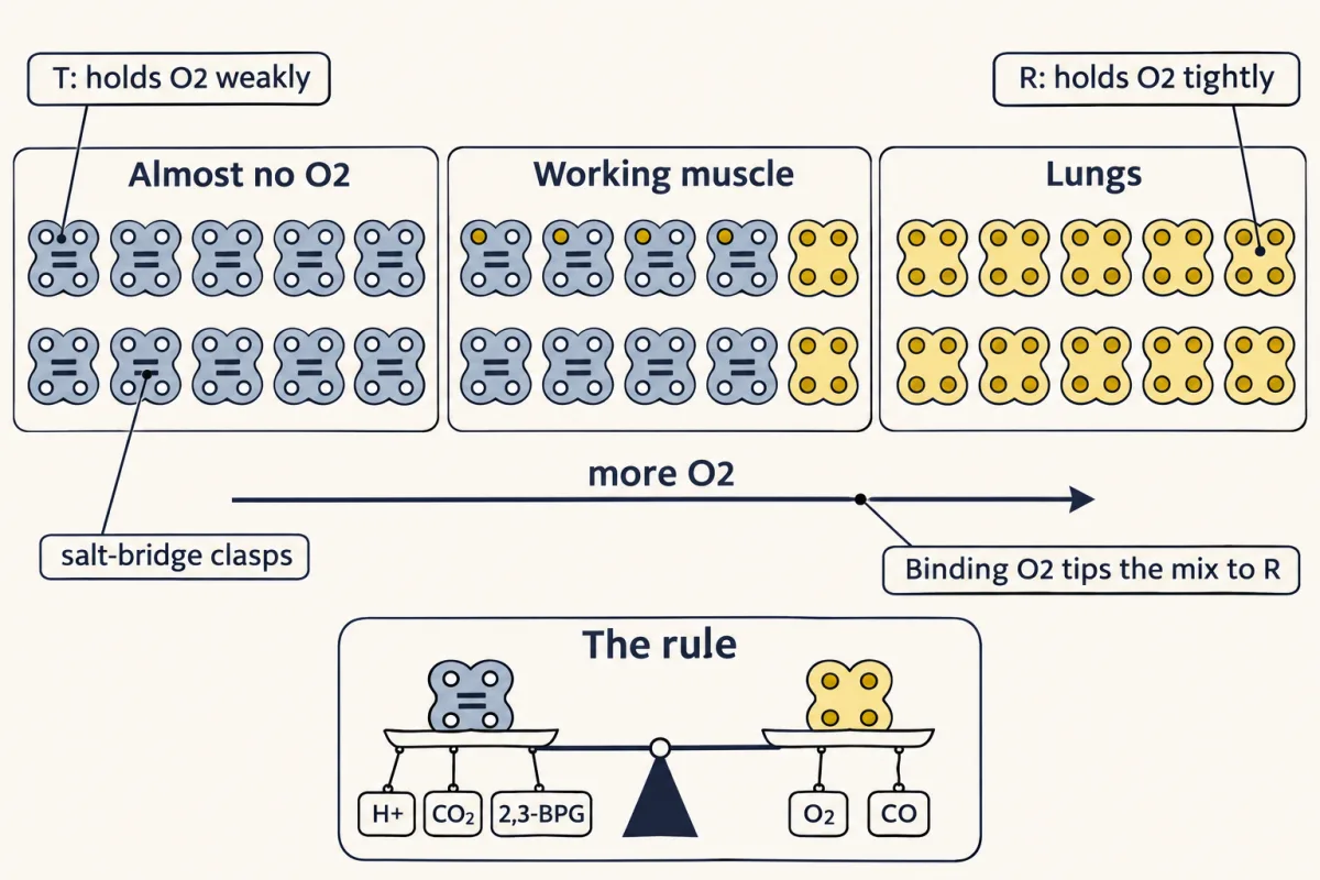
Hemoglobin is always a mix of two shapes. With no oxygen, almost every molecule is T. Because R holds oxygen far more tightly, each oxygen bound tips the mix toward R: in the lesson's illustrative fit, about a fifth of the molecules are R at 20 torr and about 96% at 100 torr. Anything that binds one shape better pulls the balance its way (inset). Counts are schematic. AI-generated illustration -
The switch inside the molecule
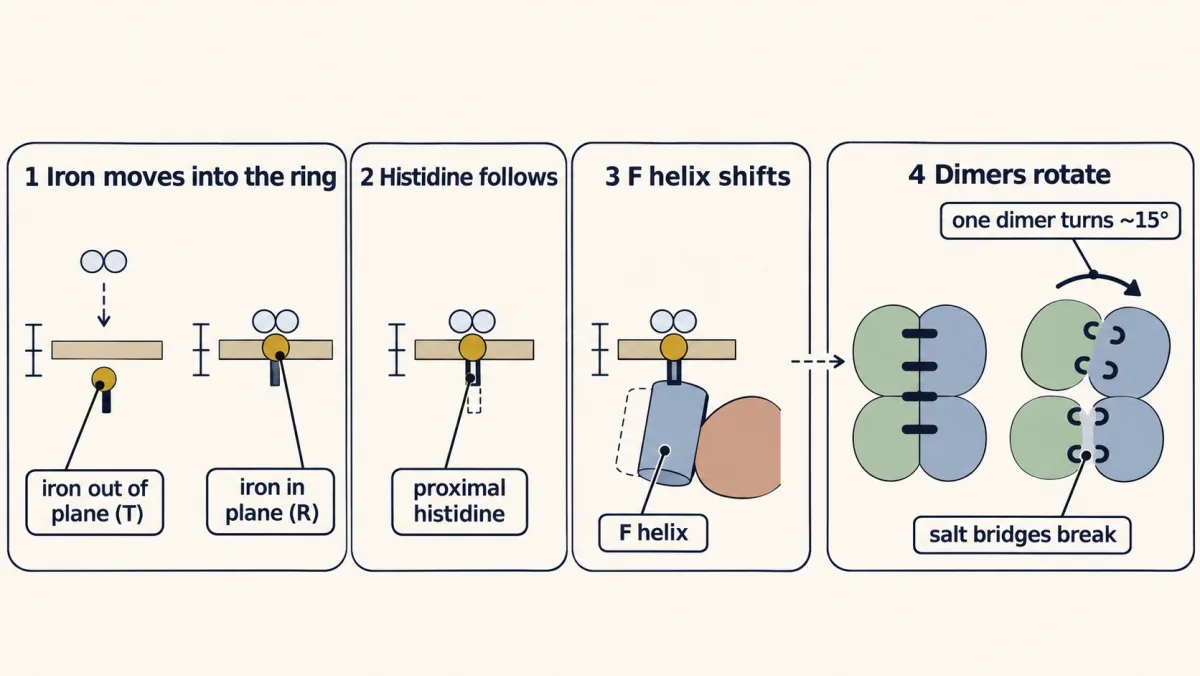
Perutz's chain from T to R. Oxygen binding lets the iron, about 0.4 Å out of the heme plane in deoxy hemoglobin, move into the plane. The proximal histidine and the F helix follow, and the movement is levered into a roughly 15° rotation of one αβ dimer against the other that breaks the T-state salt bridges. The dashed arrow marks the hedge: each iron shift raises the odds of the switch rather than forcing it. Sizes and movements are exaggerated. AI-generated illustration -
Tuning the mix: allosteric regulators
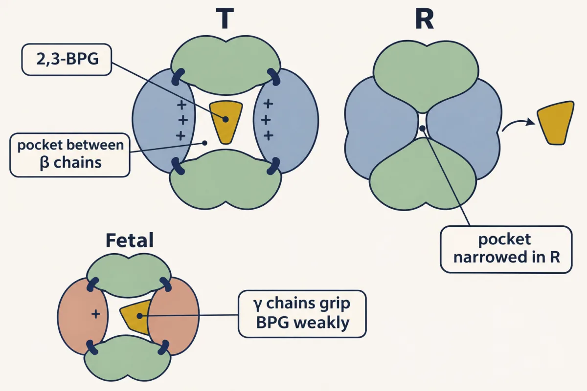
2,3-BPG is a doorstop for the T shape. It binds the wide central pocket between the β chains in T, while R narrows that pocket and binds it far more weakly, so BPG shifts the mix toward T and moves the curve right. Fetal hemoglobin's γ chains (serine instead of histidine at position 143) grip BPG weakly, so HbF stays at higher oxygen affinity. Schematic. AI-generated illustration -
When the shapes betray you: hemoglobin pathology
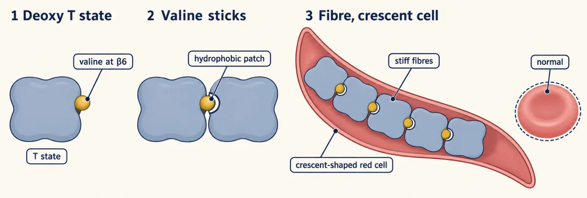
In deoxygenated hemoglobin S, valine at β6 contacts a hydrophobic patch on a neighboring hemoglobin molecule. These intermolecular contacts help assemble long, stiff fibers that deform red cells. The molecular contact is enlarged separately from the cell-scale view. AI-generated illustration -
Going deeper
Where this lesson fits
Before thisReview first
- Protein Three-Dimensional Structure: The Sequence Sets the ShapeThis course · Lesson 5
You need how a sequence folds into a shape, and how one residue change alters it, to follow the T and R shapes.
- Water, Weak Bonds, and the Generation of OrderThis course · Lesson 3
The salt bridges that hold the T shape together are weak bonds, so you need their strength and reversibility from there.
- Amino Acids: How One Side Chain Decides EverythingThis course · Lesson 4
Histidine's charge and pKa from there explain why protons stabilize the T shape in the Bohr effect.
After thisStudy next
- Oxidative Phosphorylation: The Electron-Transport ChainThis course · Lesson 20
It uses the oxygen that hemoglobin delivers, as the final electron acceptor of the respiratory chain.
- Signal-Transduction Pathways: How Cells Compute With Molecular SwitchesThis course · Lesson 13
It reuses the idea that a ligand shifts a protein's balance between active and inactive shapes.
- Integration of Metabolism: How the Body Switches FuelsThis course · Lesson 30
It follows how tissues' oxygen and energy demands change with fed and fasting states.
Elsewhere on the siteFilms, simulations and method films in other courses
- Why Blood Needs an S-Shaped CarrierFilm · Organismal Biology
It shows why plasma alone carries little oxygen and hemoglobin's S-shaped curve raises the load and release.
- Osmosis chamberSimulation · Biology
Its simulation is not about this idea, so it should be skipped.
- Dose–response explorerSimulation · Cell Signaling
It lets you explore occupancy against ligand concentration, the same Kd and half-saturation idea.
Suggested by an AI model from the lessons themselves, then checked. How we use AI