Cell Signaling · Skim version
PLC, IP3, DAG and Protein Kinase C
Read the full lesson → Skim the whole course →
The guiding idea
How does a kinase know that a receptor has just cut a lipid, here and now?
One PIP2 cut sends IP3 away and leaves DAG at the membrane; conventional PKC integrates calcium targeting with DAG activation at membranes.
From the film: One Cut, Two Destinations
- One PIP2 cut sends IP3 away and leaves DAG at the membrane.
- IP3 is water-soluble, while DAG remains in the inner membrane leaflet.
- Gq activates PLC-beta; receptor phosphotyrosines dock PLC-gamma, and the receptor kinase then phosphorylates it.
- IP3 opens an ER calcium channel; it does not carry calcium itself.
From the film: How Conventional PKC Reads Both Branches
- Conventional PKC integrates calcium targeting with DAG activation at membranes.
- Calcium-bound C2 recruits conventional PKC to anionic membrane lipids.
- DAG-bound C1 strengthens membrane engagement and helps displace the pseudosubstrate.
- The response is graded: stronger and longer where calcium and DAG overlap, not an all-or-none switch.
-
One cut makes two messengers
-
Two receptor routes converge on the cut
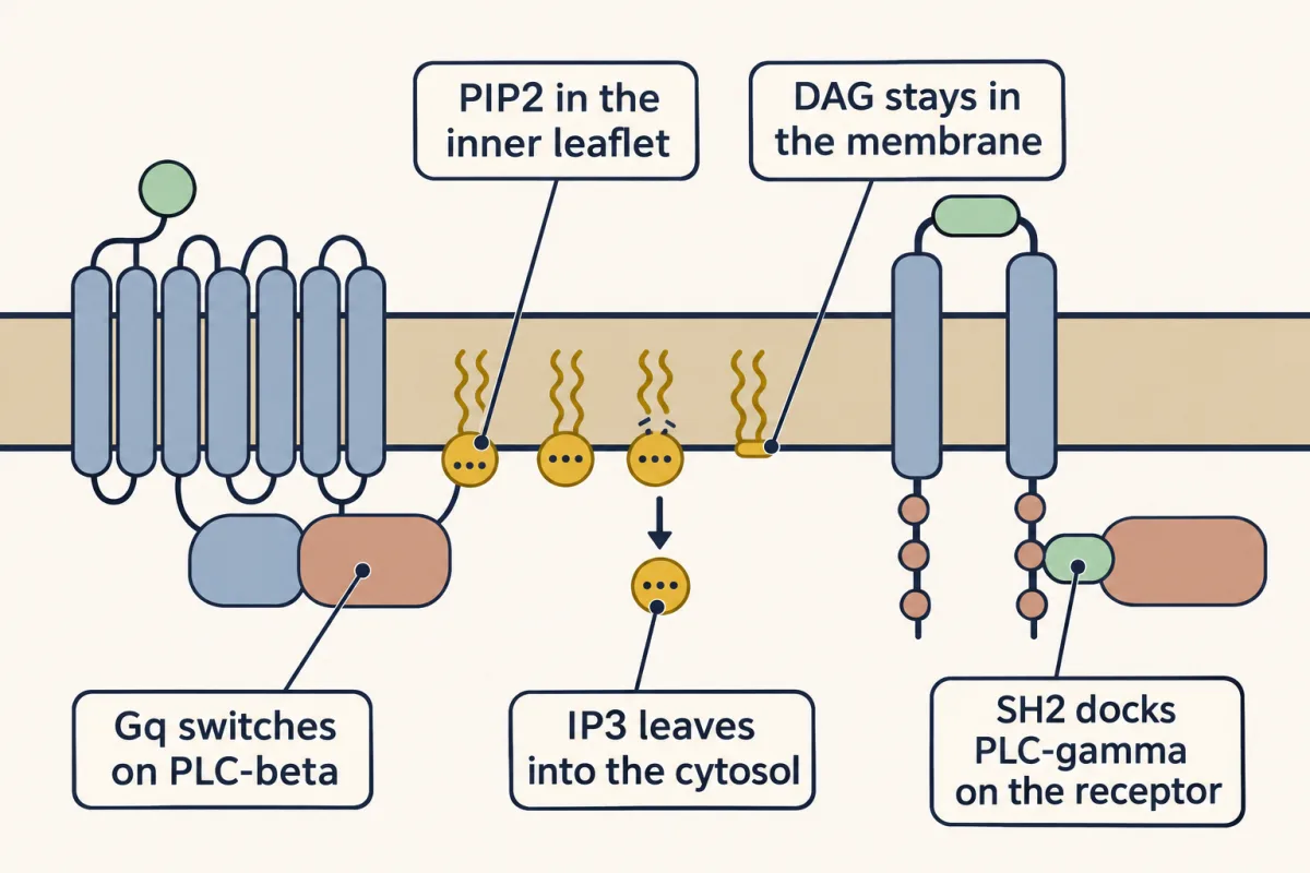
Gq activates PLC-beta, while receptor phosphotyrosines recruit PLC-gamma; either route cuts PIP2 so IP3 leaves and DAG remains in the membrane. AI-generated illustration -
How IP3 earned the name messenger
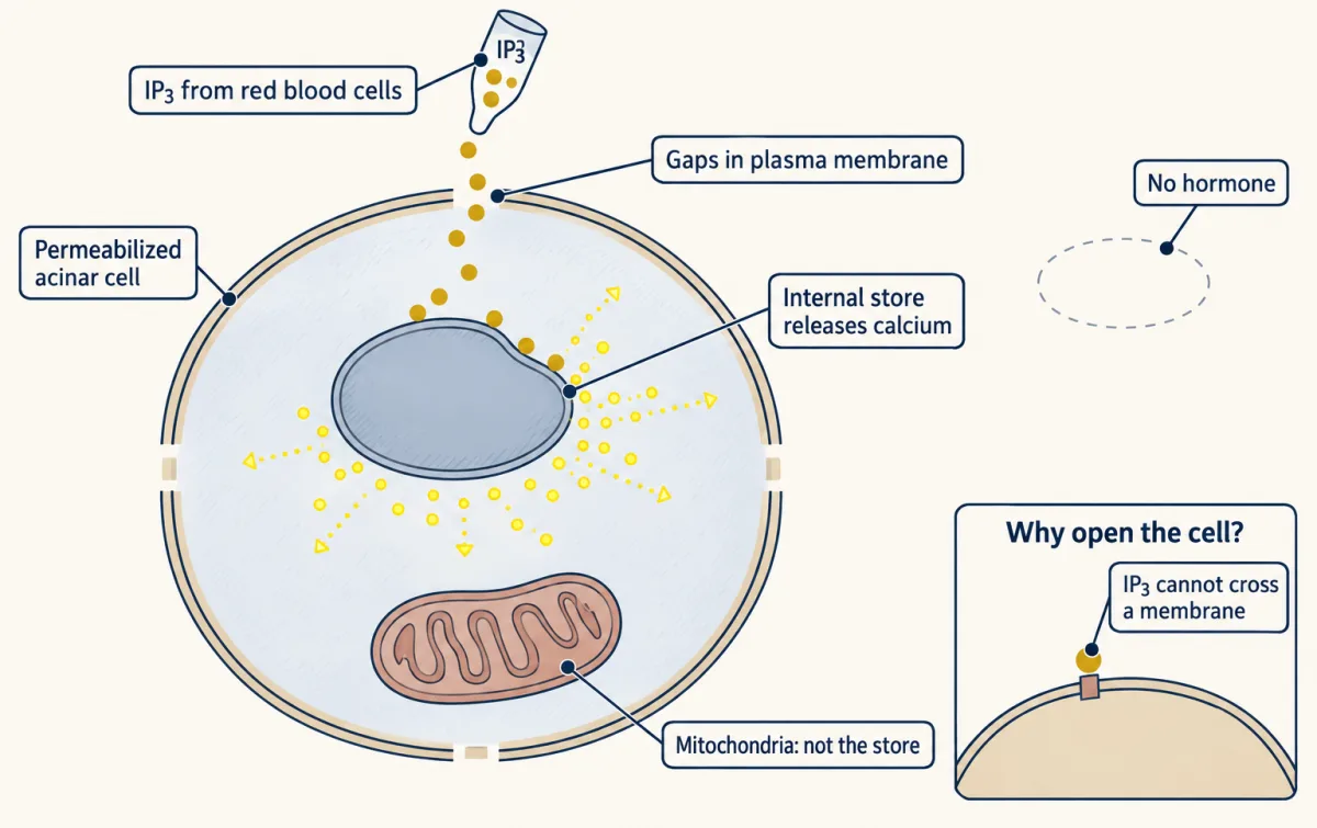
Added IP3 released calcium from a sealed nonmitochondrial store without hormone, establishing that IP3 was sufficient for the response. AI-generated illustration -
Conventional PKC joins the branches
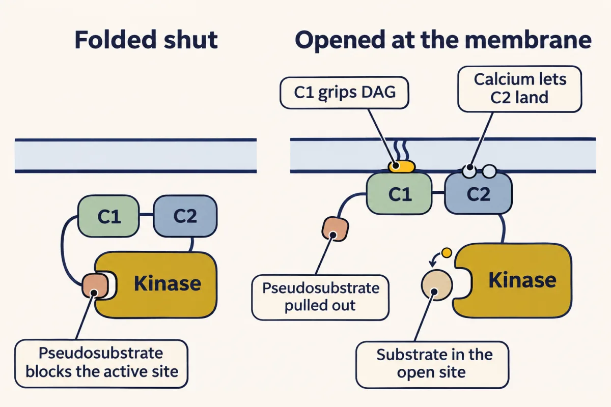
Calcium-dependent C2 binding recruits conventional PKC to an anionic membrane, where C1 can recognize DAG and membrane engagement relieves pseudosubstrate inhibition. AI-generated illustration -
A DAG mimic shows why duration matters
-
Ending and rebuilding the signal
-
Going deeper
Where this lesson fits
Before thisReview first
- GPCRs and Trimeric G ProteinsThis course · Lesson 12
You need to know how a receptor loads GTP onto Gq alpha, since that is what switches on PLC-beta.
- Protein Modules: SH2, SH3, PTB, PDZ and PHThis course · Lesson 9
PLC-gamma docks on receptor phosphotyrosines through its SH2 domains, so you need how SH2 domains read marks.
- Lipids as Anchors and AddressesThis course · Lesson 11
PIP2 is the lipid that PLC cuts, and this lesson assumes you know it as a membrane label.
After thisStudy next
- Calcium: The Messenger Kept at Arm's LengthThis course · Lesson 15
It takes up the calcium that IP3 releases from the ER and shows how the cell reads and clears it.
- Turning the Signal Off: Arrestins, RGS Proteins and PhosphodiesterasesThis course · Lesson 16
It shows how the receptor and G-protein side of this pathway is switched off.
- Receptor Tyrosine KinasesThis course · Lesson 18
It explains how receptor tyrosine kinases are activated and how they recruit PLC-gamma.
Suggested by an AI model from the lessons themselves, then checked. How we use AI