Biology · Skim version
Organelles as a System
Read the full lesson → Skim the whole course →
The guiding idea
The insulin gene never leaves a beta cell's nucleus, yet insulin leaves the cell after a meal. What carries it out, and in what order?
A eukaryotic cell sends a protein out along a route of parts, and each part can only work on what the part before it hands over.
From the film: One Cell, One Shipping Line
- A beta cell keeps the insulin gene in its nucleus and sends out a messenger RNA copy instead.
- A ribosome on the rough ER reads the messenger RNA and builds the protein, which folds inside the ER.
- Vesicles carry the protein to the Golgi apparatus, which sorts and packs it, and a vesicle fuses with the cell membrane to release it.
- Each part can only work on what the part before it hands over.
- Many steps use energy, much of it as ATP made by mitochondria.
From the film: Catching Proteins on the Move
- A short pulse of a radioactive building block marks only the proteins made during it.
- Following that marked batch over time shows where it goes in the cell.
- Each part's share of the mark peaked in turn: rough ER, then Golgi vesicles, then condensing vacuoles, then storage granules.
- The marked batch spread out as it moved: by 7 minutes, Golgi vesicles held almost as many grains as the rough ER.
- These cells make digestive enzymes, not insulin, and they gave direct evidence for the order of the stops.
-
What the first film left out
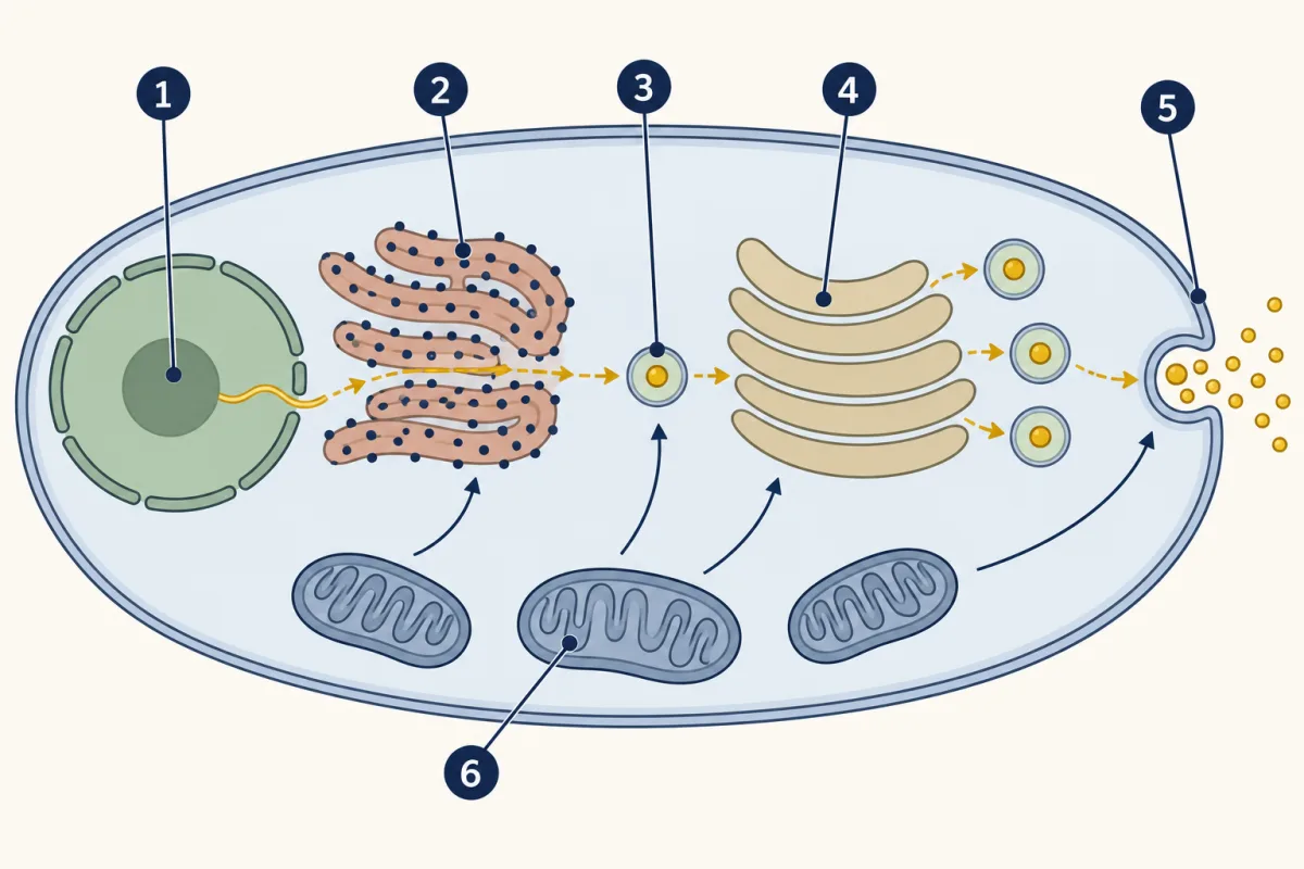
- nucleus
- rough ER
- vesicle
- Golgi apparatus
- cell membrane
- mitochondria
-
Parts on the route, and parts off it
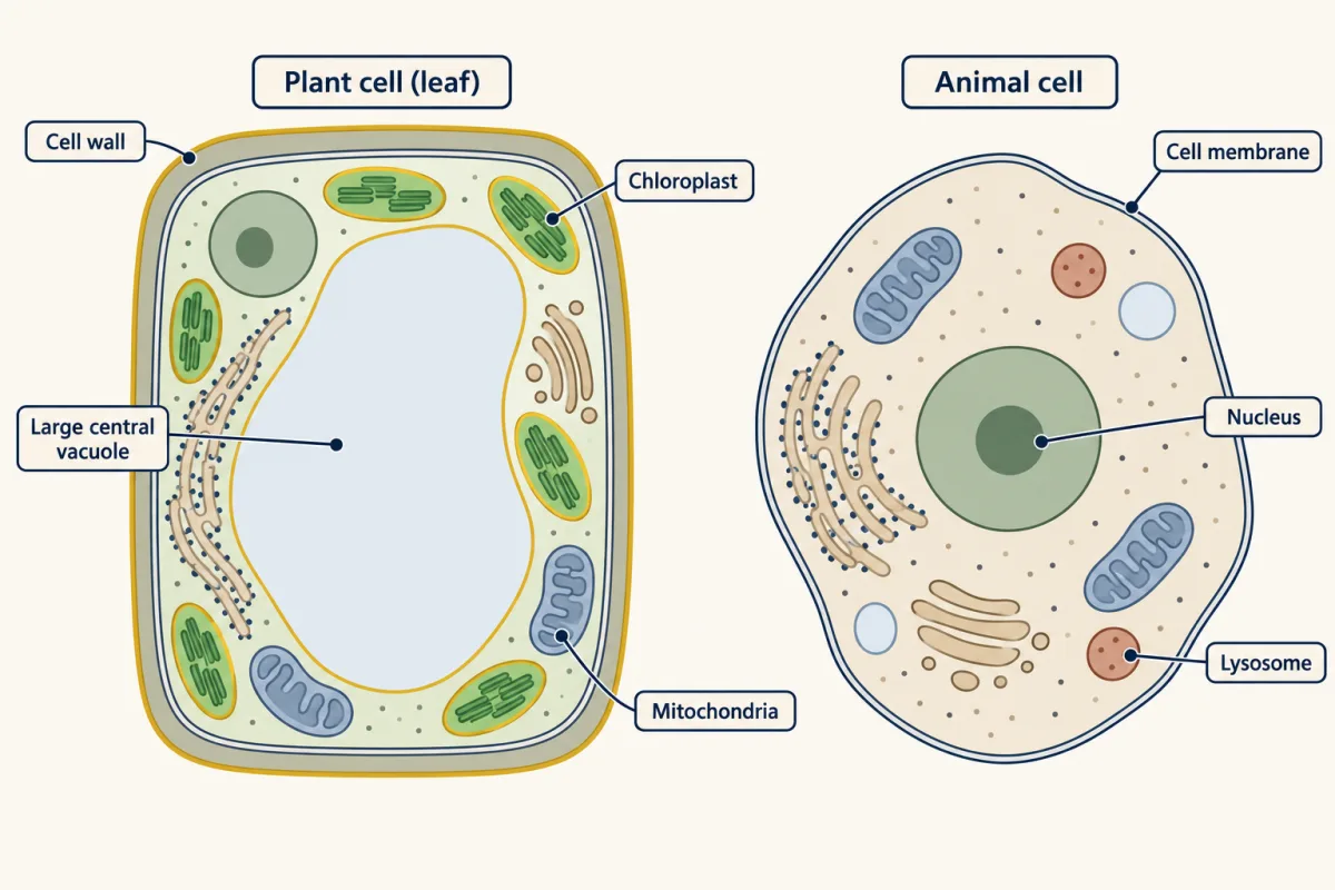
A typical leaf cell and a typical animal cell share most parts, including mitochondria. The plant cell adds a cell wall, a large central vacuole and, because it is green, chloroplasts. AI-generated illustration -
Explore: test the one idea in a model
-
A factory model, and where it breaks
-
How a marked batch really moves
-
Same jobs, simpler parts: prokaryotic cells
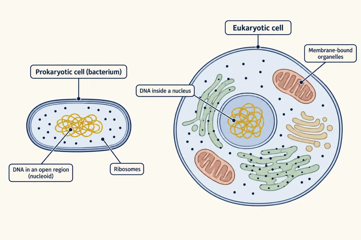
A bacterium keeps its DNA in an open region with no membrane around it and sends proteins out through channels in its membranes; a eukaryotic cell keeps most of its DNA in a nucleus and sends proteins along a route of membrane-bound parts. Not drawn to scale. AI-generated illustration -
Cells built for their work
-
Evaluate: check your thinking
-
Going deeper
Where this lesson fits
Before thisReview first
- Respiration and ATPThis course · Lesson 5
Mitochondria make the ATP that pays for steps along the route, and that lesson shows how sugar becomes ATP.
- What Am I Made Of?This course · Lesson 2
You need to know proteins are built from amino acids, since the route makes and moves proteins.
After thisStudy next
- Crossing the MembraneThis course · Lesson 8
The cell membrane is the last stop here, and this lesson shows what it lets in and out.
- From DNA to ProteinThis course · Lesson 18
It takes the messenger RNA copy and ribosome step further, showing how the code is read into a protein.
- One Cell or ManyThis course · Lesson 11
It builds on cells having different parts for different jobs, and on how cells share work.
Elsewhere on the siteFilms, simulations and method films in other courses
- Pulse-chase labellingMethod film
The marked-batch data in this lesson come from labelling proteins briefly and following where they go.
Suggested by an AI model from the lessons themselves, then checked. How we use AI