Developmental Neuroscience · Skim version
Neurulation and Neural Tube Defects
Read the full lesson → Skim the whole course →
The guiding idea
In a human embryo the neural tube has closed by the end of about the fourth week after fertilization. What happens if it doesn't? Part of the brain or spinal cord stays open, and folic acid taken before conception lowers the risk.
Narrow first, then fold: the neural folds can close only a gap their bending can cover, and the midline between them sets that gap.
From the film: Narrow First, Then Fold
- The plate narrows and lengthens first, and narrowing keeps going as the folds rise.
- A stretch of the tube that does not close stays open, and where it stays open names the defect.
- The folds can close only a gap their bending can cover, and the midline between them sets that gap.
From the film: Where in the Plate?
- The midline must become narrow enough, in time, for the folds to meet.
- Convergent extension narrows the midline, and planar cell polarity tells each cell which way is across.
- In frogs, blocking Dishevelled at the midline left the tube open, but blocking it at the sides did not.
- In mice with two copies of the loop-tail Vangl2 mutation, the tube is open from the midbrain down the spine.
From the film: Four Weeks, and a Vitamin
- Primary closure is complete by about the end of the fourth week after fertilization, about six weeks of pregnancy by the doctor's count.
- Many pregnancies are not yet recognized by then, so folic acid has to be in place before conception.
- In the first trial, folic acid cut recurrence by about 72 percent, an absolute fall of about 2.5 percentage points.
- Folic acid lowers the risk but does not remove it, and how it works is not settled.
-
From a flat plate to a tube
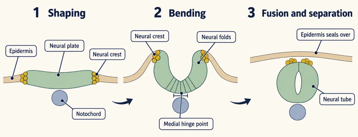
Closing the neural tube in the trunk, in cross-section: the plate creases at the hinge over the notochord, its edges rise as neural folds, meet and fuse, and the tube separates from the epidermis, which seals over it. Neural crest arises at the plate border; in the trunk it leaves around or after fusion, but in the mammalian head crest cells begin leaving before the folds fuse. Crest migration is not shown. AI-generated illustration -
Where closure fails
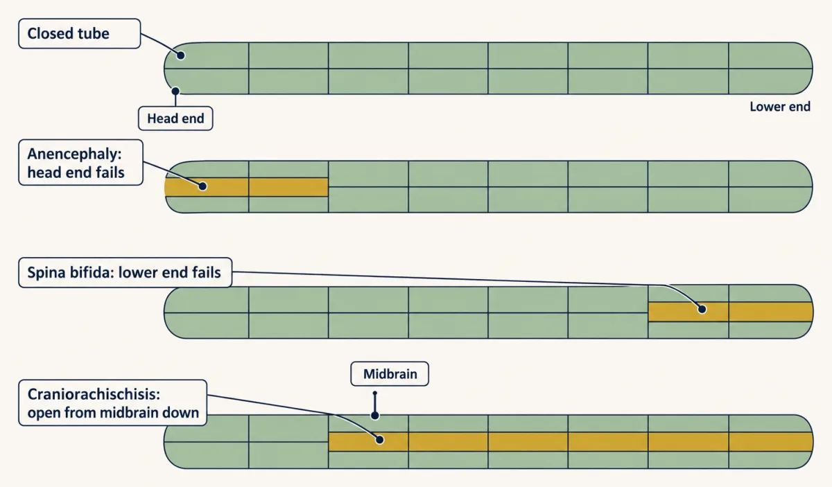
An open neural tube defect is a failure of closure, and its name depends on where the failure is: the head end in anencephaly, the lower end in open spina bifida (myelomeningocele), and the whole stretch from the midbrain down in craniorachischisis. The map is schematic. AI-generated illustration -
Narrow first, then fold
-
When the tube closes
-
The folic-acid trials: strong evidence
-
What is still open
-
Going deeper
Where this lesson fits
Before thisReview first
- Neural Induction: The Default ModelThis course · Lesson 6
Neural induction makes the ectoderm neural; this lesson starts from the neural plate that induction produces and rolls it into a tube.
- The Organizer: Spemann and MangoldThis course · Lesson 5
The organizer and notochord set up the midline plate and the tissues beneath it that the folds close over.
After thisStudy next
- The Neural Crest: Cells That Leave HomeThis course · Lesson 20
Neural crest cells arise at the plate border and leave the folds around closure, which this lesson only mentions.
- Head to Tail: Hox Genes, Retinoic Acid and RhombomeresThis course · Lesson 8
After the tube closes, its head-to-tail axis is patterned into hindbrain segments and the spinal cord.
- Ventral to Dorsal: Sonic Hedgehog and BMP in the Neural TubeThis course · Lesson 10
It uses the closed tube and the notochord below it to explain how the tube is patterned from floor to roof.
Suggested by an AI model from the lessons themselves, then checked. How we use AI