Cell Signaling · Skim version
Integrins and Rho GTPases
Read the full lesson → Skim the whole course →
The guiding idea
How does a cell hold on to what it stands on, feel how hard it is pulling, and use that grip to crawl?
Integrins tie the matrix outside a cell to the actin inside it and signal in both directions, and three Rho-family switches turn that grip into movement.
-
The idea: a receptor for something solid
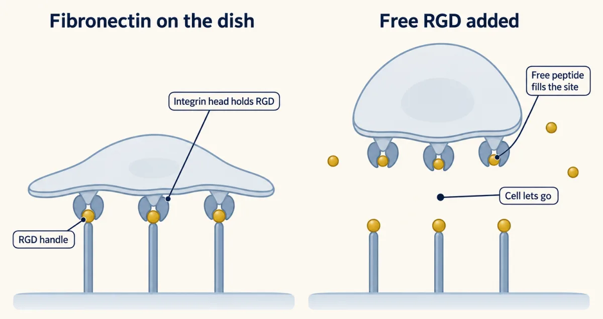
RGD is the handle that integrins hold on fibronectin. Add enough free RGD peptide to a dish and it fills the integrin sites that the matrix would have filled, so fewer cells hold on. Drawn schematically. AI-generated illustration -
A grip that is switched on from inside
-
The focal adhesion: a signalling machine that can feel a pull
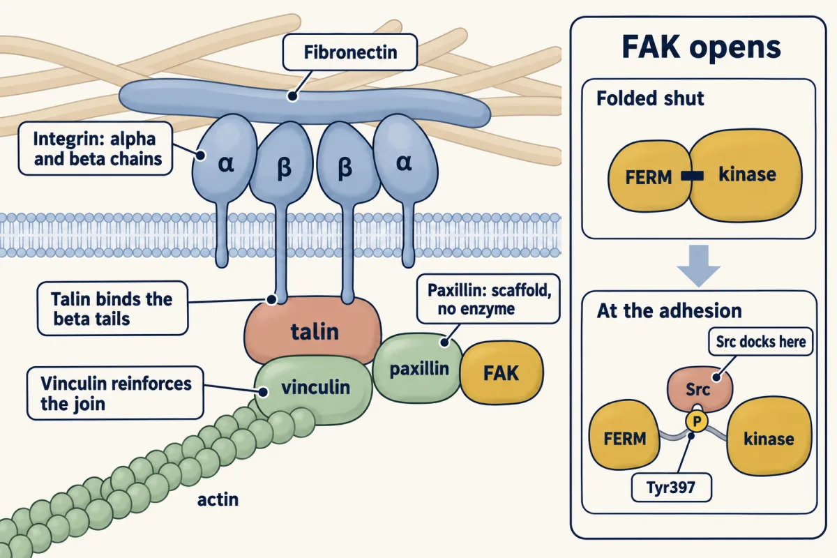
Clustered integrins collect talin, vinculin, paxillin and FAK on their short tails to make a focal adhesion. Talin binds the beta tails, and vinculin reinforces the join to actin. FAK starts folded shut; at a cluster it opens, phosphorylates Tyr397 in its linker, and Src docks there. How clustering opens it is not fully worked out. The drawing is a simplified schematic. AI-generated illustration -
Three switches for the actin skeleton
-
How a cell crawls, and what happens when it cannot
-
Going deeper
Where this lesson fits
Before thisReview first
- GTPase Switches: The TimerThis course · Lesson 7
Rho, Rac and Cdc42 work as GTP-on, GDP-off switches with GEFs and GAPs, as taught there.
- Protein Modules: SH2, SH3, PTB, PDZ and PHThis course · Lesson 9
Src docks on phosphorylated FAK through its SH2 domain, and paxillin works as a docking scaffold.
- Adaptors, Docking Proteins and ScaffoldsThis course · Lesson 10
Paxillin and Grb2 act as adaptors and scaffolds at the focal adhesion, linking an enzyme-free receptor to kinases.
After thisStudy next
- Endocytosis and Signaling EndosomesThis course · Lesson 36
Integrins and their adhesion signals are also taken into the cell and recycled, which moves where they signal.
- Antigen Receptors and CheckpointsThis course · Lesson 29
White-cell integrins are switched on from inside by receptor signals, as in your design problem, to hold other cells.
Elsewhere on the siteFilms, simulations and method films in other courses
- One Chain, Four LinksFilm · Cell Biology
It shows the chain from matrix to receptor to adaptors to a cable inside, the same link integrins make.
- How Internal Motion Becomes MovementFilm · Cell Biology
It shows how actin polymerization and integrin coupling drive forward movement at a cell's leading edge.
- Polymerization minus flowSimulation · Cell Biology
It lets you vary polymerization and rearward flow to see how adhesion coupling sets crawling speed.
- Tight Ends, Loose EndsFilm · Cell Biology
It shows how actin filament ends grow and shrink, the structures that Rho-family switches shape.
Suggested by an AI model from the lessons themselves, then checked. How we use AI