One Chain, Four Links
Read the transcript of “One Chain, Four Links”
Film transcript: One Chain, Four Links
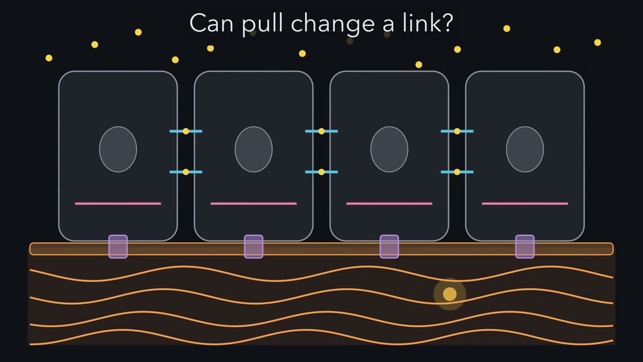
Take the calcium away, and this sheet of cells comes apart. What was the calcium holding together? Put it back, and the cells knit together again. Each cell reaches across with arms called cadherins. Calcium sits at the joints of each arm and keeps it stiff. The arm on one cell grips the arm on the next. Follow one arm into the cell. Adaptor proteins grip its tail and pass the hold inward: beta β-catenin, then alpha α-catenin. They tie it to a cable, a filament of actin. This grip is an adherens junction. So, in its simplest form, one grip is a chain of four links. A partner outside, a receptor in the membrane, adaptors, and a cable inside. Now pull on the sheet.
In this simple picture, the pull runs from cable to cable, through every link. Break any one link, and this path is cut. Now take the calcium away. Which link gives way first? The arms outside, the adaptors, or the cable?
Choose one. Calcium leaves the joints, and the arms go floppy. The outer link lets go. The adaptors and the cable are still there, but this contact can no longer pass the pull across. One broken link, and the sheet comes apart. Now look down, at the matrix, the material cells secrete around themselves. Here the receptor is an integrin, a pair of subunits, α and β. It grips a matrix protein, fibronectin, at a short tag, RGD. Inside, talin ties the integrin's tail to actin, and vinculin adds extra ties to the same cable. Notice the pattern. The receptor has changed, but the four links have not. When the cell tugs on its cable, the pull runs down into the matrix.
Two more junctions build the same chain with a different cable. Desmosomes join cell to cell. Hemidesmosomes pin a cell to the basement membrane, the thin matrix sheet beneath. In epithelia, both end on keratin, an intermediate filament, not on actin. At the chain's far end is the matrix itself. Its collagen ropes straighten under a pull and resist it strongly. But squeeze the tissue, and the ropes buckle and go slack. Ropes are poor at pushing. So the matrix also holds a gel of proteoglycans, proteins with long sugar chains. In a first picture, the chains' negative charges draw in water, and the gel swells. Squeeze it, and it pushes back. The tissue is one web of chains, from matrix ropes to cell cables.
A real junction holds many chains side by side, sharing the load. One question is left. Does the pull change the links themselves?
Take the calcium away and a sheet of epithelial cells comes apart. What was the calcium holding together?
Continue in the lesson: Adhesion and the Extracellular Matrix →
3:26 · Free, with captions and a transcript. The lesson has the full text, the questions and the sources.
More films from this lesson
How this film was made. Narration: written from this lesson's text and read by an AI narrator voice (Kokoro, af_heart). Animation: AI-generated in code (Manim) by Claude, a schematic with no drawn molecular structures. Captions come from the narration script. See something wrong? Report it.