Biochemistry · Skim version
Basic Concepts of Enzyme Action
Read the full lesson → Skim the whole course →
The guiding idea
Can an enzyme make an impossible reaction happen? No — it can only make a possible one fast.
Can an enzyme make an impossible reaction happen?
From the film
- An enzyme lowers the ridge between substrate and product; it never moves the valleys.
- Which valley is lower (ΔG) decides which way a reaction runs; the ridge height (ΔG‡) decides how fast.
- At 25 °C, every 5.7 kJ/mol taken off the barrier makes a reaction ten times faster.
- An enzyme speeds the forward and backward trips by the same factor, so the equilibrium constant stays the same.
- It lowers the top by gripping the transition state more tightly than the substrate.
-
Where a reaction ends up: ΔG, ΔG°′ and Q
-
How fast it gets there: the height of the hill
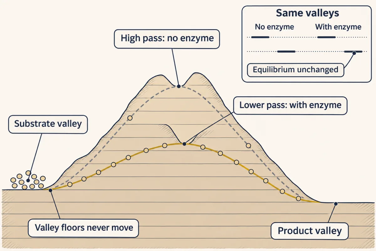
An enzyme opens a lower pass over the same ridge. The valleys, free substrate and free product, stay exactly where they were, so the equilibrium is unchanged; only the height of the crossing, and so the speed, changes. AI-generated illustration -
How an enzyme lowers the top: grip the transition state
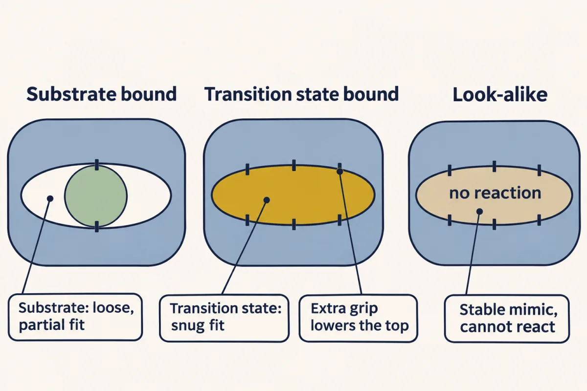
The active site is built for the transition state, not the substrate. The extra grip on the transition state is what lowers the top of the barrier, and a stable look-alike that copies its shape binds more tightly than the real substrate. AI-generated illustration -
The evidence: transition-state analogs bind tighter
-
A separate question: does the active site change shape?
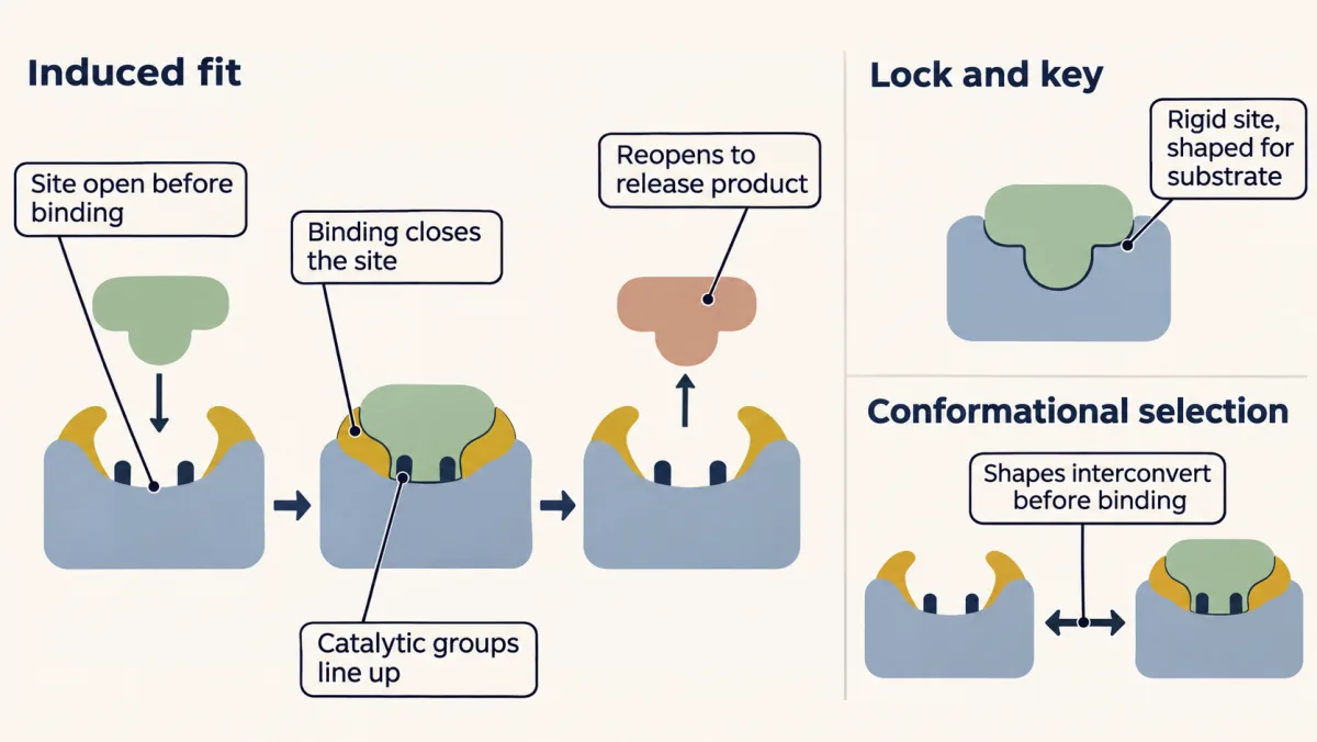
Does the active site change shape? Lock and key: a rigid site already shaped for the substrate. Induced fit: binding closes the site and lines up catalytic groups. Conformational selection: open and closed shapes interconvert first, and substrate picks the one that fits. Which shape the site fits best, the transition state, is a separate question. AI-generated illustration -
Tight binding as a drug strategy
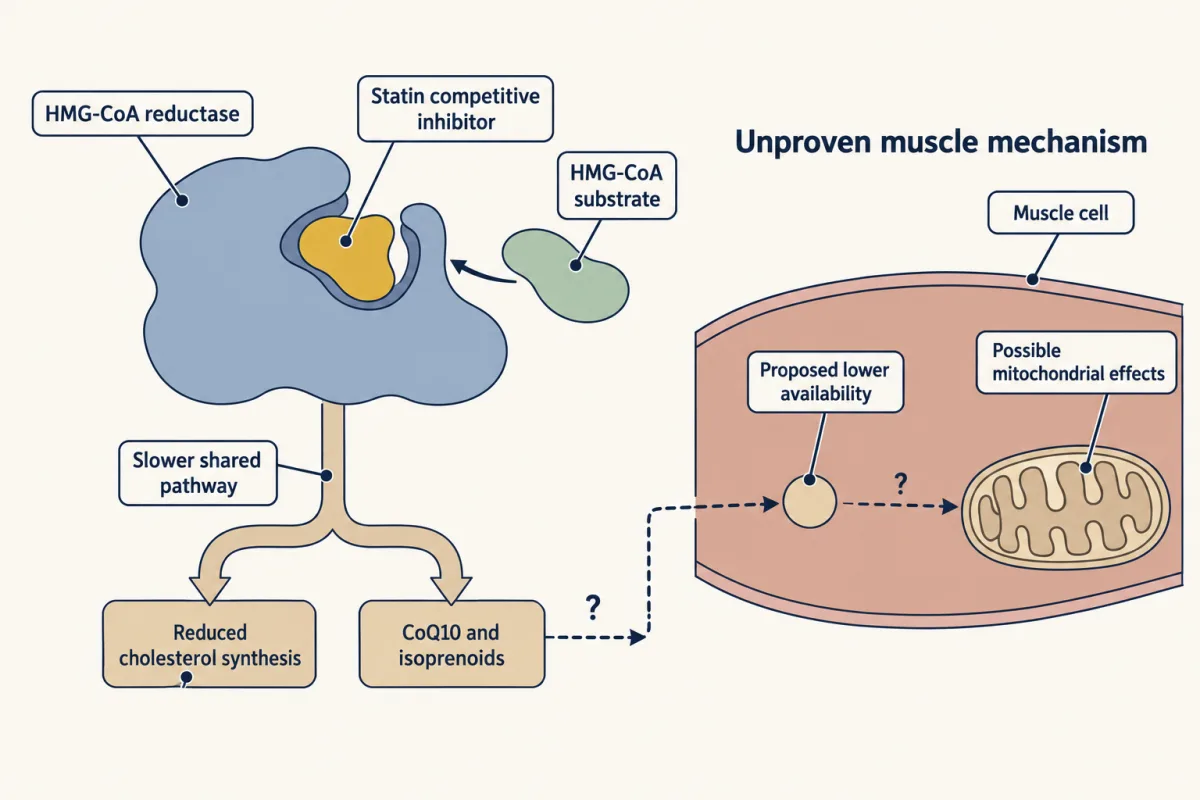
Statins competitively inhibit HMG-CoA reductase and slow the shared biosynthetic pathway. Reduced CoQ10 and isoprenoid availability with possible mitochondrial effects is one proposed, unproven explanation for muscle symptoms; true muscle damage with a large CK rise is rare. AI-generated illustration -
Naming enzymes and their helpers
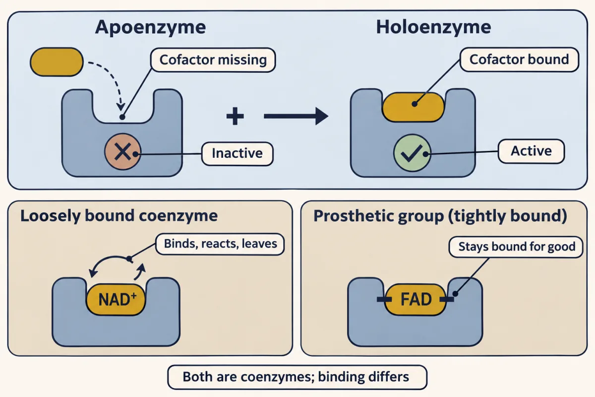
Apoenzyme (cofactor missing, inactive) and holoenzyme (cofactor bound, active). NAD⁺ and FAD are both coenzymes; what differs is how tightly they bind. NAD⁺ comes and goes, while tightly bound FAD stays put and so counts as a prosthetic group. AI-generated illustration -
Going deeper
Where this lesson fits
Before thisReview first
- Water, Weak Bonds, and the Generation of OrderThis course · Lesson 3
You need weak, reversible bonds from there to understand how an active site grips a substrate or transition state.
- Protein Three-Dimensional Structure: The Sequence Sets the ShapeThis course · Lesson 5
The active site is a cleft formed by folding, so you need how sequence sets a protein's shape.
After thisStudy next
- Enzyme Kinetics: The Michaelis–Menten ModelThis course · Lesson 8
It measures kcat and KM, the rates this lesson links to transition-state binding, and shows saturation.
- Metabolism: Basic Concepts and DesignThis course · Lesson 15
It uses ΔG, Q and coupling to ask which metabolic reactions can run in cells.
- Protein Binding and Allostery: How Hemoglobin Loads and Delivers OxygenThis course · Lesson 9
It takes binding and Kd further, showing how binding changes with shape and other ligands.
Elsewhere on the siteFilms, simulations and method films in other courses
- Speed up and slow down an enzymeSimulation · Biology
It lets you speed up and slow down an enzyme and see the catalyst come out unchanged.
- Count the Working SitesFilm · Biology
It shows the substrate fitting the active site and the enzyme working again unchanged.
- Osmosis chamberSimulation · Biology
It shows a free-energy downhill direction setting net movement, the same ΔG-decides-direction idea applied to transport.
Suggested by an AI model from the lessons themselves, then checked. How we use AI