Cell Signaling · Skim version
Calcium: The Messenger Kept at Arm's Length
Read the full lesson → Skim the whole course →
The guiding idea
How can the same calcium ion trigger contraction, secretion or gene expression without making the cell respond everywhere at once?
Cells give calcium meaning by controlling where and when free calcium rises.
From the film: How a Cell Makes a Calcium Signal
- Calcium signals begin and end by moving calcium, not changing it.
- Opening a channel lets an existing gradient raise free cytosolic calcium.
- Pumps, exchangers and buffers lower free cytosolic calcium again.
- Restoring the low resting level makes the next rise noticeable.
From the film: How One Ion Carries Different Messages
- Cells give calcium meaning by controlling where and when free calcium rises.
- Buffers can keep a rise local, so a sensor beside the channel can respond while an identical one farther away does not.
- Calcium-dependent release can recruit more sites and spread a wave.
- Closely spaced spikes can build activity in a reader that keeps some activity from one spike to the next.
-
Build the rise: gradient, channels and removal
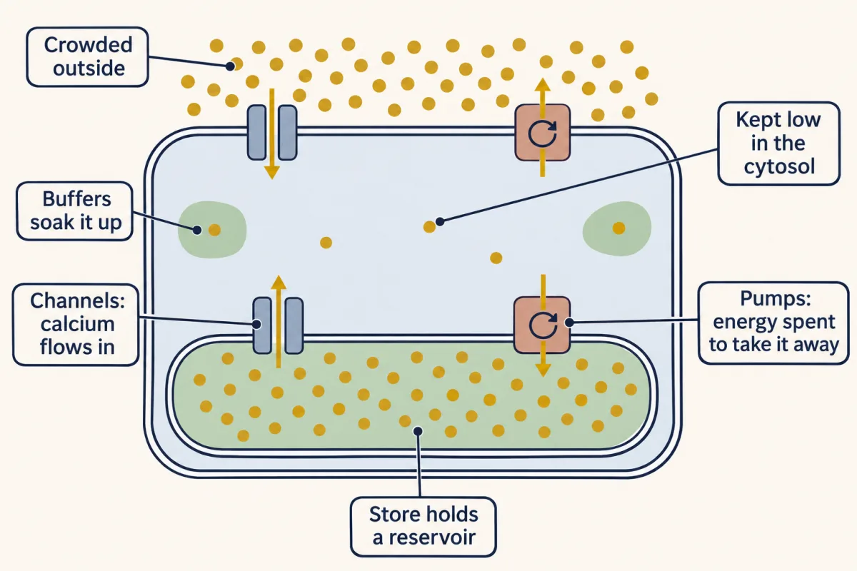
Channels raise free cytosolic calcium; pumps, exchangers and buffers lower it again. AI-generated illustration 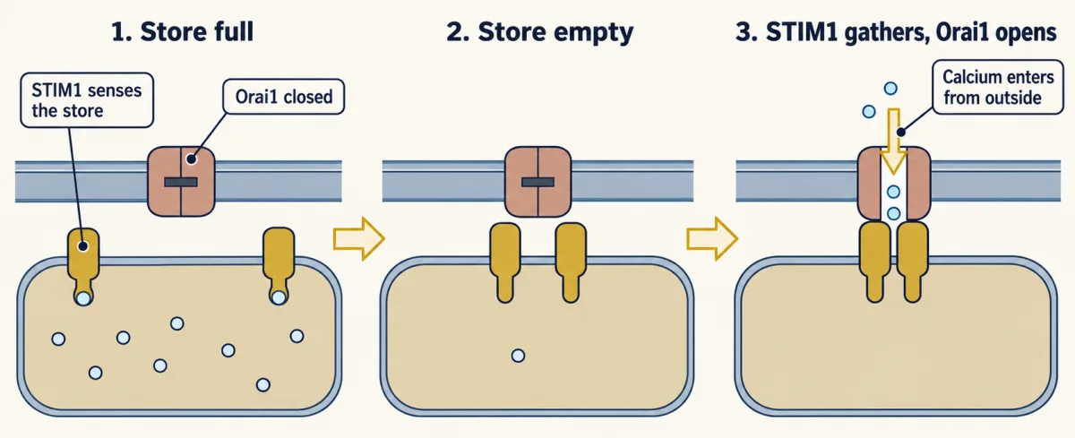
Low calcium inside the ER makes STIM1 cluster and open Orai1, coupling store depletion to entry from outside. Numbers of parts are schematic. AI-generated illustration -
How a protein reads the rise
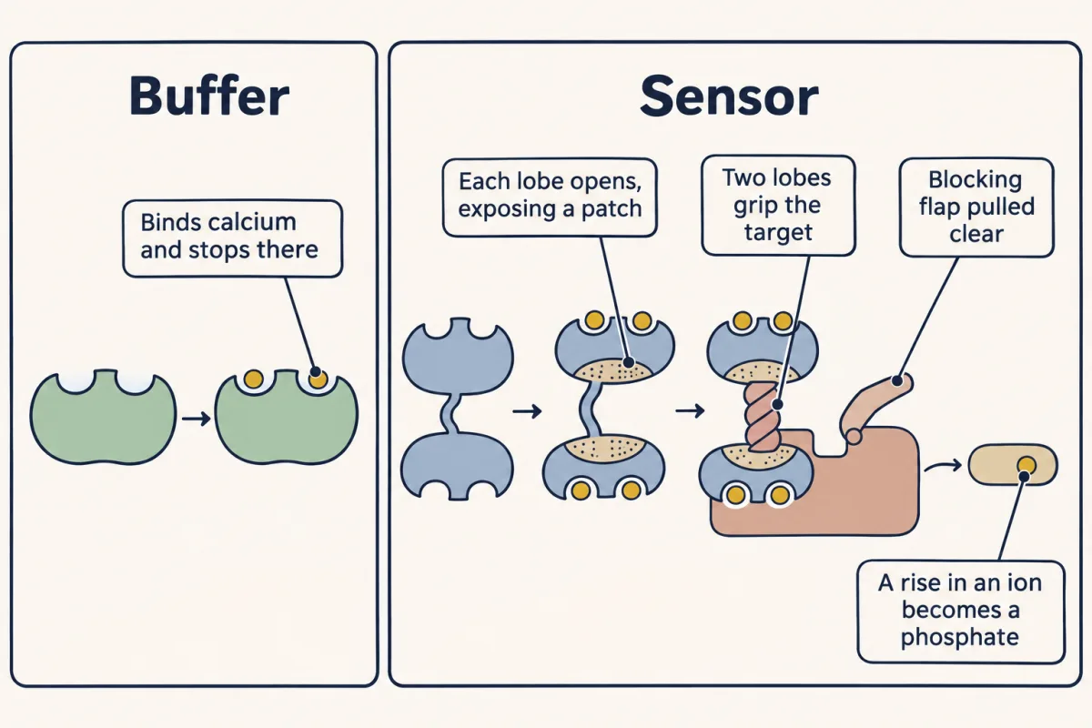
A buffer binds calcium; a sensor also changes shape and passes the signal to a target. AI-generated illustration -
Where and how often: the evidence and its limits
-
Seeing calcium without confusing the signal
-
Going deeper
Where this lesson fits
Before thisReview first
- Binding: Affinity, Occupancy and Dose–ResponseThis course · Lesson 3
You need Kd and occupancy from there to see why an EF hand fills at about a micromolar.
- PLC, IP3, DAG and Protein Kinase CThis course · Lesson 14
The IP3 made there is what opens the IP3 receptor on the store here.
- Phosphorylation: Kinases and PhosphatasesThis course · Lesson 6
CaMKII turns a calcium rise into a phosphate, which you read using kinase and phosphatase logic from there.
After thisStudy next
- Turning the Signal Off: Arrestins, RGS Proteins and PhosphodiesterasesThis course · Lesson 16
It takes the idea of switching a signal off further, with arrestins, RGS proteins and phosphodiesterases.
- The Shape of a Signal: Size, Duration, Frequency and PlaceThis course · Lesson 35
It uses calcium spike size, duration, frequency and place as examples of how a cell reads signal shape.
- Signaling to the Nucleus: CREB, AP-1 and Immediate Early GenesThis course · Lesson 32
Calcium and CaMKII signals reaching the nucleus switch on CREB and immediate early genes.
Elsewhere on the siteFilms, simulations and method films in other courses
- The length constantSimulation · Neuroscience
Resting leak and pump balance show how a steep ion gradient is held by pumps.
- Two Pushes, One HillFilm · Cell Biology
Concentration and voltage together set which way calcium flows when a channel opens.
- Blips from a Silent NerveFilm · Neuroscience
Calcium entry triggers vesicle release at the terminal, a key calcium-triggered response.
- A Tap, a Hole, a LevelFilm · Cell Biology
A pump keeps a store's gradient against leak, as in the calcium store.
Suggested by an AI model from the lessons themselves, then checked. How we use AI