Sensory Systems · Skim version
Spinal Circuits That Gate Pain
Read the full lesson → Skim the whole course →
The guiding idea
Light touch can start to hurt when a spinal gate fails. A gate can fail because the gatekeeper dies, or because the gatekeeper is still there and its signal has changed sign. In adult male rat lamina I, nerve injury was shown to do the second.
Inhibitory circuits in the spinal dorsal horn normally keep light touch from reaching pain pathways, and losing that control can make touch painful.
From the film: Who Opens the Touch Gate?
- In mice, inhibitory dorsal-horn neurons can keep touch input from recruiting mechanical-pain relays.
- Removing somatostatin-lineage relays reduces mechanical nocifensive behaviour in the assays tested.
- Ablation removes cells, while silencing can remove transmitter output from surviving cells.
- Behaviour identifies a failed gate; circuit measurements help locate the failure.
From the film: When Inhibition Changes Sign
- In adult male rats, nerve injury lowered KCC2 and shifted lamina I anion reversal.
- A synapse can remain present while its postsynaptic effect changes.
- A depolarizing anionic response is not automatically excitatory because it can still shunt current.
- Cell counts, evoked output and gramicidin recordings test different failure points.
-
Build one circuit, then test the tool
-
More than one excitatory route
-
Inhibitory populations are not interchangeable
-
The causal tests the second film did not show
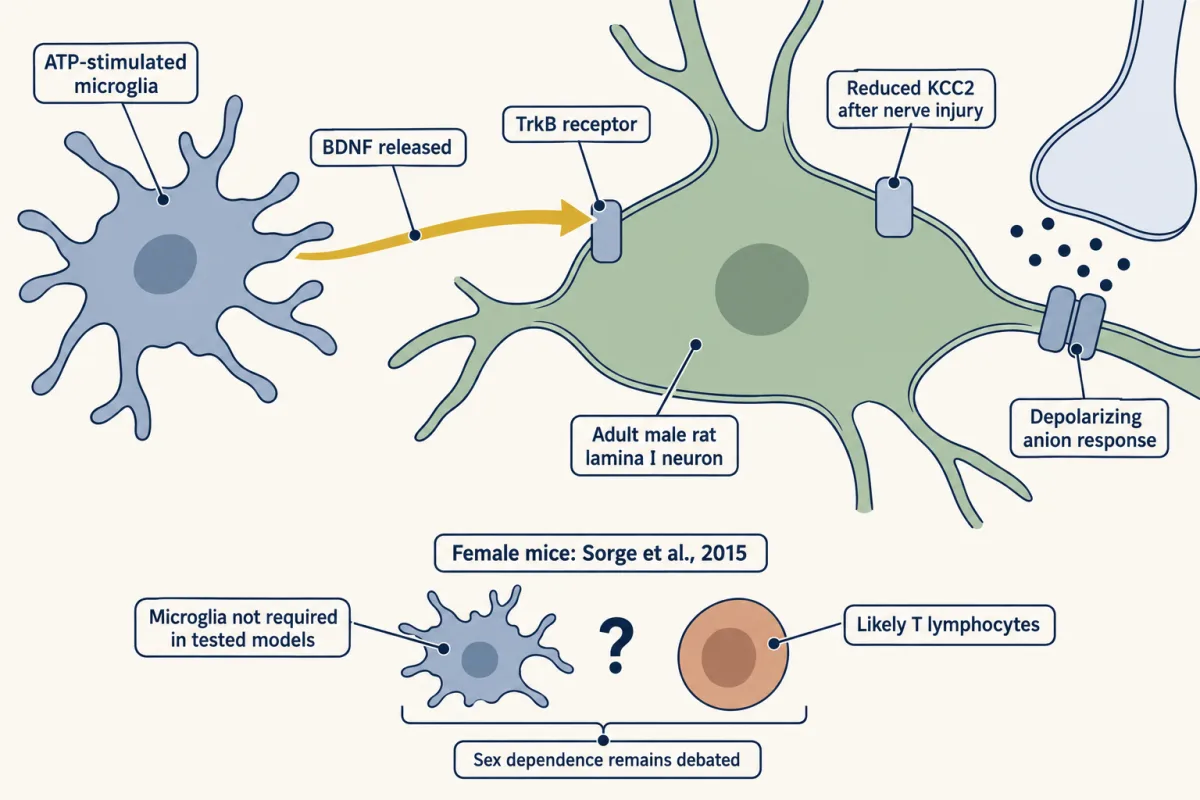
After nerve injury in adult male rats, dorsal-horn KCC2 fell, the anion gradient shifted and inhibitory synaptic currents in lamina I neurons became excitatory; ATP-stimulated microglia released BDNF, and blocking BDNF-TrkB signalling reversed the shift and the allodynia. In female mice, microglia were not required in the models tested, and adaptive immune cells, likely T lymphocytes, were implicated instead. AI-generated illustration -
The output neurons and the denominator trap
-
Going deeper
Where this lesson fits
Before thisReview first
- Where Touch Is First Processed: Dorsal Horn and Dorsal Column NucleiThis course · Lesson 8
It shows where low-threshold touch afferents end in the dorsal horn, which is the input this gate controls.
- Nociceptors and the Sodium Channels of PainThis course · Lesson 11
It covers the nociceptor input that touch must be kept from recruiting when the gate works.
- The Low-Threshold Mechanoreceptors: One Touch, Several SignalsThis course · Lesson 4
The A-beta touch afferents that the inhibitory neurons restrict are described there.
After thisStudy next
- When Pain PersistsThis course · Lesson 14
It takes disinhibition and a changed sign of inhibition further, to pain that persists after nerve injury.
- Itch: A Sense of Its OwnThis course · Lesson 13
Itch uses partly separate dorsal-horn populations, so the cell-type logic of gating carries over to it.
Elsewhere on the siteFilms, simulations and method films in other courses
- Why Capsaicin Feels HotFilm · Neuroscience
It shows how spinal pathways carry touch and pain, the circuit that is gated here.
- One More RopeFilm · Neuroscience
It shows how inhibition can shunt a cell without changing its voltage, as with depolarizing anionic responses here.
- The StandoffFilm · Neuroscience
It gives the resting balance of ion currents that a shifted anion reversal potential disturbs.
Suggested by an AI model from the lessons themselves, then checked. How we use AI