Cell Signaling · Skim version
Protein Modules: SH2, SH3, PTB, PDZ and PH
Read the full lesson → Skim the whole course →
The guiding idea
A receptor has just phosphorylated its own tyrosines. What reads the marks, and how does each reader pick its mark?
Reusable binding domains turn recognized motifs or lipids into protein connections.
From the film: How One Mark Finds Its Reader
- Reusable binding domains turn recognized motifs or lipids into protein connections.
- An SH2 domain grips phosphotyrosine and also reads the residues beside it.
- Sequence preference narrows possible partners; location, abundance and timing narrow them further.
From the film: Two Grips, One Decision
- Combining modules can make binding conditional on more than one signal.
- Src's SH2 and SH3 domains help hold its own kinase in an inactive shape.
- A multi-domain detector stays recruited only while its required targets are together.
-
Why a mark needs a reader
-
A module keeps its binding job when it moves
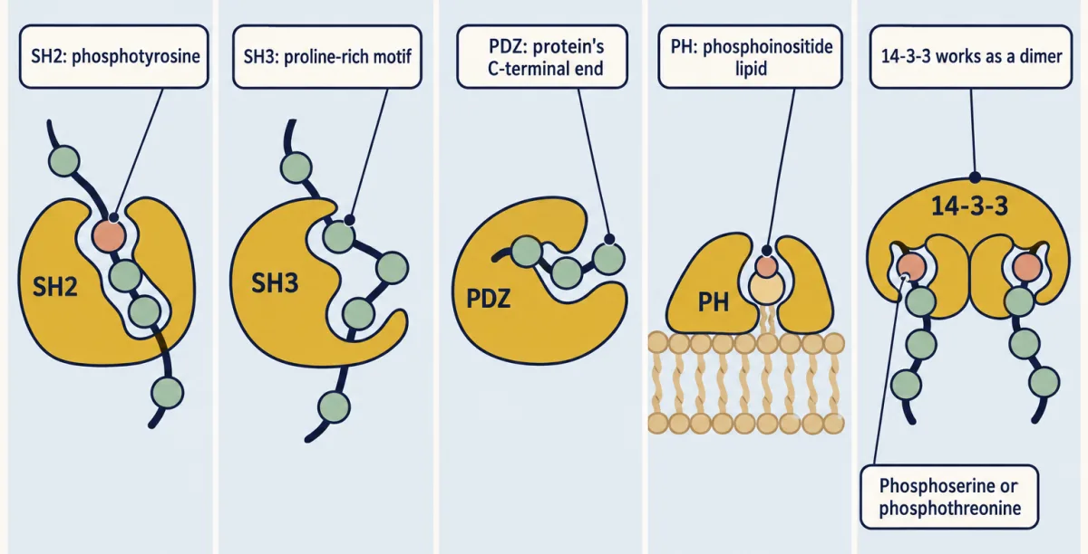
Reader proteins recognise different targets: SH2 binds phosphotyrosine and neighbouring residues, SH3 binds proline-rich motifs, and PDZ binds a free C-terminal end. Some PH domains bind phosphoinositides; 14-3-3 proteins form dimers that bind phosphoserine or phosphothreonine motifs. AI-generated illustration -
SH2 specificity: two parts of one binding surface
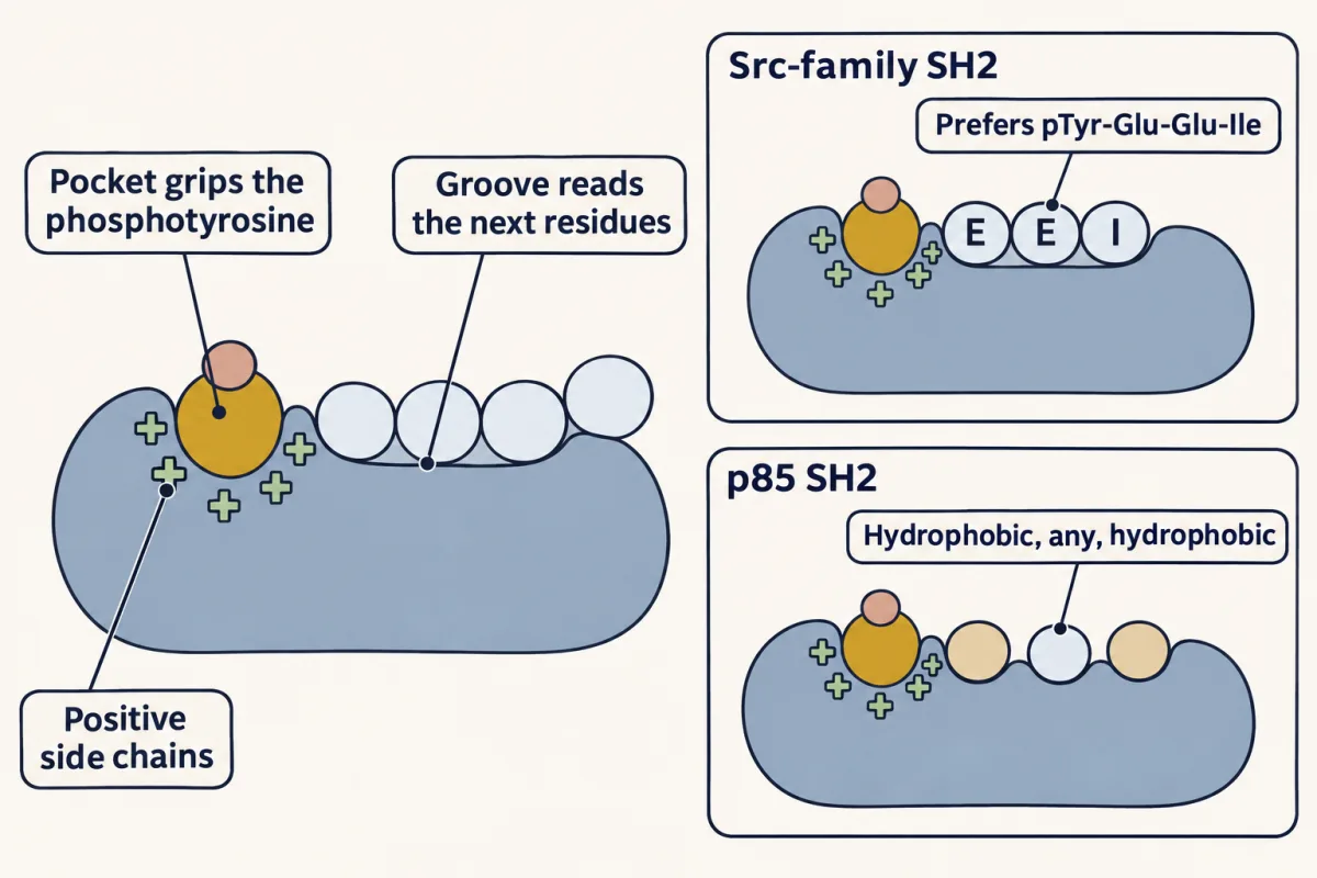
An SH2 domain has two grips: a pocket that clamps the phosphotyrosine and a groove that reads the residues just after it. The pocket alone cannot sort the crowd of phosphoproteins; the groove sets much of each domain's preference, such as pTyr-Glu-Glu-Ile for Src-family domains and hydrophobic, any, hydrophobic for p85. AI-generated illustration -
How much is bound?
-
Src: a protein that reads itself
-
The virtue of modularity: new pathways from old parts
-
Going deeper
Where this lesson fits
Before thisReview first
- Phosphorylation: Kinases and PhosphatasesThis course · Lesson 6
You need to know how kinases and phosphatases add and remove the phosphate that these domains read.
- A Field Guide to ReceptorsThis course · Lesson 4
It introduces receptor tyrosine kinases and their self-phosphorylated tails, which create the marks read here.
- Binding: Affinity, Occupancy and Dose–ResponseThis course · Lesson 3
The Kd and fraction-bound equation used here for domain grip is taught there.
After thisStudy next
- Adaptors, Docking Proteins and ScaffoldsThis course · Lesson 10
Adaptors like Grb2 are built from SH2 and SH3 modules strung together, as shown here.
- Receptor Tyrosine KinasesThis course · Lesson 18
It shows which SH2 and PTB readers dock on the activated receptor's phosphotyrosines.
- PI 3-Kinase, Akt and InsulinThis course · Lesson 20
It uses the PH domain of Akt reading PIP3 and the p85 SH2 domain docking.
Suggested by an AI model from the lessons themselves, then checked. How we use AI