Biochemistry · Glucose Homeostasis
The Pentose Phosphate Pathway: NADPH and Five-Carbon Sugars
Rewritten on : new text, figures and flashcards. Your notes and highlights stay, but the text around them may have moved.
It makes no ATP, and that is the point: NADPH and ribose-5-phosphate are the products that matter.
Pause when the reading reaches a film, and offer to play it. Saved on this device.
Watch the film now, or keep listening
Builds on:Glycolysis: The Investment-and-Payoff PathwayMetabolism: Basic Concepts and DesignGluconeogenesis: Pushing Glucose Back UphillWhy these?
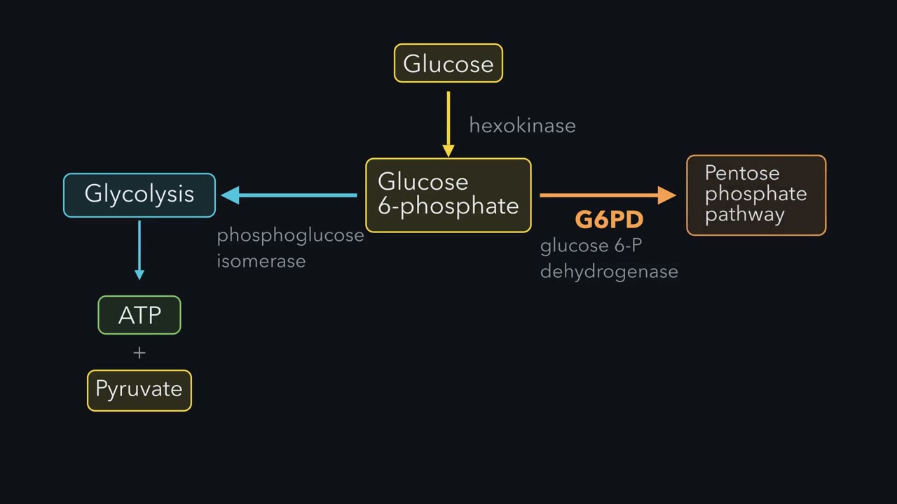 ▶ Watch Glucose's Other Road · 3:56
Read the transcript of “Glucose's Other Road”
Film transcript: Glucose's Other Road
Glucose enters and hexokinase traps it as glucose 6-phosphate, the true branch point of central metabolism. From here, the molecule faces a real choice. Phosphoglucose isomerase channels it leftward into glycolysis, where the payoff is ATP and pyruvate, with ATP the energy currency the cell spends. Alternatively, glucose 6-phosphate dehydrogenase commits it rightward into the pentose phosphate pathway, whose products are NADPH for reductive biosynthesis and ribose 5-phosphate for nucleotide synthesis. The deciding factor is simple. What does the cell need right now? Energy or building blocks? This fork captures the entire logic of why the pentose phosphate pathway exists alongside glycolysis.
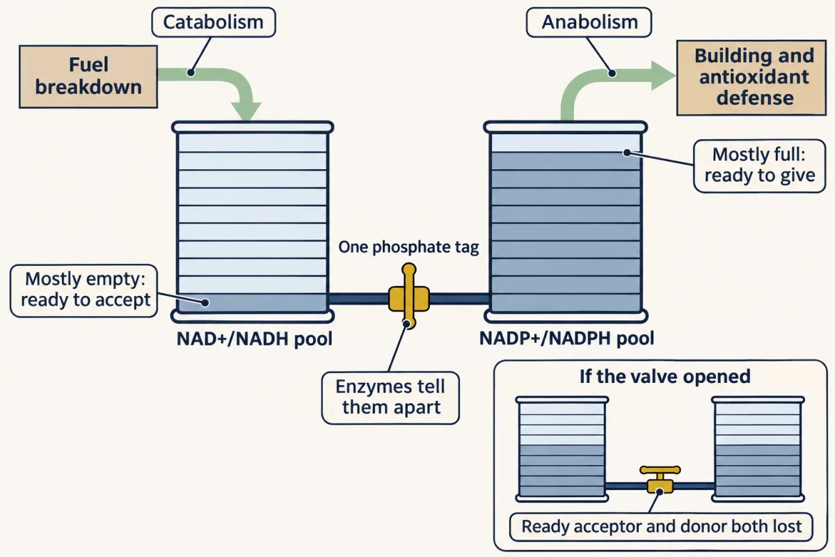
NAD and NADP look almost identical, differing by a single phosphate, yet the cell keeps them in dramatically different states for dramatically different purposes. The NAD pool, free in the cytosol, sits oxidized, roughly a thousand to one in favor of NAD⁺, +, because catabolism needs an electron acceptor ready to grab electrons from fuel. The NADP pool is the mirror image, held strongly toward NADPH, because anabolism and antioxidant defense need an electron donor on tap. Same backbone, opposite redox poise. By maintaining these as separate cytosolic pools, the cell can run breakdown and biosynthesis simultaneously without the two electron economies interfering with each other.
Reactive oxygen species are oxygen-derived molecules, many with an unpaired electron, that attack and damage proteins, lipids, and DNA. The cell's frontline defense is glutathione, which neutralizes these threats by taking the oxidative hit so other molecules don't have to. But once glutathione absorbs that hit, it becomes oxidized and spent, linked into the GSSG dimer. To get it back into the reduced protective GSH form, the cell spends NADPH. And where does that NADPH come from? The pentose phosphate pathway, a major source. So G6PD supports antioxidant defense. Lose that enzyme, and cells struggle to regenerate glutathione, so oxidative damage can run unchecked, red blood cells most of all.
The non-oxidative phase is best understood as carbon bookkeeping carried out by two enzymes, transketolase and transaldolase. Track the carbons. First, transketolase takes two five-carbon sugars and produces a three-carbon and a seven-carbon sugar. Then transaldolase converts that three plus seven into a six and a four-carbon sugar. Finally, transketolase acts again, turning a four and a five into a six and a three. Sum it all and the net result is clean. Three five-carbon sugars become two six-carbon sugars and one three-carbon sugar. That is how excess pentoses are recycled back into glycolytic intermediates when the cell needs energy more than ribose.
The pentose phosphate pathway is not one fixed route, but four operating modes, and the cell selects among them based on what it needs. When it needs ribose for nucleotides but little NADPH, it runs only the non-oxidative phase. When it needs both NADPH and ribose in balance, it runs the oxidative phase straight through. When it needs lots of NADPH but little ribose, it routes ribose back into glycolytic intermediates and recycles them. And when it needs NADPH plus ATP, it pushes carbon all the way into pyruvate. One pathway, four behaviors, each a different combination of glycolysis, gluconeogenesis, and the shunt.
A lymphocyte dividing to fight an infection, a liver cell turning sugar into fat, and a red blood cell hit by an oxidant drug all want different things from glucose, and one pathway serves all three.
The dividing cell needs ribose-5-phosphate, the sugar every nucleotide is built from. The liver cell needs NADPH, the reducing power for building molecules. The red cell needs NADPH from it above all, and little ribose, to keep its antioxidant defenses loaded. None of them needs ATP from this pathway, and it makes none.
The pathway starts at a fork. At glucose-6-phosphate, phosphoglucose isomerase pulls toward glycolysis and ATP; glucose-6-phosphate dehydrogenase pulls toward the pentose phosphate pathway and NADPH. Dividing cells need the second road twice over: nucleotides are built on ribose, and turning ribonucleotides into DNA building blocks spends NADPH. The pathway is one of several NADPH sources, but your red blood cells depend on it as their main one, and that is where this lesson ends up.
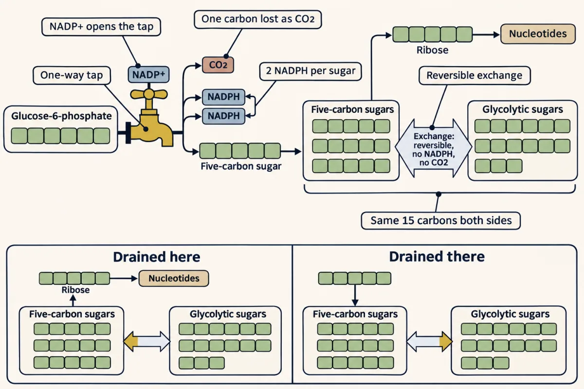
The one idea to carry through this lesson. Every NADPH the pathway makes comes from a one-way tap. Each glucose-6-phosphate that goes through it gives 2 NADPH, 1 CO₂ and 1 five-carbon sugar, and NADP⁺ is what opens the tap. The rest of the pathway is a reversible exchange that makes no NADPH and loses no carbon. It only converts five-carbon sugars to and from glycolytic sugars: 3 C5 ⇌ 2 C6 + 1 C3. So the tap sets how much NADPH the cell gets. The exchange closes the gap between the tap’s fixed 2-to-1 output and the ribose the cell actually needs, and it runs in whichever direction that gap is being drained.
The rule covers the cytosolic pathway in animal cells. Other NADPH sources, and slower controls on how much G6PD a cell makes, sit outside it (Going deeper).
Why NADPH, not NADH
NAD and NADP differ by one phosphate group, and that tag is enough for enzymes to tell them apart. Both carriers hold electrons with almost the same strength. What differs is how full each pool is. In the cytosol the free NAD⁺/NADH pool sits at roughly 1000:1 in favor of the oxidized form (inside mitochondria it is far more reduced), so it is kept mostly empty, ready to accept electrons. That is what catabolism wants: glycolysis and the citric acid cycle are constantly pulling electrons off fuel. The NADP⁺/NADPH pool sits the other way, far toward the reduced form, so it is kept mostly full, ready to give electrons. That is what anabolism wants: fatty-acid synthesis, nucleotide synthesis and glutathione regeneration all spend reducing power.
If enzymes could not tell the two apart, electrons would flow from the full pool to the empty one, like water through a pipe joining a full tank to an empty one, until both sat at the same level, and the cell would lose both an acceptor ready for catabolism and a donor ready for biosynthesis. Keeping them apart lets breakdown and building run at once (Going deeper, “Same voltage, different fill”).
The tap: the oxidative phase
The first three steps are a one-way street.
Glucose-6-phosphate dehydrogenase (G6PD) oxidizes C1 of glucose-6-phosphate, which turns the sugar’s ring into a cyclic ester called a lactone (6-phosphoglucono-δ-lactone), and reduces the first NADP⁺ to NADPH. This is the committed, rate-limiting step, and it is the regulated one. Lactonase then opens the strained lactone ring to give 6-phosphogluconate. 6-phosphogluconate dehydrogenase makes the second NADPH and, in the same step, removes a carbon as CO₂, leaving the five-carbon ribulose-5-phosphate.
G6P + 2 NADP⁺ + H₂O → ribulose-5-P + 2 NADPH + 2 H⁺ + CO₂
No ATP is made. Each step runs downhill in the cell, the lactone hydrolysis strongly so, and once the carbon has left as CO₂ there is no route back.
The valve. G6PD is product-inhibited by NADPH, so the main throttle on the tap is NADP⁺ concentration: feedback written into the active site lets the pathway answer moment-to-moment demand directly.
Predict. A red blood cell meets an oxidant drug. Its glutathione reductase, the enzyme that restores its antioxidant glutathione, starts spending NADPH fast, on fighting oxidative damage. What happens to NADP⁺, and so to the rate of G6PD?
Check your prediction
NADP⁺ rises as NADPH is spent. Less NADPH means less product inhibition, so G6PD speeds up and the tap opens. The demand itself opened the tap.
The mismatch
The tap’s 2-to-1 output is fixed, and real cells rarely need exactly that. The liver cell making fat needs a great deal of NADPH and little ribose. The dividing cell may already have plenty of NADPH and need ribose fast.
Predict. A liver cell making fat runs its tap hard and needs almost no ribose. What has to happen to all the five-carbon sugar the tap is making?
Check your prediction
It has to be turned back into glycolytic sugar, or it piles up. The dividing cell has the opposite problem: with its tap nearly shut, its ribose must come from somewhere else. Both need a route between five-carbon and glycolytic sugars that runs both ways.
The exchange: the non-oxidative phase
This phase is that route, and unlike the tap, every step is reversible.
Two routing enzymes set up the sugars. Phosphopentose isomerase turns ribulose-5-phosphate into ribose-5-phosphate, the aldose that exits toward nucleotide synthesis. Phosphopentose epimerase turns ribulose-5-phosphate into xylulose-5-phosphate, its epimer, which differs at a single stereocenter. Then three transfer reactions move carbons between sugars:
- Transketolase (the “molecular scissors”) snips a two-carbon unit off xylulose-5-P and attaches it to ribose-5-P: C5 + C5 → C3 (glyceraldehyde-3-P) + C7 (sedoheptulose-7-P). It runs on thiamine pyrophosphate, the same cofactor pyruvate dehydrogenase uses.
- Transaldolase moves a three-carbon unit: C7 + C3 → C6 (fructose-6-P) + C4 (erythrose-4-P). It needs no external cofactor: an active-site lysine forms a Schiff base with the substrate, exactly the mechanism aldolase uses in glycolysis.
- Transketolase again: C4 (erythrose-4-P) + C5 (a second xylulose-5-P, also from the epimerase) → C6 (fructose-6-P) + C3 (glyceraldehyde-3-P).
Net: three C5 sugars (one ribose-5-P and two xylulose-5-P, all made from ribulose-5-P) become two fructose-6-P and one glyceraldehyde-3-P, or the reverse. Check the carbons: 3 × 5 = 15, and 2 × 6 + 3 = 15. Both products are glycolytic intermediates.
What decides the direction? Nothing has to. These reactions sit close to equilibrium, so they run toward whichever end is being used up. When nucleotide synthesis pulls ribose-5-phosphate away, the exchange runs backward to replace it. When the tap piles up five-carbon sugar the cell has no use for, the exchange runs forward to clear it.
The film at the top of the page names only transketolase and transaldolase, the enzymes that move carbon; the isomerase and epimerase set up their substrates. Its forward exchange feeds glycolysis for energy, but the same forward run also serves a cell that wants only NADPH (next section).
Walk the interactive below; each metabolite carries its carbon count, so you can check every cut.
regulated step
- Part
- Tap (steps 1–3): one-way; per G6P, 2 NADPH + 1 CO₂ + one five-carbon sugar
The committed, rate-limiting step. Oxidizes C1 of glucose-6-phosphate and reduces NADP⁺ to the first NADPH. This is the door into the pathway, and the cell holds it shut when NADPH is plentiful.
Regulation. NADPH product-inhibits G6PD, so NADP⁺ is what opens the tap: spend NADPH and the step speeds up.
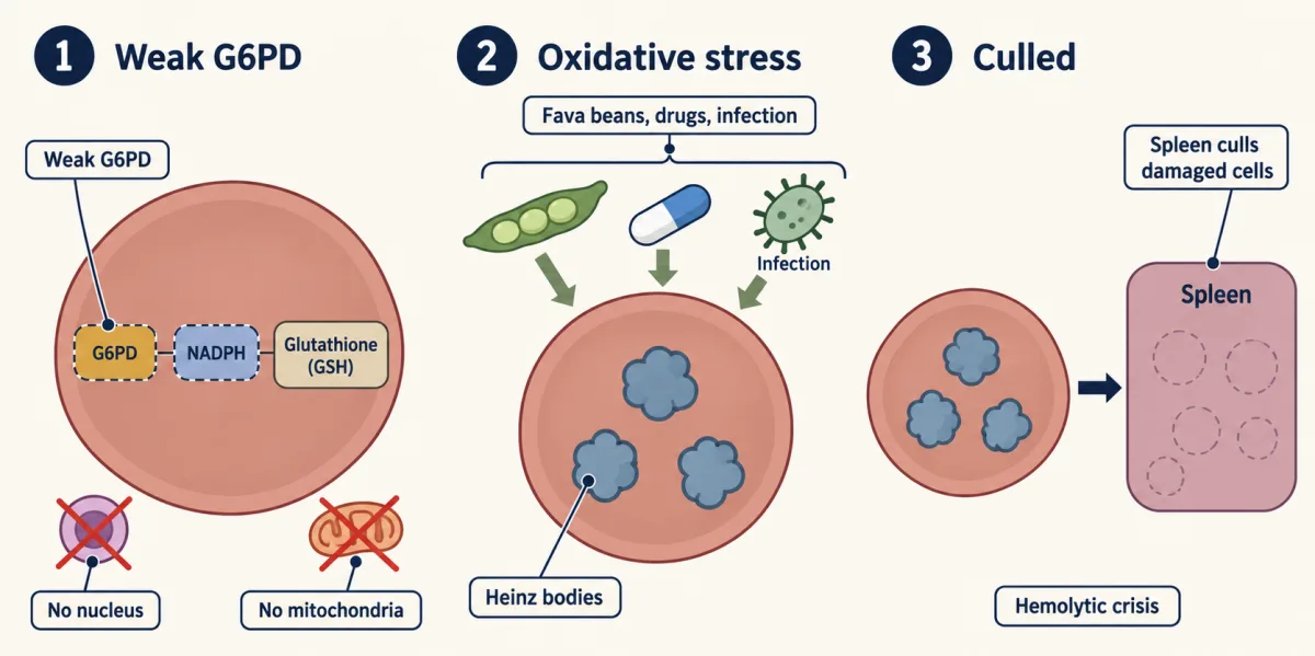
Clinical. G6PD deficiency affects ~400 million people. With G6PD weak, red cells cannot regenerate NADPH, glutathione falls, hemoglobin oxidizes into Heinz bodies, and the damaged cells burst or are removed by the spleen — a hemolytic crisis triggered by fava beans, certain drugs, or infection. Deficient red cells also seem to resist the malaria parasite, which is thought to be why the trait persists where malaria is endemic.
Hydrolyzes the strained lactone ring open to the linear acid. A quiet housekeeping step that also occurs slowly on its own, but the enzyme makes it fast and clean.
Oxidative decarboxylation: makes the second NADPH and releases C1 as CO₂. Losing that carbon, after the downhill oxidation and lactone hydrolysis, is what locks the oxidative phase in one direction — you can run a sugar uphill, but you cannot put a CO₂ back.
Clinical. This step releases CO₂ without consuming O₂, but PPP flux alone does not raise the body's CO₂/O₂ ratio, because the NADPH is later spent on oxygen-using reactions or on making fat. Tracers that follow glucose C1 are how PPP flux is actually measured.
Ketose → aldose. This is the exit toward biosynthesis: ribose-5-phosphate is the sugar every nucleotide is built from (DNA's deoxyribose is made from it later), so this branch feeds DNA, RNA, ATP, NAD⁺, and coenzyme A.
Clinical. Ribose-5-phosphate is the precursor of PRPP, the activated sugar that both purine and pyrimidine synthesis require — the reason dividing cells lean on this pathway.
Inverts the configuration at a single carbon (C3). Ribulose and xylulose are epimers, identical except at that one stereocenter, but transketolase needs xylulose, so the small change matters.
- Part
- Exchange (steps 4–8): reversible, no NADPH, no CO₂; needs three five-carbon sugars, so three turns of the tap
Molecular scissors: snips a two-carbon ketol unit off xylulose and welds it onto ribose. Requires thiamine pyrophosphate (TPP), the same cofactor pyruvate dehydrogenase uses. Carbon check: 5 + 5 = 3 + 7.
Clinical. TPP comes from vitamin B1 (thiamine). In thiamine deficiency (Wernicke-Korsakoff, classically in alcoholism) transketolase activity falls — the assay for it is a clinical test of thiamine status.
Transfers a three-carbon dihydroxyacetone unit using an active-site lysine that forms a Schiff base with the substrate, the same trick aldolase uses in glycolysis. No external cofactor. Carbon check: 7 + 3 = 6 + 4.
Transketolase again, second cut, moving another two-carbon unit so both end products are ordinary glycolytic intermediates. Carbon check: 4 + 5 = 6 + 3. Net of the non-oxidative phase: three C5 sugars become two F6P and one G3P.
The tally shows one turn of the tap. Steps 6–8 use three five-carbon sugars, so a full forward exchange needs three turns: 3 G6P → 6 NADPH + 3 CO₂ → 2 fructose-6-P + 1 glyceraldehyde-3-P. The exchange itself added no NADPH and lost no carbon.
Four demands, four tallies
The tap and the exchange cover every case, the four textbook modes. Start with Mode 2, the only demand the tap meets on its own.
Mode 2: both, in the tap’s own ratio. If the cell wants 2 NADPH for every ribose, the tap alone does the job: one glucose-6-phosphate gives 2 NADPH, 1 CO₂ and, through the isomerase, one ribose-5-phosphate. The exchange does nothing.
Mode 1: ribose, with plenty of NADPH already. A dividing cell that needs nucleotides fast can be in this spot. With NADPH high, the tap is nearly shut. The ribose has to come from the exchange run backward, out of fructose-6-P and glyceraldehyde-3-P pulled from glycolysis.
Predict. The cell needs 6 ribose-5-phosphate and makes no NADPH. How many glucose-6-phosphate does it need, and does it lose any carbon as CO₂?
Check your prediction
Five, and no CO₂. Six ribose hold 30 carbons, and five glucose-6-phosphate hold 30. Four of them become fructose-6-phosphate. The fifth is split by the first steps of glycolysis into two glyceraldehyde-3-phosphate, which costs one ATP (spent by phosphofructokinase-1). Then the exchange runs backward twice: 4 C6 + 2 C3 → 6 C5. Net: 5 glucose-6-phosphate + 1 ATP → 6 ribose-5-phosphate.
Mode 3: NADPH, almost no ribose. A liver or fat cell making fatty acids is in this spot (they also draw on other NADPH sources). The tap runs hard, and every five-carbon sugar it makes is surplus. The exchange runs forward and turns that surplus into fructose-6-phosphate and glyceraldehyde-3-phosphate. Phosphoglucose isomerase and the enzymes of gluconeogenesis then put those back together as glucose-6-phosphate, which can go through the tap again. A red blood cell under oxidant attack has the purest NADPH-only demand, but it has no gluconeogenesis: it returns its fructose-6-phosphate to the tap and sends the three-carbon sugar down glycolysis, a blend of Modes 3 and 4.
Predict. Six glucose-6-phosphate go through the tap, and all the five-carbon sugar is recycled. How many NADPH come out, and how many glucose-6-phosphate come back?
Check your prediction
Twelve NADPH, and five glucose-6-phosphate come back. The tap gives 6 × 2 = 12 NADPH, 6 CO₂ and six five-carbon sugars (30 carbons). The exchange turns those into four fructose-6-phosphate and two glyceraldehyde-3-phosphate, and the two three-carbon sugars join into a fifth; all five become glucose-6-phosphate again. Net: one whole glucose-6-phosphate is taken apart into 6 CO₂, and the cell collects 12 NADPH. That is the most NADPH one glucose can give.
Mode 4: NADPH and ATP. As in Mode 3, but the fructose-6-phosphate and glyceraldehyde-3-phosphate go down glycolysis instead of back up, leaving no net ribose. Three glucose-6-phosphate give 6 NADPH and 3 CO₂, and their leftover sugars give 5 pyruvate, 5 NADH and a net 8 ATP (five three-carbon sugars earn 10 ATP; phosphofructokinase-1 spends 2 splitting the two fructose-6-phosphate). That ATP is glycolysis’s; the pentose phosphate pathway itself still makes none.
| Mode | What the cell needs | Tap | Exchange | Net per batch |
|---|---|---|---|---|
| 1 | Ribose, no NADPH | nearly shut | backward | 5 G6P + 1 ATP → 6 ribose-5-P |
| 2 | 2 NADPH per ribose | runs | idle | 1 G6P → 1 ribose-5-P + 2 NADPH + 1 CO₂ |
| 3 | NADPH, no ribose | runs hard | forward, back to G6P | 6 G6P through the tap, 5 back: net 1 G6P → 12 NADPH + 6 CO₂ |
| 4 | NADPH and ATP | runs | forward, into glycolysis | 3 G6P → 6 NADPH + 3 CO₂ + 5 pyruvate + 5 NADH + 8 ATP |
These are limits, not settings. A real cell blends them, and the blend shifts minute by minute as NADP⁺ opens or closes the tap and as nucleotide synthesis pulls on ribose. Pick a demand below and watch which parts run.
- Net output
- —
- Where you see it
- —
- Net per batch
- —
The same enzymes, run forward or backward in different proportions, let one pathway serve four very different cellular needs.
The tap (G6PD onward) is one-way; NADP⁺ opens it. The exchange (transketolase, transaldolase) is reversible and runs toward whichever end is being drained.
The red cell: when the tap breaks
NADPH is what keeps glutathione reduced. Glutathione is a small tripeptide (γ-Glu-Cys-Gly) with a reactive cysteine thiol, and it is the cell’s frontline antioxidant. Glutathione peroxidase spends reduced glutathione (GSH) to neutralize peroxides, turning it into the oxidized disulfide GSSG; glutathione reductase then spends NADPH to turn GSSG back into GSH.
A mature red blood cell needs little ribose, has no nucleus and no mitochondria, and gets its NADPH mainly from the PPP, so its whole glutathione cycle leans on the tap.
That is why a fava bean can be dangerous. Glucose-6-phosphate dehydrogenase (G6PD) deficiency, the most common human enzyme deficiency, affecting roughly 400 million people, weakens the tap itself. The gene is on the X chromosome, so males are affected more often, and its many variants differ in severity. Fava beans, certain antimalarial and sulfa drugs, and infection all push oxidative stress past what a weakened cell can handle. The cascade: G6PD↓ → NADPH↓ → GSH↓ → reactive oxygen accumulates → hemoglobin oxidizes and clumps into Heinz bodies → the damaged cells burst in the circulation or are removed by the spleen → hemolytic crisis.
Predict. After a fava meal, NADP⁺ rises in a G6PD-deficient red cell, just as it would in a normal one. Why doesn’t the tap open enough?
Check your prediction
The signal is there, but the tap itself is weak: there is too little working G6PD to answer it. A red cell has no nucleus, so it cannot transcribe more enzyme, and it has little else to fall back on.
If the deficiency is dangerous, why is it so common? It is thought to persist because it offers some protection against Plasmodium falciparum, the malaria parasite: deficient red cells seem to be a poorer home for it, and the allele’s prevalence broadly overlaps the historical range of malaria. It is a balanced polymorphism, the textbook companion to sickle-cell trait, though how strong the protection is, and in whom (males with one deficient X, or heterozygous females), still differs between studies. The practical lesson: screen for G6PD before prescribing certain oxidant drugs.
Where the NADPH goes next
Many dividing tumor cells run glycolysis hard (the Warburg effect) and the PPP hard, because they need NADPH for lipid and nucleotide synthesis and for surviving their own oxidative stress. Lose p53 function (TP53 is the most frequently mutated gene in human cancer) and a brake on this NADPH economy can come off (Going deeper). Next we turn to fats: first how cells take fatty acids apart, then how they build them, spending the NADPH this pathway makes.
Going deeper
Other NADPH sources
In many cells, malic enzyme, isocitrate dehydrogenase and folate metabolism make NADPH too. Liver and fat tissue making fatty acids get a substantial share of theirs from malic enzyme as well, which is why the red cell is the cleaner example of an NADPH-only demand, even though it can recycle only part of its carbon.
Same voltage, different fill
To put a number on the difference in fill, take 1000:1 oxidized for NAD and about 1:100 for NADP, close to classic estimates for rat-liver cytosol. The NADP couple is then about 150 mV more reducing in the cytosol: RT/2F × ln 10⁵ ≈ 154 mV at 37 °C.
The tally for any demand
For a demand of n NADPH and r ribose-5-phosphate, the tap must run n/2 times, and the exchange must run forward by (n/2 − r)/3 turns. That is zero only when n = 2r (Mode 2); a negative answer means it runs backward (Mode 1: n = 0, r = 6 gives −2).
Why the oxidative phase does not raise the respiratory quotient
The oxidative phase itself uses no O₂, but that does not by itself raise the body’s ratio of CO₂ exhaled to O₂ consumed (the respiratory quotient), because the NADPH it makes is later spent on reactions that use oxygen or on building fat. An RQ above 1 signals net fat synthesis, which spends PPP NADPH.
Long-term control of G6PD, and how p53 acts
Over longer times, signals such as NRF2 and modifications such as SIRT2 deacetylation also change how much glucose-6-phosphate dehydrogenase (G6PD) the cell has and how active it is. The tumor suppressor p53 can restrain G6PD by binding it and blocking its assembly into the active dimer. p53 touches the pathway in other ways too (it induces TIGAR, which can push glucose into the PPP), so its net effect depends on the cell.
Your notes
Select any text in the lesson to highlight it or add a note to it. Your highlights and notes are saved in this browser and collected in your notebook.
This browser is not letting the site save anything (a private window or strict privacy settings), so highlights and notes will be lost when you leave the page.
How we measure it
Respiratory quotient (RQ = CO2/O2)
Measure the ratio of CO2 exhaled to O2 consumed and you can estimate which fuel the body is mainly burning. Pure carbohydrate oxidation gives RQ ≈ 1.0; fat gives ≈ 0.7. An RQ above 1.0 signals net conversion of carbohydrate to fat: CO2 is released while oxygen-poor fat is stored. The PPP supplies part of the NADPH for that fat-making, but its own CO2 does not raise the RQ, because the NADPH it makes is later spent on reactions that use oxygen or on that net synthesis. RQ is a whole-body fuel readout, not a PPP assay.
Isotopic carbon tracing
Feed cells glucose labeled at a specific carbon (13C or 14C on C1 vs C6) and follow where the atom lands. Because the oxidative phase removes C1 of glucose as CO2 while glycolysis would keep it, the position of the released label tells you what fraction of glucose ran the PPP versus glycolysis. This is the classic way the relative flux through the two branches has been measured in different tissues, though carbon recycling through the non-oxidative phase complicates the reading.
Enzyme assay for G6PD (the clinical screen)
Glucose-6-phosphate dehydrogenase activity is measured by following NADPH production spectrophotometrically (NADPH absorbs at 340 nm; NADP+ does not). A slower rise in A340 (absorbance at 340 nm) in a patient's red cells is the standard diagnostic for G6PD deficiency — the same initial-velocity logic from enzyme kinetics, used to decide whether certain oxidant drugs are safe to prescribe.
Glutathione redox ratio (GSH/GSSG)
The ratio of reduced glutathione to its oxidized disulfide form is a readout of a cell's antioxidant reserve, and it is held high by NADPH, largely from the PPP. Measuring GSH/GSSG (by HPLC or enzymatic recycling assays) reports whether NADPH supply, from the PPP and other sources, is keeping up with oxidative demand — it collapses when NADPH supply fails.
Practice questions on this lesson (3) → MCAT-style, original, each with an explanation
Where this lesson fits
Before thisReview first
- Glycolysis: The Investment-and-Payoff PathwayThis course · Lesson 16
You need glucose-6-phosphate as the branch point, and glycolysis, to follow where the exchange phase sends its sugars.
- Metabolism: Basic Concepts and DesignThis course · Lesson 15
It explains why NAD and NADP pools are kept differently filled, and how cells couple energy-releasing steps to building steps.
- Gluconeogenesis: Pushing Glucose Back UphillThis course · Lesson 17
Mode 3 uses gluconeogenesis enzymes to turn fructose-6-phosphate and glyceraldehyde-3-phosphate back into glucose-6-phosphate.
After thisStudy next
- Fatty Acid Synthesis: Building the Chain Two Carbons at a TimeThis course · Lesson 26
Fat building spends the NADPH this pathway makes, so you see where that reducing power goes.
- Nucleotide Metabolism: Build It, Recycle It, or Pay in Uric AcidThis course · Lesson 29
Ribose-5-phosphate from here is turned into PRPP, the start of building nucleotides.
- Integration of Metabolism: How the Body Switches FuelsThis course · Lesson 30
It shows how hormones and fuel state switch pathways like this one up or down.
Suggested by an AI model from the lessons themselves, then checked. How we use AI
Finish line
Test yourself
8 questions
Practice and MCAT-style, with explained answers.
Play it · about 2 min
Wire the Cell: Red blood cell
No mitochondria. Make ATP and the NADPH that protects the cell from oxidants.
Play →Next · Lesson 25
Fatty Acid Degradation: The β-Oxidation Spiral
Fat comes apart two carbons at a time, and that is how a fasting body keeps its tissues running when glucose is scarce.
Continue →Also in the arcade for this lesson: Wire the Cell: A cell about to divide