Developmental Neuroscience · Skim version
Neural Induction: The Default Model
Read the full lesson → Skim the whole course →
The guiding idea
Pull a square of frog skin-to-be apart into single cells, and it becomes neural. Why? Because BMP signaling, which makes skin, falls in those cells. In the embryo, the organizer's secreted proteins make it fall.
BMP says skin: in early frog animal caps, turning its signal down far enough lets competent cells become neural, provided their other required inputs, including early FGF signaling, are intact.
From the film: The Map and the Question
- Ectoderm can become skin or nervous system, and an animal cap left intact becomes epidermis.
- BMP says skin: in early frog animal caps, turning its signal down far enough lets competent cells become neural, provided other needed inputs such as FGF are intact.
- BMP's effect comes down to one dial, the activity of Smad1 in the nucleus.
From the film: Break the Receptor, Add the Ligand
- In frog animal caps, jamming a receptor with a dominant-negative copy makes neural tissue, with no mesoderm detected that could have induced it.
- Blocking a receptor, not adding a signal, made neural tissue.
- Adding BMP4 protein back to separated cap cells restores epidermis and keeps neural genes off.
From the film: One Dial
- In these four cap conditions, where Smad1 output was high the cells made skin, and where it was low they made neural tissue.
- Separated cells still have signaling BMP receptors, but MAPK activity can damp Smad1, so dilution is probably not the reason they turn neural.
- The rule names the dial, Smad1 in the nucleus, and not BMP in the fluid.
From the film: The Organizer's Catchers
- The organizer makes at least three proteins that catch BMP outside the cell: Noggin, Chordin and Follistatin.
- Noggin protein turns intact caps neural, with anterior markers, without dorsal mesoderm.
- In Xenopus tropicalis embryos, depleting all three catchers lost the neural plate, so the frog embryo needs them; early FGF signaling is needed too.
From the film: A Fly Turned Over
- The fly's Dpp and Sog are the counterparts of the frog's BMP4 and Chordin, and frog Chordin works in the fly and fly Sog in the frog.
- In both animals, nervous system forms where the antagonist keeps the BMP signal low.
- The fly's map is inverted relative to the frog's: Dpp is dorsal and the nerve cord forms ventrally.
From the film: Where the Rule Stops
- The rule says neural, not where: Noggin-treated caps made only anterior markers.
- In chick, Chordin outside the future neural plate did not induce early neural markers, so lowering BMP is not enough on its own there; early FGF signaling and Wnt state matter.
- In mouse, embryos lacking both chordin and noggin still form neural tissue but have severe forebrain defects.
-
The layout
-
Three cap experiments
-
One dial
-
The organizer’s catchers
-
The same contest in a fly, upside down
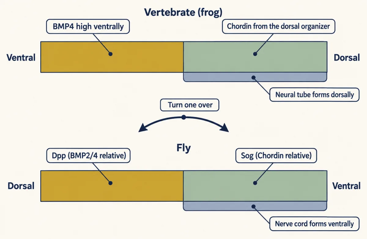
The fly's Dpp and Sog are the counterparts of the frog's BMP4 and Chordin, but their sides are swapped. In the vertebrate, BMP4 signaling is high ventrally and the neural tube forms dorsally; in the fly, Sog in ventrolateral neuroectoderm keeps Dpp signaling low, and this ectoderm gives rise to the ventral nerve cord. The drawing is simplified: the ventral-most fly cells are mesoderm. AI-generated illustration -
Where the rule stops
-
Putting it together
-
Going deeper
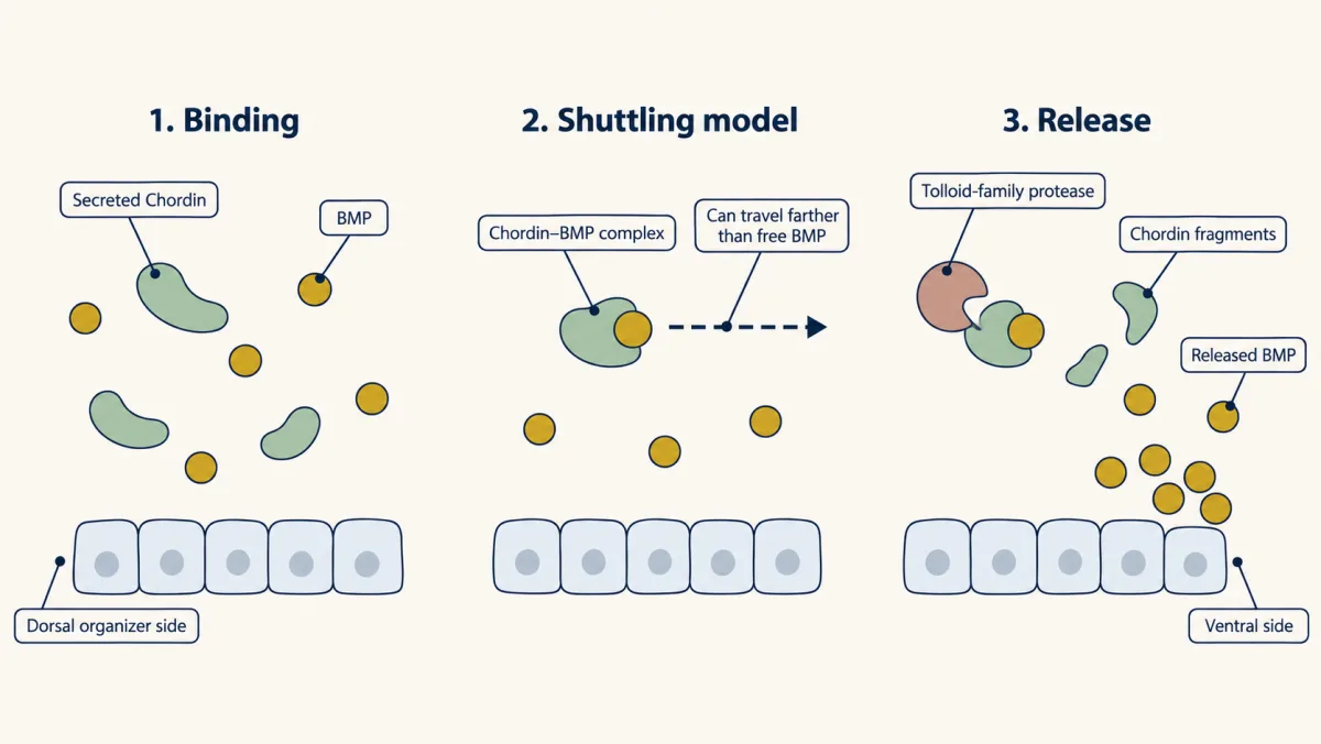
In the model, a Chordin-BMP complex travels farther than free BMP. Where a Tolloid-family protease cuts Chordin the BMP is set free, piling up on the ventral side while signaling stays low dorsally. AI-generated illustration
Where this lesson fits
Before thisReview first
- The Organizer: Spemann and MangoldThis course · Lesson 5
The organizer graft and its power to change nearby ectoderm is the starting point for asking what it sends.
After thisStudy next
- Head to Tail: Hox Genes, Retinoic Acid and RhombomeresThis course · Lesson 8
Neural identity is separate from head-to-tail position, and this lesson takes up how posterior fates are set.
- Ventral to Dorsal: Sonic Hedgehog and BMP in the Neural TubeThis course · Lesson 10
It uses BMP as a graded signal once the neural tube exists, extending the BMP dial to dorsal patterning.
- Proneural Genes and Lateral InhibitionThis course · Lesson 14
It continues the neural-versus-other choice inside neural ectoderm, now between neuron and neighbor cell.
Elsewhere on the siteFilms, simulations and method films in other courses
- RNAi and antisense knockdownMethod film
The lesson's depletion of Noggin, Chordin and Follistatin relies on knockdown and checking specificity.
- β-catenin levelSimulation · Cell Signaling
Not available
Suggested by an AI model from the lessons themselves, then checked. How we use AI