Biology · Skim version
Living or Not? Viruses and Bacteria
Read the full lesson → Skim the whole course →
The guiding idea
A cold virus on a light switch does nothing on its own for hours, yet inside your nose it gets cells to build new copies of it. Is it alive?
Bacteria are living cells; viruses are not cells and are classed nonliving.
From the film: Who Can Act Alone?
- Bacteria are living cells; viruses are not cells and are classed nonliving.
- The characteristics of life organize evidence, but no single checked box decides the case.
- A bacterium is a cell that uses energy, grows, responds and divides when conditions suit it.
- A virus is not a cell and has no metabolism of its own.
- Inside a host cell, viral genes use the cell's machinery to make new virus parts.
From the film: Parts Reveal the Difference
- Bacteria are living cells; viruses are not cells and are classed nonliving.
- A bacterium has a membrane, cytoplasm, ribosomes and DNA in a nucleoid.
- A virus has DNA or RNA in a capsid, sometimes inside an envelope.
- Bacteria can get energy from carbon compounds, light or chemicals.
- Size does not decide whether something is living.
-
Explore: run the living-or-not checklist
-
Explain: what counts as alive
-
Explain: parts reveal the difference
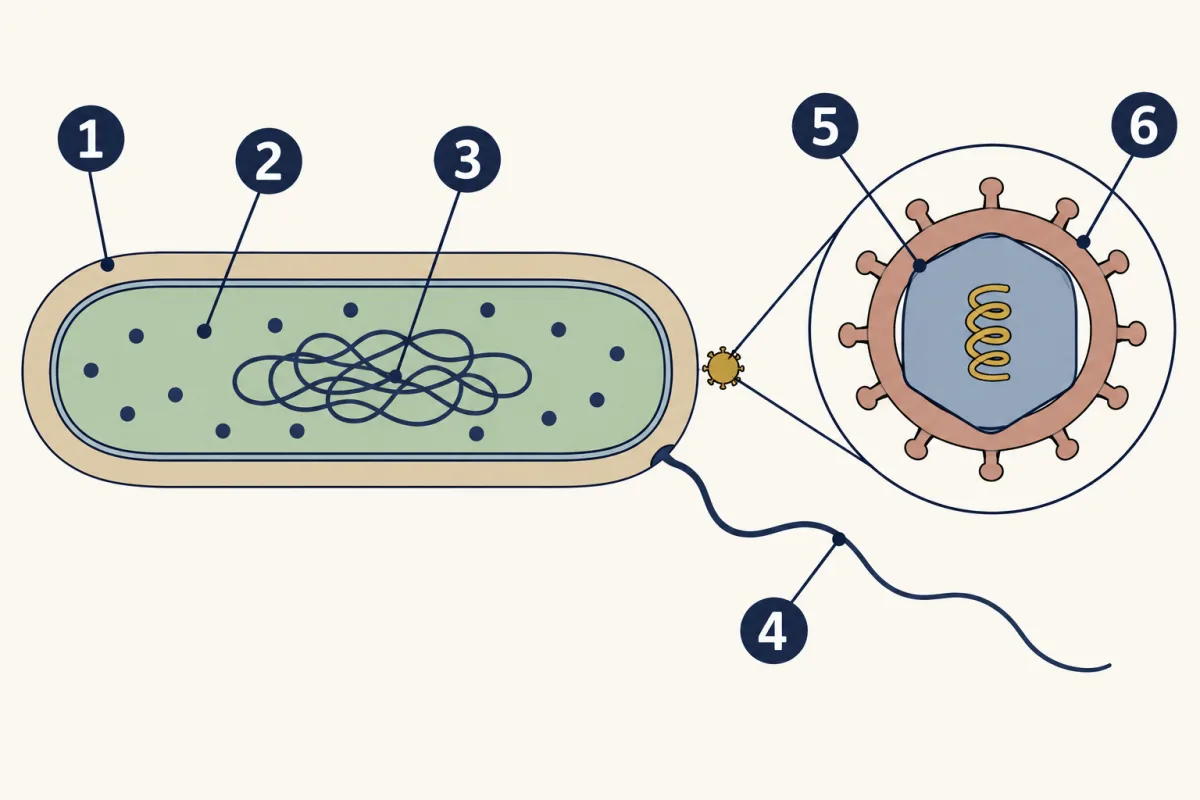
- cell wall
- ribosomes
- nucleoid
- flagellum
- capsid
- envelope
-
Explain: three bacterial energy patterns
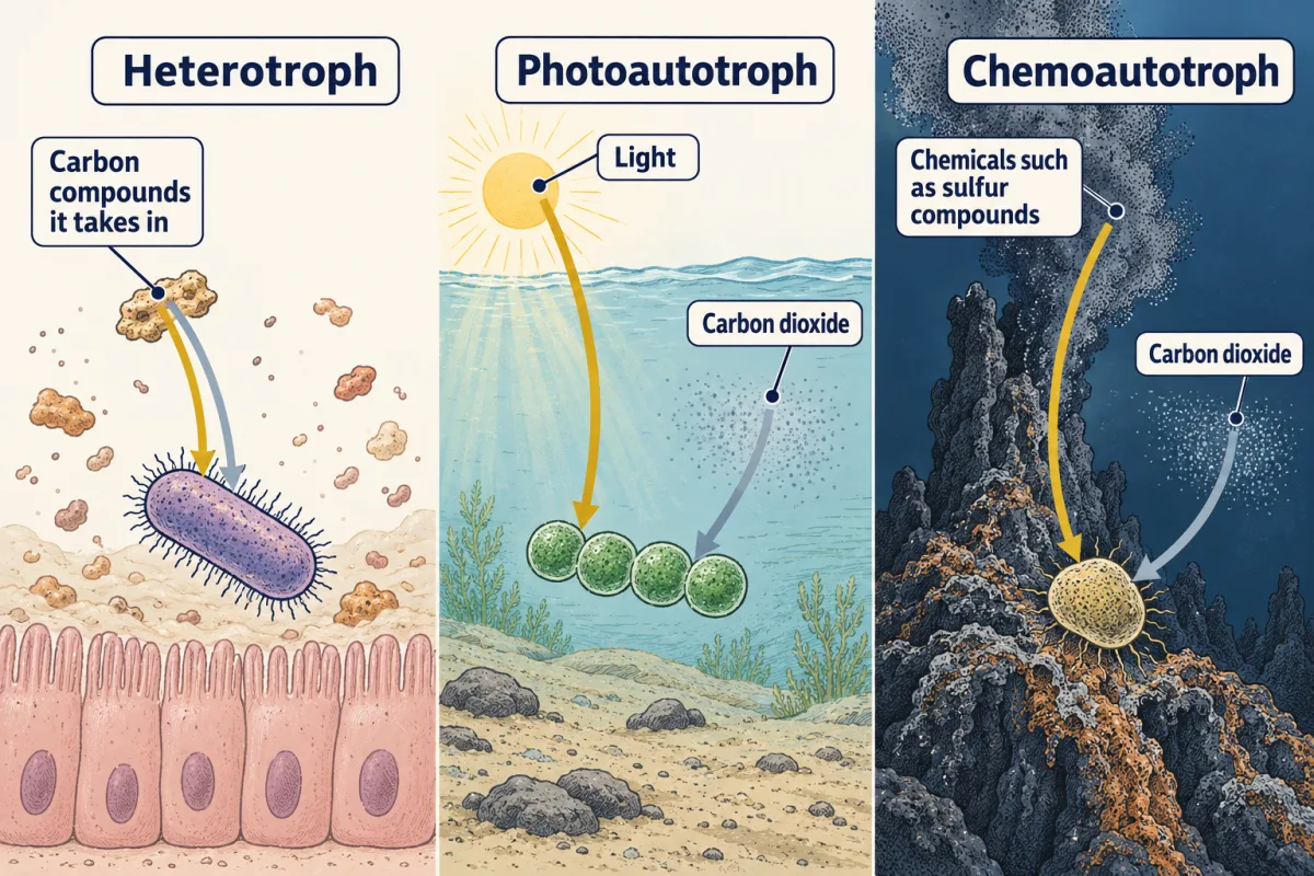
Bacteria sorted by how they get energy: gold arrows show the energy source and grey-blue arrows the carbon source. Photoautotrophs and chemoautotrophs both take their carbon from carbon dioxide. AI-generated illustration -
Elaborate: compare real sizes
-
Evaluate: check your thinking
Where this lesson fits
Before thisReview first
- One Cell or ManyThis course · Lesson 11
It compares prokaryotes and eukaryotes, which you need to sort bacteria from your own cells.
- The Cell Theory and Its EvidenceThis course · Lesson 6
The rule that living things are made of cells is what separates bacteria from viruses here.
- Respiration and ATPThis course · Lesson 5
You need to know how cells get energy to sort bacteria into energy groups and to understand metabolism.
After thisStudy next
- How Microbes MultiplyThis course · Lesson 13
It shows how bacteria divide and how a virus makes copies inside a host cell.
- Microbes in Bodies and EcosystemsThis course · Lesson 14
It puts the bacterial energy groups to work in soil and water ecosystems.
- Germs, Vaccines and AntibioticsThis course · Lesson 15
It uses the difference between bacteria and viruses to explain why antibiotics work on one and not the other.
Elsewhere on the siteFilms, simulations and method films in other courses
- Set Up: Alive or Not?Film · Biology Together
It shows the basic checklist of what living things do, which this lesson applies to viruses and bacteria.
Suggested by an AI model from the lessons themselves, then checked. How we use AI