Biochemistry · Skim version
Lipid Synthesis: Building Cholesterol from Acetyl-CoA
Read the full lesson → Skim the whole course →
The guiding idea
Skin cells from children whose blood is flooded with cholesterol keep making more of it. Why would a cell bathed in cholesterol keep building it?
Cells get cholesterol two ways, by building it from two-carbon acetyl-CoA and by taking up LDL through the LDL receptor, and one gauge, the cholesterol level in the ER membrane read by SCAP, sets both through SREBP-2.
From the film
- A liver cell gets cholesterol by making it or importing LDL.
- In liver cells, ER cholesterol controls both supply lines through SCAP and SREBP-2.
- Statins block HMG-CoA reductase and indirectly raise LDL receptors.
- In most familial hypercholesterolemia, too few working LDL receptors slow clearance.
-
The spine: condense, assemble, cyclize
-
Where the throttle sits
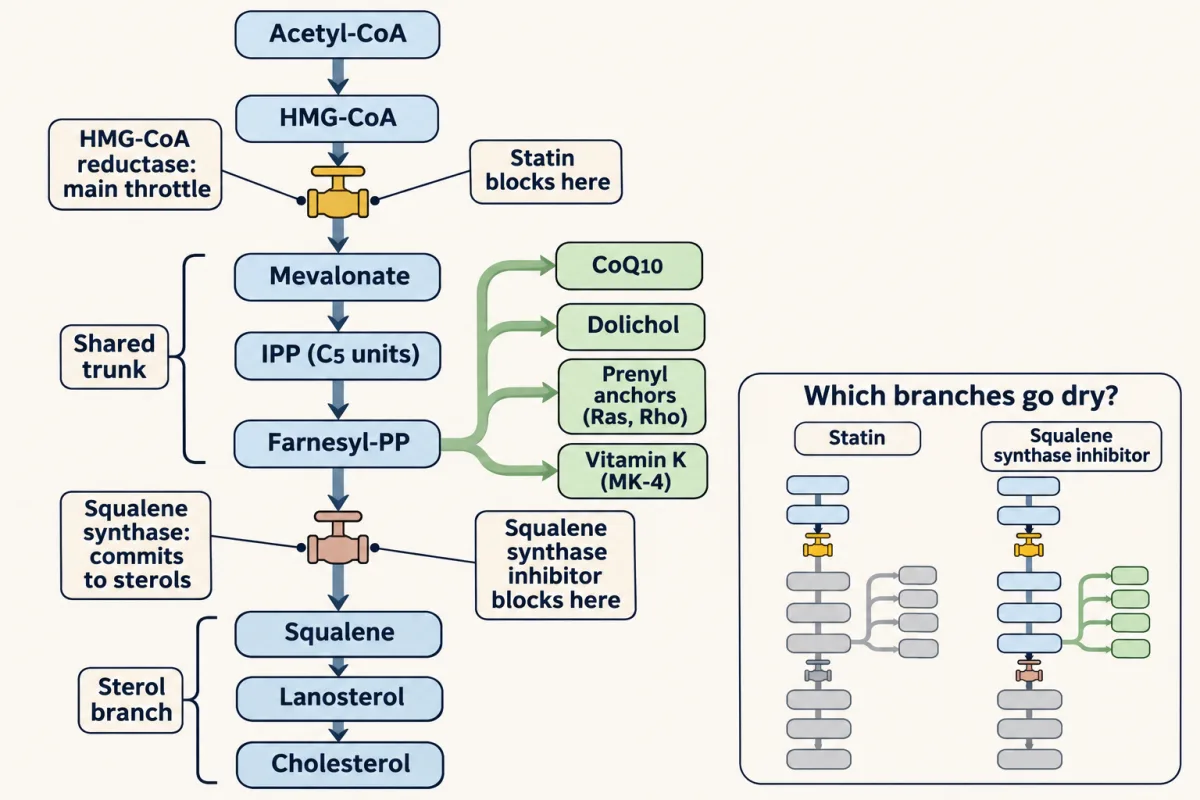
Mevalonate feeds a shared trunk. HMG-CoA reductase, the main throttle, sits at the top, so a statin cuts the whole isoprenoid supply, CoQ10 and the prenyl anchors included; squalene synthase commits carbon to sterols only, so blocking it spares the side branches. AI-generated illustration -
How LDL gets in
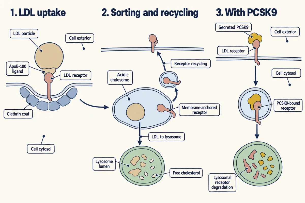
The LDL receptor reads ApoB-100, the pit pinches off, the receptor recycles and the LDL goes on to the lysosome; PCSK9 sends the receptor to the lysosome instead, so fewer receptors return to the surface. AI-generated illustration -
One gauge, two supply lines
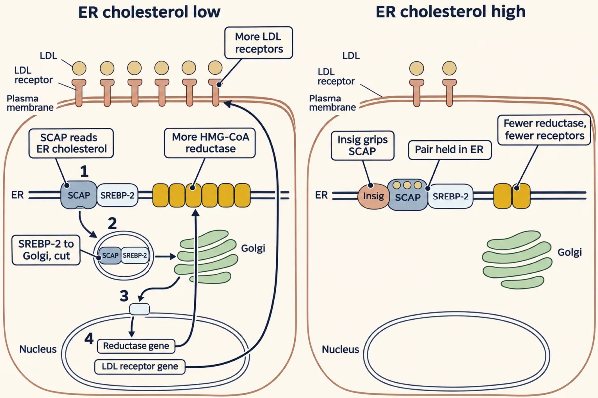
One gauge sets both supply lines. When ER cholesterol is low, SCAP escorts SREBP-2 to the Golgi, where it is cut and freed to turn on the reductase and the LDL-receptor genes together; when it is high, cholesterol binds SCAP, Insig grips SCAP, and both lines are turned down. Insig and cholesterol act on SCAP, not on SREBP-2. AI-generated illustration -
Same gauge, more drugs
-
What cholesterol becomes
-
Going deeper
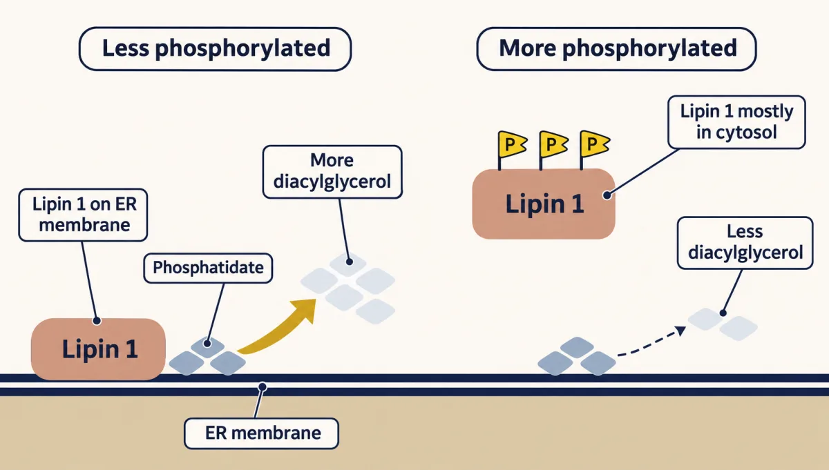
Lipin 1 localization changes its access to substrate at the ER membrane: less phosphorylated Lipin 1 associates more with the membrane and makes more diacylglycerol; more heavily phosphorylated Lipin 1 is mostly cytosolic and makes less. The diacylglycerol supplies both storage fat and membrane phospholipids. AI-generated illustration
Where this lesson fits
Before thisReview first
- Metabolism: Basic Concepts and DesignThis course · Lesson 15
You need the energy economy of ATP and NADPH from there to count the cost of building cholesterol.
- Membranes: The Impermeable Sheet That Builds ItselfThis course · Lesson 12
You need membrane structure from there to see where the reductase and SCAP sit and why cholesterol matters.
- Fatty Acid Synthesis: Building the Chain Two Carbons at a TimeThis course · Lesson 26
You need acetyl-CoA as the cytosolic two-carbon building block and NADPH as the reducing power from there.
After thisStudy next
- Integration of Metabolism: How the Body Switches FuelsThis course · Lesson 30
It shows how fed and fasted hormones switch these cholesterol and fat pathways up or down across the body.
Elsewhere on the siteFilms, simulations and method films in other courses
- A Loop and a One-Way TripFilm · Cell Biology
It shows the LDL receptor looping back while its LDL goes one way to the lysosome.
- Binding or entry?Simulation · Cell Biology
It lets you separate LDL binding from LDL entry, which is the defect in familial hypercholesterolemia.
- An Enzyme Guards the ReceptorFilm · Physiology
It follows cholesterol into the adrenal steroid hormones and the enzymes that shape them.
- Where the Reader WorksFilm · Cell Biology
It shows how a signal sequence sends a protein to its compartment, as SCAP sends SREBP-2 to the Golgi.
Suggested by an AI model from the lessons themselves, then checked. How we use AI