Sensory Systems · Skim version
The Hair Cell's Transduction Channel
Read the full lesson → Skim the whole course →
The guiding idea
A gene is required for hearing, and its protein sits at the right place. What else must be shown before it counts as the channel?
The mean current from a hair bundle is I = N × i × P_open, so losing a protein that any factor needs silences the cell without saying which factor was lost.
From the film: One Current, Three Factors
- The mean current from a hair bundle is the product of the number of working channels, the current through one open channel and the open probability.
- A lost current cannot tell you which of those three factors was removed.
- Evidence about permeation, a residue reached in a working channel and activity of the protein on its own speaks more directly to the pore, and the case rests on several rungs together.
From the film: Change It While It Works
- Loss and rescue of TMC1 and TMC2 put them in the pathway but cannot separate a pore from its helpers.
- A point mutation that changes which ions pass, as in the Beethoven mouse, narrows the candidates, though a partner beside the pore can also change what passes.
- A reagent that alters permeation in a channel already carrying current rules out altered synthesis or delivery, but the native partners were still present.
From the film: A Pore Inside a Complex
- The native worm TMC-1 complex solved by cryo-EM held two copies each of TMC-1, CALM-1 and TMIE, and the human complex in that paper was a model.
- In mice, without CIB2 or TMIE, TMC1 was not detected in the stereocilia, and TMIE is needed for more than getting TMC1 there.
- The current you measure belongs to the complex, not to TMC1 alone.
-
What a transduction current looks like
-
One current, three factors
-
The ladder: what each rung rules out
-
Requirement and rescue
-
Change the protein, not its amount
-
Change it while it works: acute accessibility
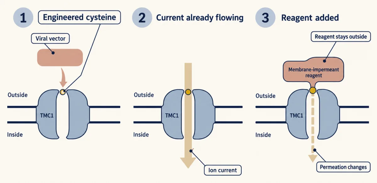
Pan and colleagues (2018) expressed cysteine-substituted TMC1 in Tmc1/Tmc2 double-null mouse cochlear hair cells. After current was established, an extracellular, membrane-impermeant cysteine-reactive reagent changed permeation and inhibited current in responsive constructs. This supports TMC1 as a pore-forming component; native partners remained present, and TMC1 was not tested alone. AI-generated illustration -
Without the hair cell: purified and transferred proteins
-
A pore inside a complex: TMIE, LHFPL5, CIB2 and a worm structure
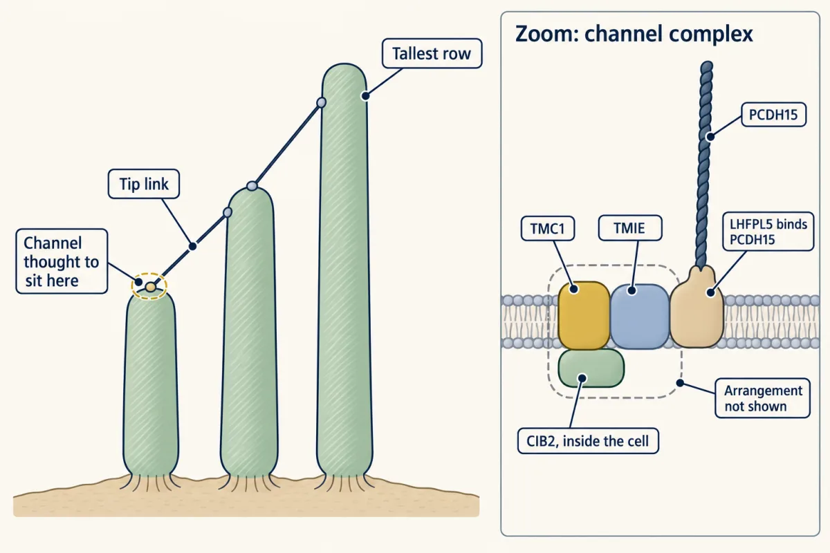
The transduction channel is thought to sit at the lower end of a tip link. Mouse cochlear studies implicate TMC1, TMIE, LHFPL5 and CIB2; LHFPL5 binds the tip-link protein PCDH15, and CIB2 is essential for TMC1/TMC2 localization. The component inventory shown here does not specify their native arrangement or how force reaches the pore. AI-generated illustration -
Adaptation: the current sags during a held push
-
What the ladder settles, and what it does not
-
Going deeper
Where this lesson fits
Before thisReview first
- Piezo Channels and the Physics of ForceThis course · Lesson 2
It compares candidate mechanically gated channels and the force-to-current logic that this lesson tests against the criteria for a pore.
- Five Ways to Turn a Stimulus into a CurrentThis course · Lesson 1
It introduces the current through a sensory cell and how a stimulus changes channel opening, which the I = N × i × P_open reading builds on.
After thisStudy next
- The Cochlear Amplifier and Otoacoustic EmissionsThis course · Lesson 23
It takes hair-bundle mechanics further, using the transduction current here to drive prestin-based amplification and otoacoustic emissions.
- Hidden Hearing Loss, Gene Therapy and RegenerationThis course · Lesson 24
Gene therapy for deafness depends on knowing which complex members the transduction current needs, as shown here.
Elsewhere on the siteFilms, simulations and method films in other courses
- Where a tone peaksSimulation · Neuroscience
It shows how pushing a hair bundle opens channels, the basic response this lesson dissects.
- Nernst calculatorSimulation · Cell Signaling
It uses the Nernst equation, which you need to get the driving force for a unitary conductance.
Suggested by an AI model from the lessons themselves, then checked. How we use AI