Developmental Neuroscience · Skim version
The Growth Cone: The Tip That Steers the Axon
Read the full lesson → Skim the whole course →
The guiding idea
Actin at the front of a growth cone often streams backward even while the tip moves forward, and in the standard assay it turns toward a cue only 5 to 10% stronger on one side. How?
A growing axon reaches its partner because the growth cone at its tip senses guidance cues and steers toward or away from them.
From the film: The Tip That Reads a Difference
- A growth cone steers by sensing guidance cues that attract or repel it.
- In the standard assay, the difference in cue across the width of the growth cone gives the direction, not the overall amount.
- The tip has microtubules in its centre and actin at its edge, in filopodia and lamellipodia.
From the film: Engine and Clutch
- On a flat surface, the edge advances by the actin added at the front minus the actin carried back.
- Adhesion to the surface, the clutch, holds back part of the backward flow.
- Grip can speed the edge just by slowing the flow, without faster building.
From the film: A Small Difference, a Turn
- A turn sticks on the side where microtubules move in and build new axon.
- Microtubules can start a turn themselves, working through actin.
- Isolated growth cones can turn in a dish without a cell body, though some cues need protein made at the tip.
-
The layout
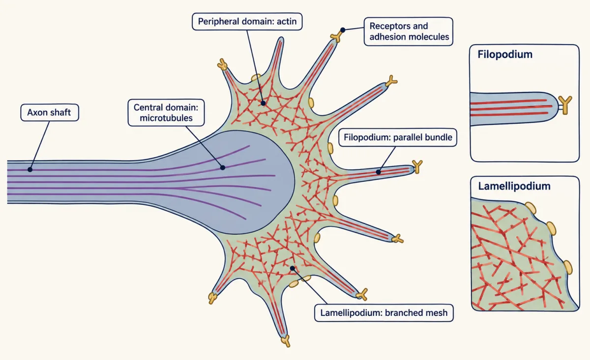
A growth cone in culture spreads into a flat fan at the end of the axon. Microtubules fill the central domain; actin fills the peripheral domain, bundled in the filopodia and branched in the lamellipodia between them, with receptors and adhesion molecules on the surface. AI-generated illustration -
Puzzle 1: how the edge advances
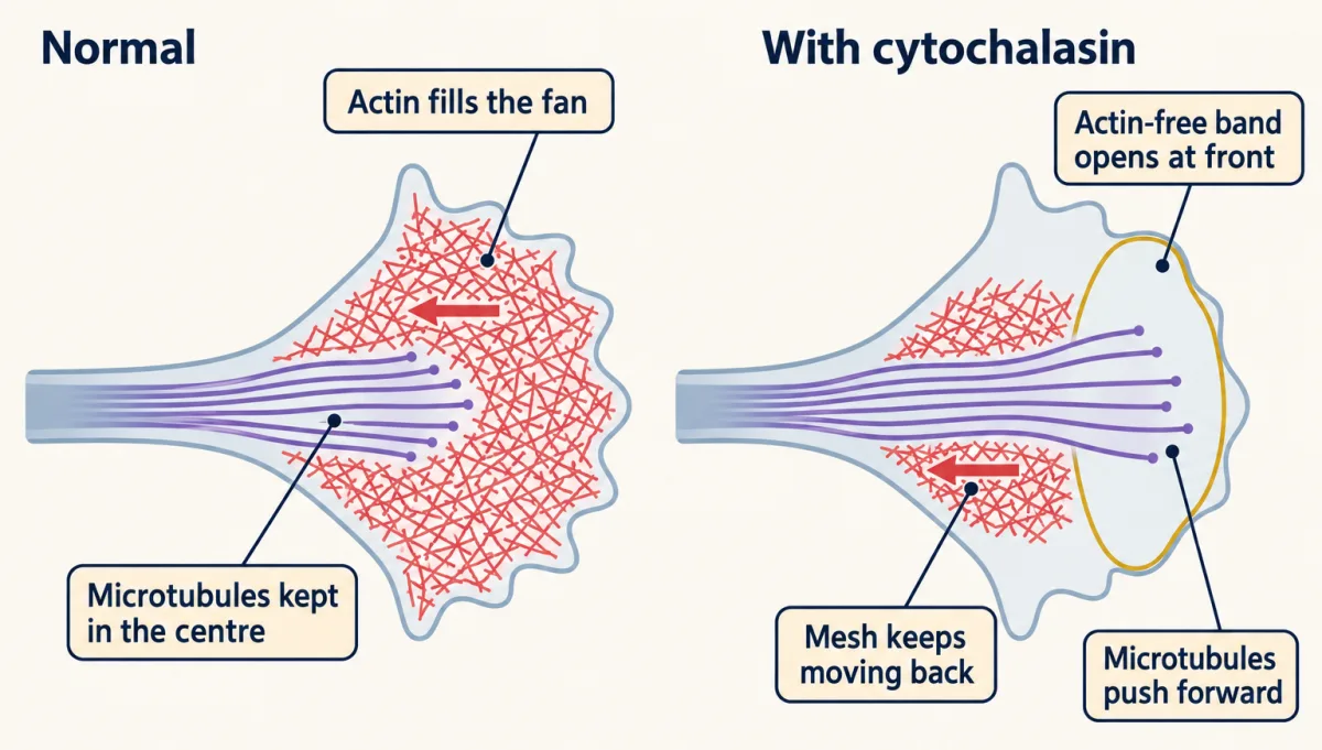
In Aplysia growth cones, cytochalasin stops actin subunits adding at the leading edge. An actin-free band opens at the front and widens while the remaining network keeps moving back, and microtubules push forward into the emptied band. AI-generated illustration -
Puzzle 2: how a small difference turns the axon
-
Puzzle 3: can the tip decide on its own?
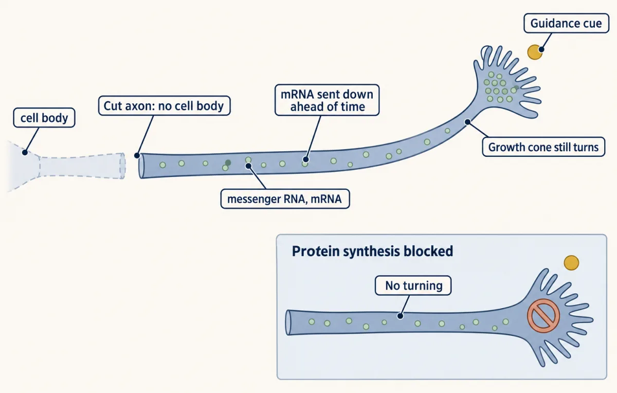
In Campbell and Holt’s 2001 assay, severed growth cones from Xenopus embryonic retinal neurons still turned in gradients of netrin-1 or Sema3A, but inhibiting local protein synthesis blocked those turning responses. The schematic shows the netrin-1 case. Growth cones contain messenger RNA supplied before severing and can translate it locally. This requirement depends on the cue and neuron: Roche and colleagues (2009) found that chick sensory growth cones still turned toward NGF when protein synthesis was blocked. AI-generated illustration -
Going deeper
Where this lesson fits
Before thisReview first
- Seeing Neurons: Golgi, Cajal and the Neuron DoctrineThis course · Lesson 2
Cajal's Golgi-stained fibres and the idea that neurons are separate cells set up the growth cone's discovery and Harrison's test here.
After thisStudy next
- Pioneers, Guideposts and the Logic of Guidance CuesThis course · Lesson 22
It asks which cues the growth cone reads and how pioneer axons use them, taking the steering idea into the embryo.
- Netrin and DCC: One Cue, Two AnswersThis course · Lesson 23
It shows how one cue attracts or repels depending on the receptor, the signalling step this lesson leaves open.
- Topographic Maps: Sperry's Eye and Eph GradientsThis course · Lesson 26
It uses the gradient-sensing growth cone to explain how axons find their place in an ordered map.
Elsewhere on the siteFilms, simulations and method films in other courses
- Tight Ends, Loose EndsFilm · Cell Biology
It explains how filament ends grow or shrink, which sets the actin and microtubule behaviour at the tip.
- How Internal Motion Becomes MovementFilm · Cell Biology
It shows polymerization minus backward flow at a leading edge, with integrin coupling cutting slip, as in the clutch.
- A Grip Switched On From InsideFilm · Cell Signaling
It shows how an integrin grip switched on from inside links the cell to its surface, as in the clutch.
Suggested by an AI model from the lessons themselves, then checked. How we use AI