Developmental Neuroscience · Skim version
Mapping the Cortex: Areas and Arealization
Read the full lesson → Skim the whole course →
The guiding idea
Add one secreted protein to the back of a mouse embryo's cortex and a second, partial whisker map can appear there.
Position draws the cortex's area sketch and input finishes it: in the mouse, graded signals such as FGF8 and transcription factors such as Emx2 help set where the primary-area borders fall.
From the film: A Sheet with No Borders
- The adult cortex has areas, but the embryonic sheet shows no borders for them.
- In the mouse, the sheet is already patterned and making neurons before thalamic axons arrive.
- Thalamic axons reach the developing mouse cortex around embryonic day 14 to 15, while neurons are still being made.
From the film: Two Answers, a Year Apart
- The protomap proposes that the outline of the areas is laid down in the progenitors; the protocortex proposes that it comes mainly from arriving thalamic axons.
- Both predictions came true in part: the map is drawn twice, sketched by the sheet and refined by its inputs.
- In newborn mice, areas are read through proxies such as marker genes, thalamic connections and the barrel field.
From the film: A Lamp at the Front
- In the mouse, extra FGF8 at the front moved the barrel field back, and blocking FGF signalling moved it forward.
- A new FGF8 source at the back gave a partial second barrel field.
- In the working model, a border falls where a graded level crosses a threshold, so changing the dose slides the borders.
From the film: The Cells' Own Record
- Position is recorded in progenitors as levels of transcription factors such as Emx2 and Pax6, which run in opposite directions front to back.
- Losing Emx2 moved the borders back, and raising it moved them the other way with total cortical area unchanged, so Emx2 allocates territory.
- Pax6 is the cautionary case: losing it shifted markers but raising it barely moved the map, so its role is unsettled.
From the film: Input Finishes the Drawing
- In mice whose thalamic axons never reach the cortex, marker borders were still normal at birth.
- Given new input, grafted visual cortex formed barrels and rewired ferret auditory cortex built an orientation map.
- Input also sharpens the sketch, as in the split between primary visual cortex and the higher-order visual areas.
From the film: One Rule, and Where It Stops
- Position draws the sketch; input finishes it.
- The strongest evidence for a factor is loss and gain moving borders in opposite directions; a missing converse result does not rule a factor out.
- The rule covers primary-area borders front to back in the mouse, not total cortical growth, the maps inside an area, or area sizes in primates or humans.
-
The layout
-
Two answers, and what each predicts
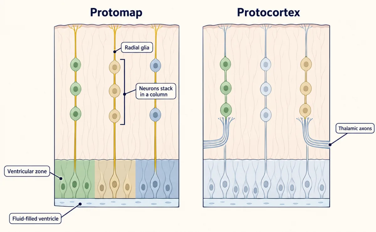
Two historical hypotheses for where cortical areas come from. Protomap: progenitors at each patch of the ventricular zone already differ, and their neurons stack in a column above them along radial glia. Protocortex: the young sheet is much alike and thalamic axons shape each part. Both were partly right; colours are schematic. AI-generated illustration -
A lamp at the front: FGF8
-
The cells’ own record of position
-
Input finishes the drawing
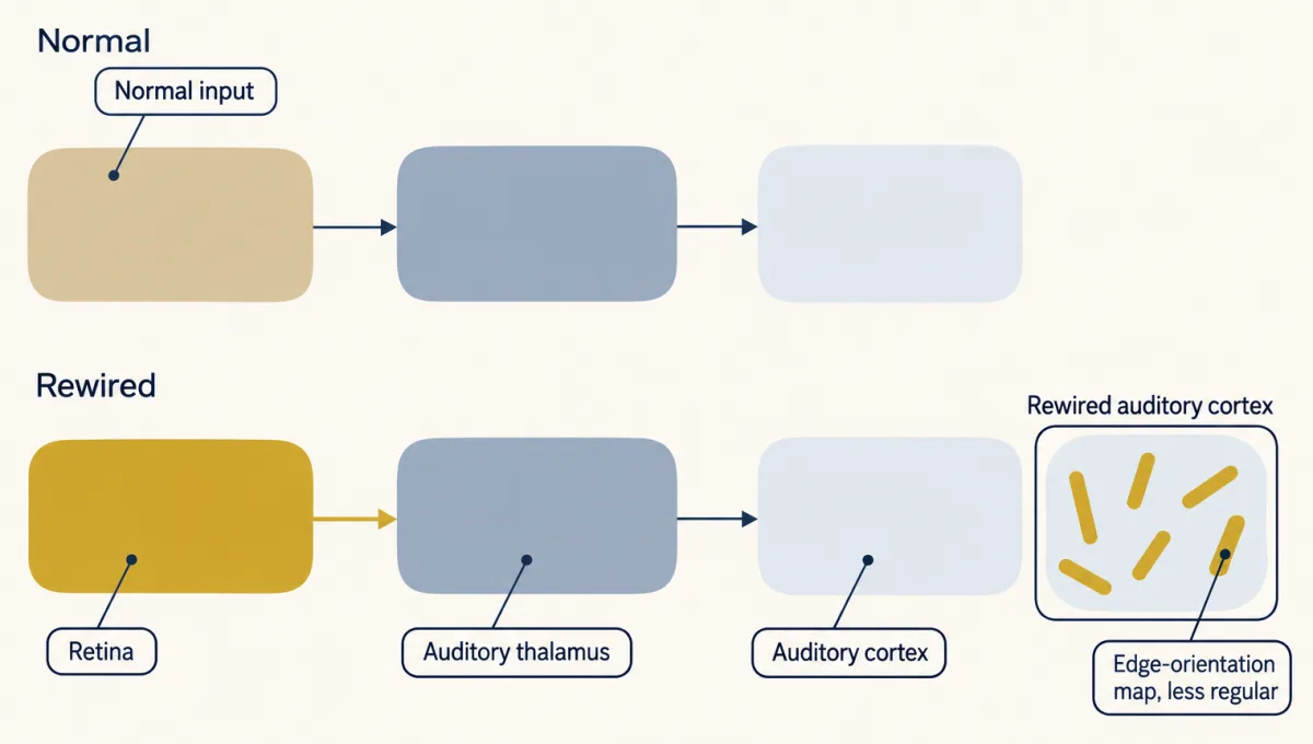
In ferrets rewired as newborns, retinal axons grew into the auditory thalamus and auditory cortex received visual activity. In adults its neurons formed groups sharing preferred edge orientations, in a map like visual cortex but less orderly. Pathway and tuning symbols are schematic; bars stand for stimulus preference, not cell shape. AI-generated illustration -
Putting it together
-
Going deeper
Where this lesson fits
Before thisReview first
- Ventral to Dorsal: Sonic Hedgehog and BMP in the Neural TubeThis course · Lesson 10
It teaches how a secreted signal's level sets position along an axis, the logic FGF8 and Emx2 gradients reuse here.
- Local Organizers: The Midbrain-Hindbrain BoundaryThis course · Lesson 9
It shows a localized FGF8 source acting as an organizer, which this lesson moves to the cortex's front edge.
- Transcription Factor Codes: Building a Motor NeuronThis course · Lesson 11
It shows transcription factor levels recording position and setting fate, as Emx2 and Pax6 do for areas.
After thisStudy next
- The Ventricular Zone: Radial Glia as Stem CellsThis course · Lesson 13
It covers the progenitors and radial glia whose columns carry the position record that sketches the area map.
- Birth Order and Fate: Temporal Identity and the Inside-Out CortexThis course · Lesson 16
It extends how cortical progenitors make neurons over time, which adds a timing axis to the area map.
- Topographic Maps: Sperry's Eye and Eph GradientsThis course · Lesson 26
It takes up map formation by gradients of graded cues, here for axon targets in a map inside an area.
Elsewhere on the siteFilms, simulations and method films in other courses
- Where is the lesion?Simulation · Neuroscience
It shows how one primary visual area represents the visual field, which is the kind of area this lesson maps.
Suggested by an AI model from the lessons themselves, then checked. How we use AI